Method For Rapid Detection And Evaluation Of Cultured Cell Growth

Inactive Publication Date: 2012-03-08
WILSON DAVID F
View PDF2 Cites 1 Cited by
  • Summary
  • Abstract
  • Description
  • Claims
  • Application Information

AI Technical Summary

Benefits of technology

[0013]The present invention provides a new, enhanced method for readily sampling individual colonies of growing cells for further analysis. The method comprises immobilizing a population of cells of at least one microorganism in an aqueous gelling medium comprising a dissolved oxygen-quenchable phosphorescent compound that is no

Problems solved by technology

Growth in liquid culture, however, is less useful for rapid identification of a slow-growing microorganism, particularly if there is a mixture of microorganisms and one seeks to identify individual clonal colonies.
The longer a microorganism is cultured, the greater the risk of contamination, usually by a fast-growing bacteria, yeast or fungus.
Fast-growing microorganisms tend to out-compete slow-growing microorganisms and overgrow the culture, obscuring the slow-growing microorganisms.
Furthermore, in such a mixture of microorganisms, one can no longer identify and select individual cells for isolated growth.
Plating slow-growing microorganisms, however, still requires long incubation periods to detect growth by conventional means, increasing the risk of contamination over time and increased handling.
However, the detection method of the '214 patent utilize a redox indicator in the medium, meaning that the method is not commercially practical because the amount of redox indicator that is required to demonstrate growth of the microorganism may also be toxic to the cells, and / or such methods require an inordinate amount of care to avoid toxicity and prevent false negative results.
Furthermore, like all other prior art methods, when the '214 method is used to detect the growth of slow-growing cells in culture, the cells require several weeks in culture before microbial cell growth is demonstrated.
To date, with the exception of the inventor's own work, none of the available detection methods provide rapid and reliable detection of cell growth in culture in a matter of only a day or two.
However, the typical volume of liquid culture media used to grow the cells in standard plates or vessels in the '741 patent requires the use of substantial volumes of reagent and marker materials, and while more rapid than other methods, there is a need to further decrease the times needed to provide reliable readings.
Moreover, because liquid culture techniques are used, the '741 patent does not permit the rapid identification and isolation of individual clonal colonies.
However, the complexity of this method and the requirement of using specialized equipment, including an oxygen microsensor, makes it unsuitable for rapid growth detection purposes.
However, this method suffers from the same drawbacks as the other prior art methods.
Although a fluorescent indicator is used, because the cells are not immobilized, the disclosed methods cannot provide a means for isolating an individual cell or colony from the culture media.

Method used


Image

  • Method For Rapid Detection And Evaluation Of Cultured Cell Growth
  • Method For Rapid Detection And Evaluation Of Cultured Cell Growth
  • Method For Rapid Detection And Evaluation Of Cultured Cell Growth

Examples


example 1

[0120]Toxicity evaluation for individual growth or metabolism indicators in accordance with the invention was conveniently carried out as follows. Phosphor powder, Pd-meso-tetra (4-carboxyphenyl) porphyrin with two layers of glutamate dendrimer, was dissolved in five milliliters of distilled, deionized and filter-sterilized water and filtered through an 0.2 micron filter to provide a filter-sterilized solution with a concentration of 8 mM and a pH of 7.4. Three dilutions were made from the 8 mM solution to create stock solutions, such that an equal volume of each stock solution was used to dilute to the final concentration in the culture medium. The final concentrations tested were 4, 8 and 16 μM, respectively. Controls were supplied with the same volume of sterile water in lieu of a phosphor dilution.

[0121]Each of final phosphor dilution (1:500, 1:1000 and 1:2000, respectively) was prepared in duplicate. The paired volumes were inoculated with two different concentrations of Mycoba...

example 2

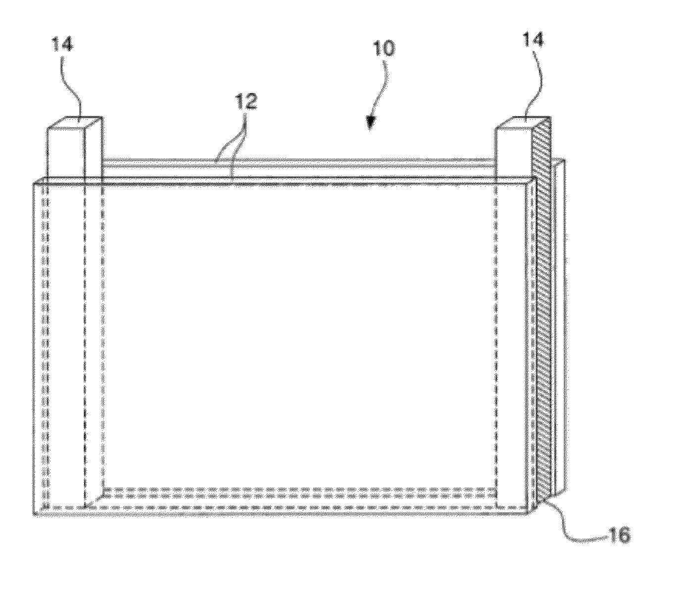
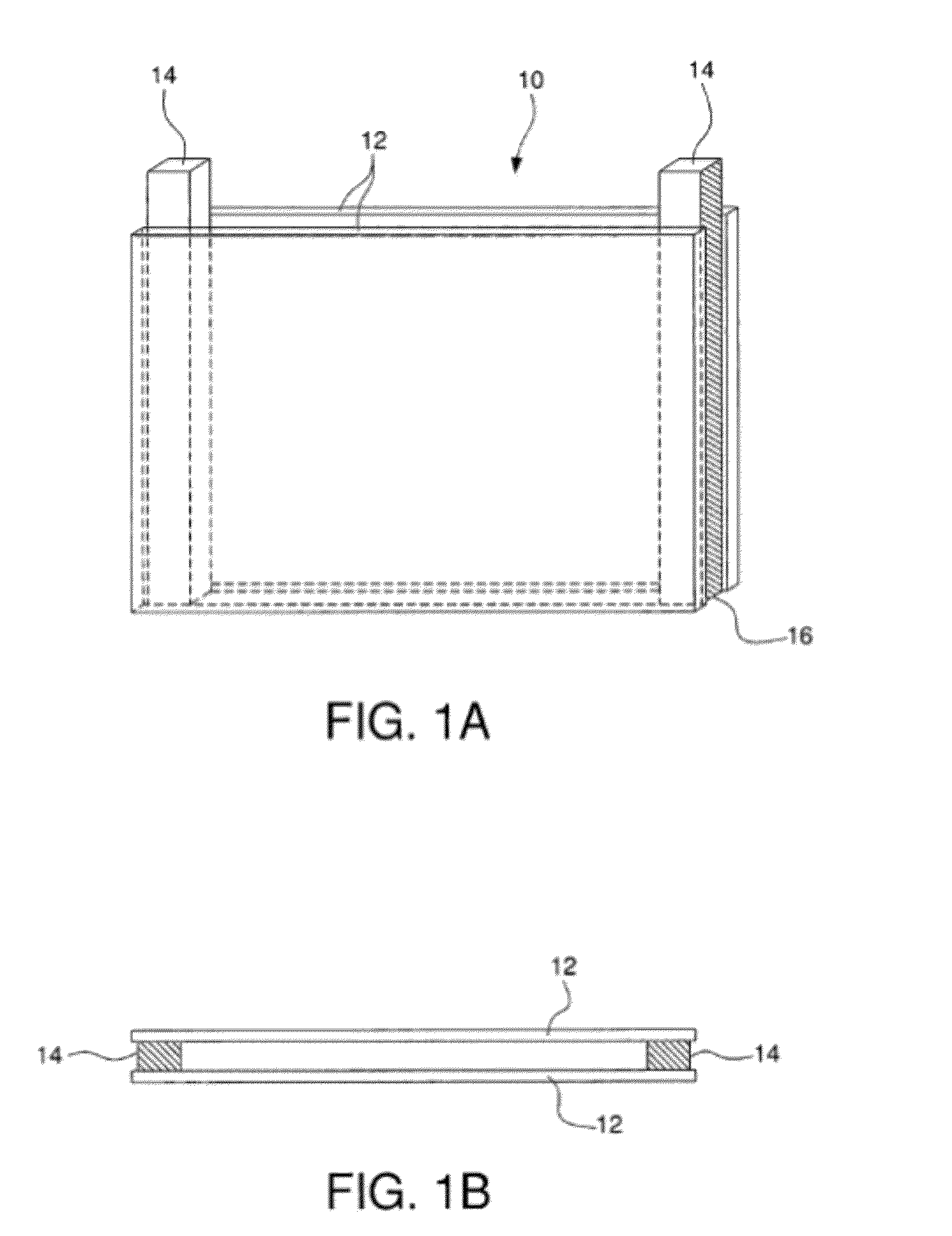
[0123]A slab gel hollow form was assembled using two 3 inch by 4 inch (7.6 cm×10 cm) glass plates separated by 0.5 mm thick spacers to form a rectangular space. Agarose was added to a final concentration of 1% agarose (wt / vol) to liquid culture medium (pH 7.2) containing physiological saline, casein hydrolysate and glucose. The mixture was heated to near boiling for about 30 minutes to make the gelling culture medium. The gelling culture medium was allowed to cool to about 40° C., then phosphor and bovine serum albumin (BSA) was added. The phosphor, Oxyphor G2, a Pd-tetra (4-carboxyphenyl) tetrabenzoporphyrin dendrimer (Dunphy et al., Anal. Biochem. 310:191-198 (2002)), was added to a final concentration of 2 micromolar. The BSA, which was not sterilized, was added to a final concentration of 1% (wt / vol).

[0124]The phosphors useful in the instant invention generally are not heat labile. However, Oxyphor G2 requires BSA, which is heat labile, for binding to give the phosphor a quenchi...

example 3

[0128]In contrast to the exemplified system above, providing a rapid method for measuring cells already patented in a thin, flat culture of cells suspended in a gel-based growth medium between two plates of a culture chamber, an alternative chamber was developed in which one of the two plates was constructed with holes or grooves in a predetermined pattern. See, FIG. 5. The plate having an array of holes or grooves forming the one side of the chamber was formed with small holes or grooves, preferably between 0.2 and 1.5 mm in depth and width, although both larger and smaller well sizes may be used. When properly constructed, such an array of wells consists of an array of, for example, 150×150 wells. This provided a total of 22,500 wells placed in a precisely determined geometry. For wells with a 0.5 mm×0.5 mm×0.5 mm dimensions, this fits on an approximately 10 cm by 10 cm square sample chamber.

[0129]Construction can be of any number of suitable materials, including those stated abov...


PUM

PropertyMeasurementUnit
Volumeaaaaaaaaaa
Depthaaaaaaaaaa
Antimicrobial propertiesaaaaaaaaaa

Abstract

Provided is a method and growth chamber for the rapid and accurate detection of growth and metabolism of a cellular microorganism in isolation within one or a plurality of wells containing a population of microorganisms in a non-liquid, culture medium, wherein the cells are distributed at not greater than one cell per well at plating. Further provided is a gelled culture medium containing a non-toxic, water-soluble, phosphorescent compound which measures oxygen content (partial oxygen pressure) of a microorganism also contained therein, by oxygen-dependent quenching of phosphorescence; or the gel contains a fluorescent pH indicator that demonstrates growth of the microorganism by pH-dependent intensity change or wavelength shift in the emission spectrum.

Description

CROSS REFERENCE TO RELATED APPLICATIONS[0001]This application is a Continuation-in-Part of U.S. patent application Ser. No. 12 / 504,346, filed on Jul. 16, 2009, which is a Divisional Application of U.S. patent application Ser. No. 11 / 334,236, filed on Jan. 18, 2006, now, U.S. Pat. No. 7,575,890, issued Aug. 18, 2009, which is incorporated herein in its entirety.FIELD OF THE INVENTION[0002]The present invention provides a method for rapidly detecting and monitoring growth of cells immobilized within a gelled culture material using oxygen-quenchable phosphorescent compounds or fluorescent pH indicators.BACKGROUND OF THE INVENTION[0003]Growth characteristics vary widely from one cell or microorganism to another. For example, it has been estimated that relatively-rapidly growing Mycobacteria require approximately one week to demonstrate growth, whereas relatively more slowly-growing tuberculosis agents, such as M. tuberculosis, M. bovis and M. avium, which are also known to appear in AID...

Claims


Application Information

Patent Timeline
IPC IPC(8): C40B30/06C40B60/12
CPCA61K31/407G01N33/84A61K31/555
Inventor WILSON, DAVID F.
Owner WILSON DAVID F
Why Eureka
Try Eureka