Ultrasound patch

a technology of ultrasound patch and wearable embodiment, which is applied in the field of wearable ultrasound patch, can solve the problems of non-intrusiveness and inability to wear wearable embodiments

Inactive Publication Date: 2012-03-15
GARY & MARY WEST HEALTH INST
View PDF11 Cites 115 Cited by
  • Summary
  • Abstract
  • Description
  • Claims
  • Application Information

AI Technical Summary

Benefits of technology

[0010]The adhesive ultrasound patch, placed appropriately on a subject, enables continuous monitoring of the heartbeat without interfering with the subject's routine activities. Another example is nonintrusive monitoring of patients with known at-risk lesions in the carotid artery prior to, or post-heart surgery by placing the patch on the neck. Yet another application is for detection of the on-set of a stroke with electronic spatial scanning techniques while the patch is attached to the neck to monitor the lesions in the carotid artery. Another application is for continuous fetus heartbeat monitoring (especially for high risk pregnancies) when the patch is adhered to mother's belly. In addition, equipping the patch with dry electrodes enables recording the EMG signals associated with uterine contraction to enable synchronous monitoring of fetal heart rate and uterine contraction [23]. Therapeutic and rehabilitation examples would be placement of the patch over target nerves or painful joints (rheumatoid arthritis) for stimulation using the incident ultrasound wave energy. These measurements and diagnostics are enabled by using one or more ultrasound transducers. Transducer array architectures are often desired because they provide redundancy, ensuring functionality even if a subset of the transducer elements fails.

Problems solved by technology

While ultrasound techniques are utilized in modern ultrasound devices [12], [18] but a nonintrusive, wearable embodiment is not available.

Method used


Image

  • Ultrasound patch
  • Ultrasound patch
  • Ultrasound patch

Examples


Embodiment Construction

[0019]Common issues with ultrasound devices are the larger the size, the higher the power consumption and cost. In addition to the miniaturized size and ease of use of the proposed patch, the applications that this patch address are compelling in the sense that they do not necessarily require a high-resolution image, but rather characterizing the conditions of the tissue or medium under investigation or just providing ultrasound energy for therapeutic, rehabilitation, and assisting in the healing process.

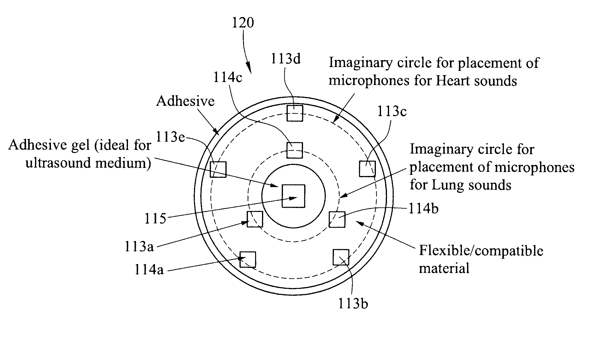
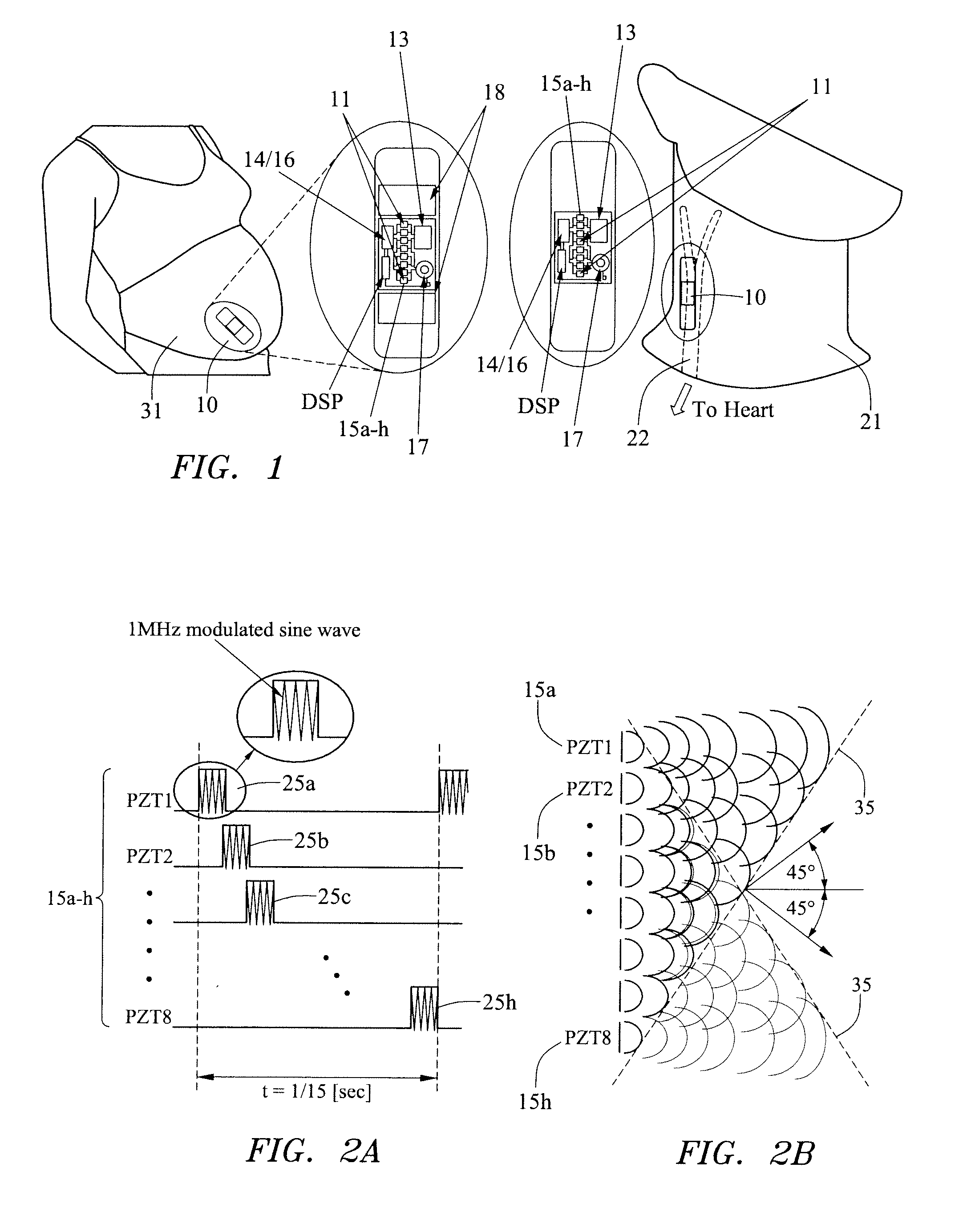
[0020]In FIG. 1, an adhesive-patch embodiment is shown. The ultrasound device 10 is placed on a designated location and held in place tightly through a special bio-compatible adhesive. Strategic use of the adhesive optionally eliminates the need for application of a gel. For example, in case of monitoring arteries (FIG. 1, right hand side), continuous-wave Doppler ultrasound uses a processing technique to measure the speed and direction of blood flow in the monitored area. It first ...


PUM


Abstract

A wearable patch is provided for use on the body which preferably comprises an ultrasound sensor array, a transmission system coupled to the ultrasound sensor array adapted to provide signal information for ultrasound transmission into the body, and a receiver system coupled to the ultrasound sensor array adapted to receive signal information from the reflected ultrasound signal received from the body. A control circuitry is coupled to the transmission system and the receiver system. The patch is preferably provided with a wireless communication system to permit external control and or communication. Applications range from diagnostics and monitoring, to rehabilitation and wound healing.

Description

RELATED APPLICATION STATEMENT[0001]This application claims priority to and benefit of U.S. Provisional Application Ser. No. 61 / 327,875, filed Apr. 26, 2010, entitled “Ultrasound Patch”, the content of which is hereby incorporated by reference in its entirety as if fully set forth herein.FIELD OF THE INVENTION[0002]The present invention relates to methods, apparatus and systems for providing a wearable ultrasound solution in form of a BandAid-type patch for use on a body. More particularly, it relates to a wearable ultrasound patch that may be used for diagnosis, monitoring and rehabilitation of various organs and parts of the body.BACKGROUND OF THE INVENTION[0003]Ultrasound waves are utilized in many different fields, typically as a tool to penetrate a medium to measure its reflection signature. In medicine, imaging ultrasound devices are usually used for diagnostic medical imaging of internal organs, muscles, tendons and even fetus inside the uterus [1]. Other existing medical appl...

Claims


Application Information

Patent Timeline
Patent Type & Authority Applications(United States)
IPC IPC(8): A61B8/00A61B5/11A61B5/01A61N7/00
CPCA61B5/02411A61B8/565A61B5/4325A61B5/4362A61B8/02A61B8/0866A61B8/0883A61B8/0891A61B8/4236A61B8/4427A61B8/4472A61B2560/0412A61N7/00A61B8/06A61B8/488A61B8/5207A61B5/04882A61B5/391
Inventor LAHIJI, ROSA R.ROHAM, MASOUDMEHREGANY, MEHRAN
Owner GARY & MARY WEST HEALTH INST
Why Eureka
Try Eureka