Microfluidic Isolation of Tumor Cells or Other Rare Cells from Whole Blood or Other Liquids

a microfluidic and tumor cell technology, applied in the field of tumor cell or other rare cell isolation from whole blood or other liquids, can solve the problem of side walls being exposed to ultraviolet radiation

Active Publication Date: 2012-04-26
BOARD OF SUPERVISORS OF LOUISIANA STATE UNIV & AGRI & MECHANICAL COLLEGE
View PDF6 Cites 30 Cited by
  • Summary
  • Abstract
  • Description
  • Claims
  • Application Information

AI Technical Summary

Benefits of technology

[0050]We also evaluated shear forces on captured cells, to determine whether flow-induced shear could either detach the cells from the walls or damage the cells. Using a simple model of the forces involved, we calculated that the linear flow velocity required to detach the EpCAM-expressing cells from a PMMA wall decorated with anti-EpCAM antibodies would be on the order of 102 to 104 cm s−1, depending on the degree to which

Problems solved by technology

We note that the side walls of the channels can be exposed with a conventional mask and UV irradiation, because the light i

Method used


Image

  • Microfluidic Isolation of Tumor Cells or Other Rare Cells from Whole Blood or Other Liquids
  • Microfluidic Isolation of Tumor Cells or Other Rare Cells from Whole Blood or Other Liquids
  • Microfluidic Isolation of Tumor Cells or Other Rare Cells from Whole Blood or Other Liquids

Examples


examples 1-21

Fabrication of Prototype HTMSU, and Isolation and Detection of Low Abundance MCF-7 Breast Cancer Cells in Whole Blood

Materials and Methods

example 1

HTMSU Fabrication

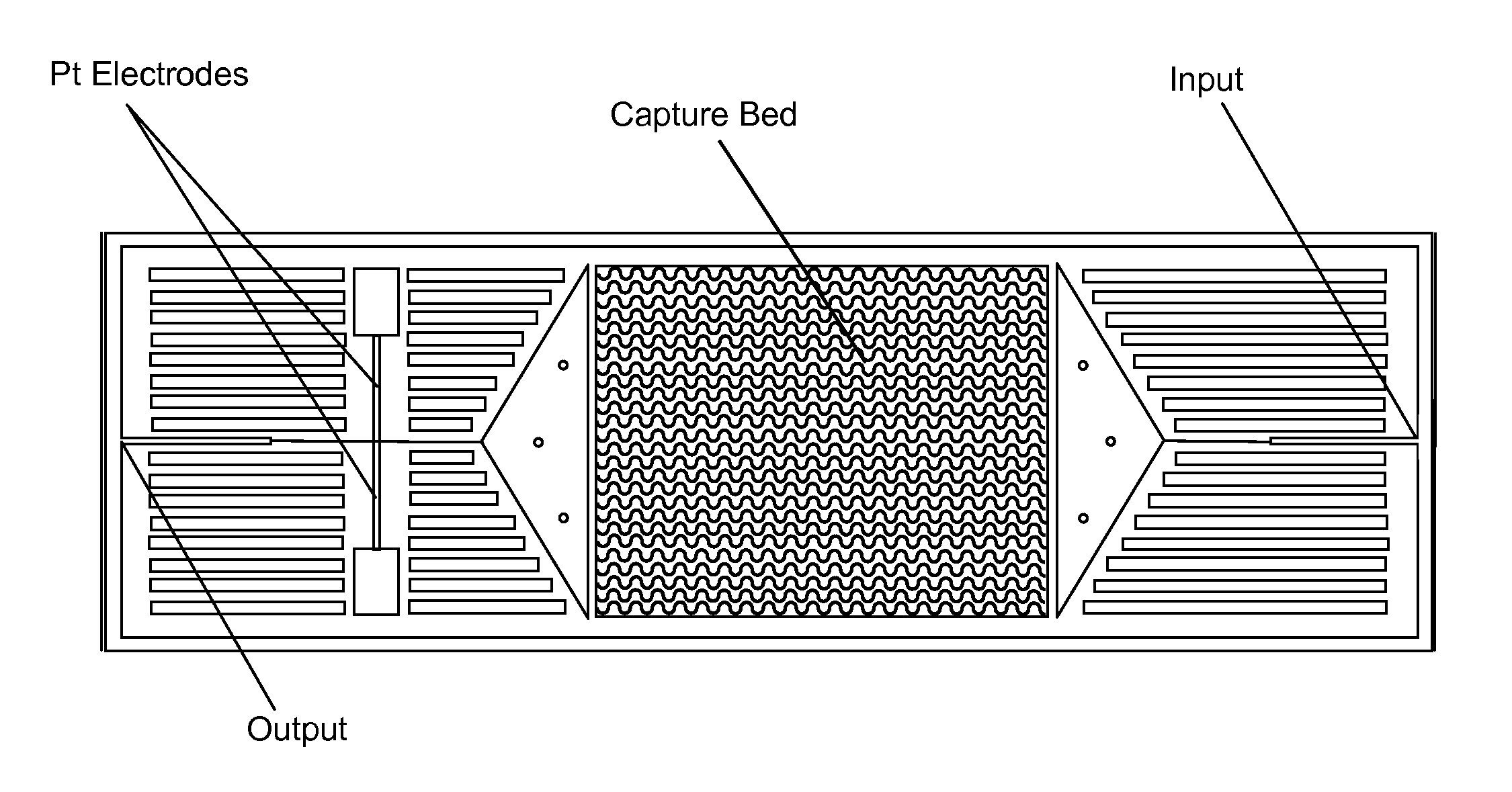
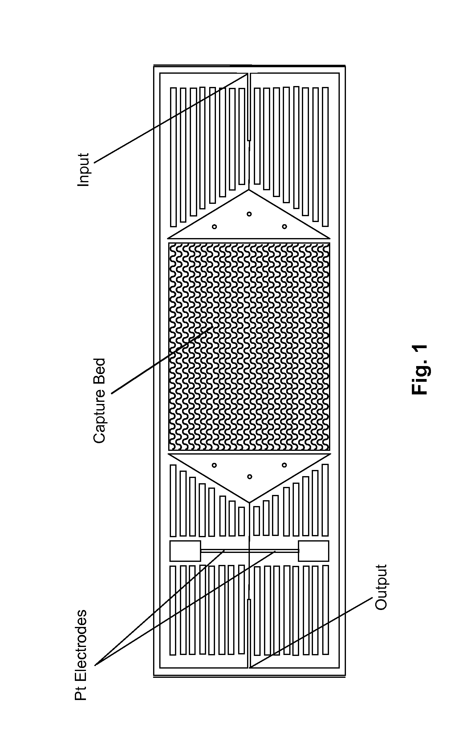
[0029]FIG. 1 depicts schematically a prototype HTMSU in accordance with the present invention. The prototype HTMSU has been successfully fabricated and tested. The prototype device contained 51 high-aspect-ratio, sinusoidal, parallel channels that shared a common input port and a common output port. Devices were replicated from a master mold using hot embossing techniques that are otherwise known in the art. See A. Adams et al., “Highly efficient circulating tumor cell isolation from whole blood and label-free enumeration using polymer-based microfluidics with an integrated conductivity sensor,”J. Am. Chem. Soc., vol. 130, pp. 8633-8641 (2008), and the supporting material that is available at pubs.acs.org for additional details concerning the fabrication of the master mold and the micro-replication procedures, the complete disclosures of which are incorporated by reference. The substrate used in the prototype HTMSU was poly(methyl methacrylate) (PMMA), which was cho...

example 2

Antibody Immobilization

[0030]Antibodies were immobilized in a two-step process. First, the UV-surface-modified HTMSU device was loaded with a solution containing 4.0 mg / mL of 1-ethyl-3-[3-dimethylaminopropyl]carbodiimide hydrochloride (EDC), 6.0 mg / mL of N-hydroxysuccinimide (NHS) in 150 mM 2-(4-morpholino)-ethane sulfonic acid at pH=6 (MES, Fisher Biotech, Fair Lawn, N.J.); and buffered saline (Sigma-Aldrich, St. Louis, Mo.) for 1.0 hr to form a succinimidyl ester intermediate. The EDC / NHS solution within the device was then hydrodynamically replaced with a 1.0 mg / mL solution of monoclonal anti-EpCAM antibody (R&D Systems Inc., Minneapolis, Minn.) in 150 mM PBS at pH=7.4 (Sigma-Aldrich, St Louis, Mo.), which was allowed to react for 4 hours. The device was then rinsed with a solution of PBS (pH=7.4) to remove any unbound anti-EpCAM antibodies. For additional details concerning these procedures, see the Adams et al. Supporting Information (2008), hereby incorporated by reference.


PUM

PropertyMeasurementUnit
Speedaaaaaaaaaa
Electrical conductivityaaaaaaaaaa
Flow rateaaaaaaaaaa

Abstract

Microdevices are disclosed to efficiently, accurately, and rapidly isolate and enumerate rare cells, such as circulating tumor cells, from liquids such as whole blood. The system employs multiple parallel meandering channels having a width on the order of 1-2 cell diameters. The microdevices can be produced at low-cost, may readily be automated, and in many instances may be used without pre-processing of the sample. They may be used to isolate and enumerate rare cells, including for example the detection and diagnosis of cancers, cancer staging, or evaluating the effectiveness of a therapeutic intervention, or detecting pathogenic bacteria. The device may optionally be used to nondestructively capture and later to release target cells.

Description

[0001]The benefit of the 16 May 2008 filing date of U.S. provisional patent application Ser. No. 61 / 053,727 is claimed under 35 U.S.C. §119(e) in the United States, and is claimed under applicable treaties and conventions in all countries.[0002]The development of this invention was partially funded by the United States Government under grant 1R33-CA099246-01 awarded by the National Cancer Institute of the National Institutes of Health; and also under grant EPS-0346411 awarded by the National Science Foundation. The United States Government has certain rights in this invention.TECHNICAL FIELD[0003]This invention pertains to the isolation of tumor cells or other rare cells from whole blood or other liquids.BACKGROUND ART[0004]There is an unfilled need for improved techniques to isolate and detect circulating tumor cells and other rare cells.[0005]Most cancer-related mortalities result from metastasis. Cancer cells can be transported from the primary tumor by the circulatory system or ...

Claims


Application Information

Patent Timeline
IPC IPC(8): C12N5/078B01L99/00C12Q1/06C12M1/34C12N5/09
CPCG01N33/49B01L3/5027G01N33/4915B01L2300/0645B01L2300/0816B01L2300/0864B01L2300/0883G01N2015/1006G01N15/1459G01N15/1484A61P35/00
Inventor SOPER, STEVEN A.MURPHY, MICHAEL C.FENG, JUNEMCCARLEY, ROBIN L.ADAMS, ANDRE A.
Owner BOARD OF SUPERVISORS OF LOUISIANA STATE UNIV & AGRI & MECHANICAL COLLEGE
Why Eureka
Try Eureka