Ultra sensitive method for in situ detection of nucleic acids

a nucleic acid analyte and ultra-sensitive technology, applied in the field of nucleic acid chemistry and biochemical assays, can solve the problems of poor access to the target sequence of the detection probe, difficult application of ish techniques in clinical settings, and inability to detect nucleic acids in situ, etc., to achieve high detection accuracy, low background, and high signal intensity

Active Publication Date: 2012-04-26
ADVANCED CELL DIAGNOSTICS INC
View PDF3 Cites 62 Cited by
  • Summary
  • Abstract
  • Description
  • Claims
  • Application Information

AI Technical Summary

Benefits of technology

[0013]The present invention combines the signal amplification method of RNAscope® with the general ISH signal amplification methods into a ultra sensitive method to detect, quantify and identify one or more target nucleic acid in a sample. The RNAscope® assay offers exceptional specificity owning to its unique paired capture probe design. By adding general ISH signal amplification methods such as the TSA-based signal amplification to the RNAscope® assay, the combined method achieves great signal intensity and low background. The signal amplification methods and system disclosed in this invention are relatively simple to use and produce consistent result. The claimed method can be easily adapted for use in routine clinic diagnostic procedures.
[0014]In a preferred embodiment of the present invention, the specificity and sensitivity of the disclosed signal amplification method are balanced. The balance is achieved by a moderate reduction of the fold of signal amplification in RNAscope®. In one embodiment of the present invention, one preamplifer is designed to bind between 1 to 16 amplifiers. In a preferred embodiment, one preamplifer is designed to bind between 2 to 10 amplifiers. In another more preferred embodiment, one preamplifer is designed to bind between 2 to 5 amplifiers.

Problems solved by technology

However, the application of ISH techniques can be limited by their inability to detect DNA or RNA targets with low copy numbers in cells due to lack of sensitivity and specificity.
It is especially challenging to apply ISH into the clinical setting where formalin fixation and paraffin embedding (FFPE) is the most commonly used method worldwide to store clinical tissue samples.
The FFPE method preserves tissue morphology well but the cross-linking fixation by formaldehyde can cause poor accessibility of the target sequence to the detection probe, chemical or physical interaction of the probe molecules with other molecules or structures, and damage to nuclear acids, especially mRNAs.
However, RNAscope® alone still cannot reliably detect some low copy genes in retrospective Formalin-Fixed, Paraffin-Embedded (FFPE) tissue sections where RNA is significantly degraded.
Also, single RNA molecule cannot to be visualized at 40× magnification using current RNAscope® technology.
All of these signal amplification methods have been used in ISH with different degrees of success but usually the signal intensity and / or signal to noise ratio are not good enough for routine diagnostic pathology.
In the case of ISA, in particular, when used alone in ISH, produces a high degree of background noise.
The day-to-day and laboratory-to-laboratory consistency is also less than desirable.

Method used


Image

  • Ultra sensitive method for in situ detection of nucleic acids
  • Ultra sensitive method for in situ detection of nucleic acids
  • Ultra sensitive method for in situ detection of nucleic acids

Examples


example 1

RNAscope®+General ISH Signal Amplification Assay

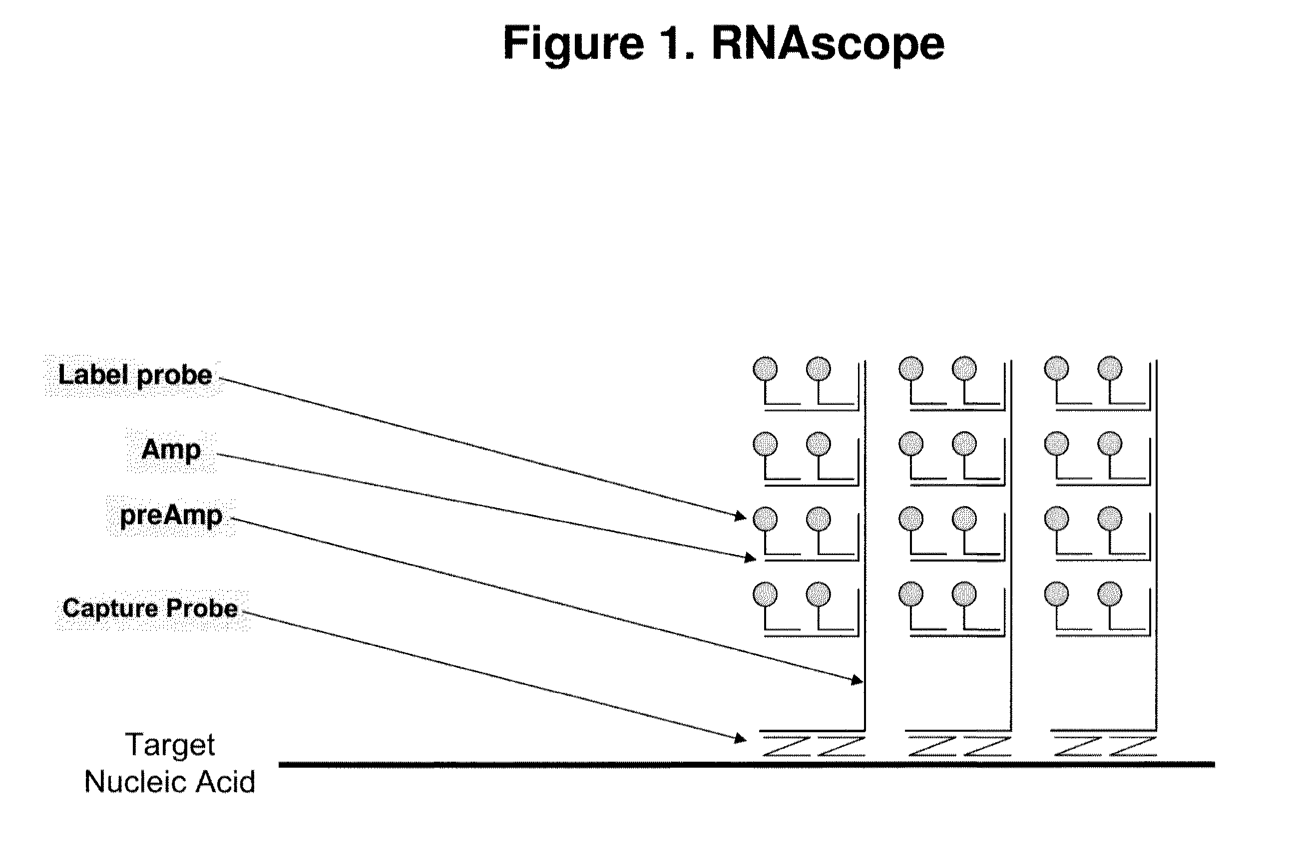
[0093]The basic assay procedure can be done within a day and generally includes the following steps. A sample containing a plurality of cells suspected of comprising a target nucleic acid HPRT1 is provided. After being fixed and permeablized, cells either fixed on a solid support or in suspension are hybridized to the following series of oligonucleotide probes. First, a set of capture probes is hybridized to the target RNA inside the cells. Next, preamplifier molecules are hybridized to the capture probes, providing a bridge for the hybridization of amplifier molecules. Multiple amplifier molecules are further hybridized to a preamplifier, e.g., up to 20 amplifiers to each preamplifier. Multiple label probes are then hybridized to an amplifier, e.g. up to 20 label probes to each amplifier.

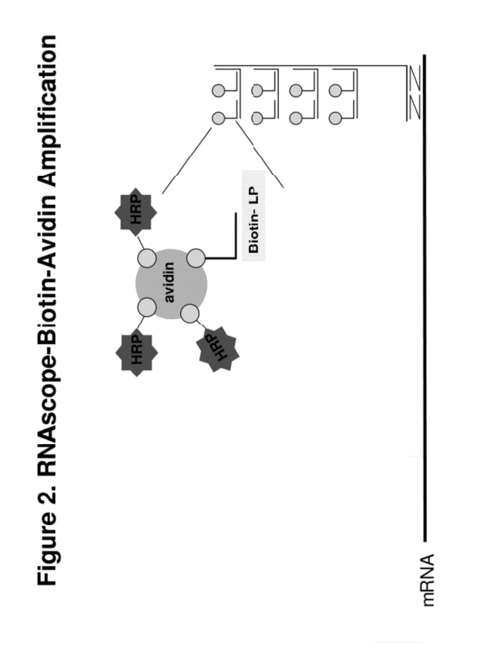
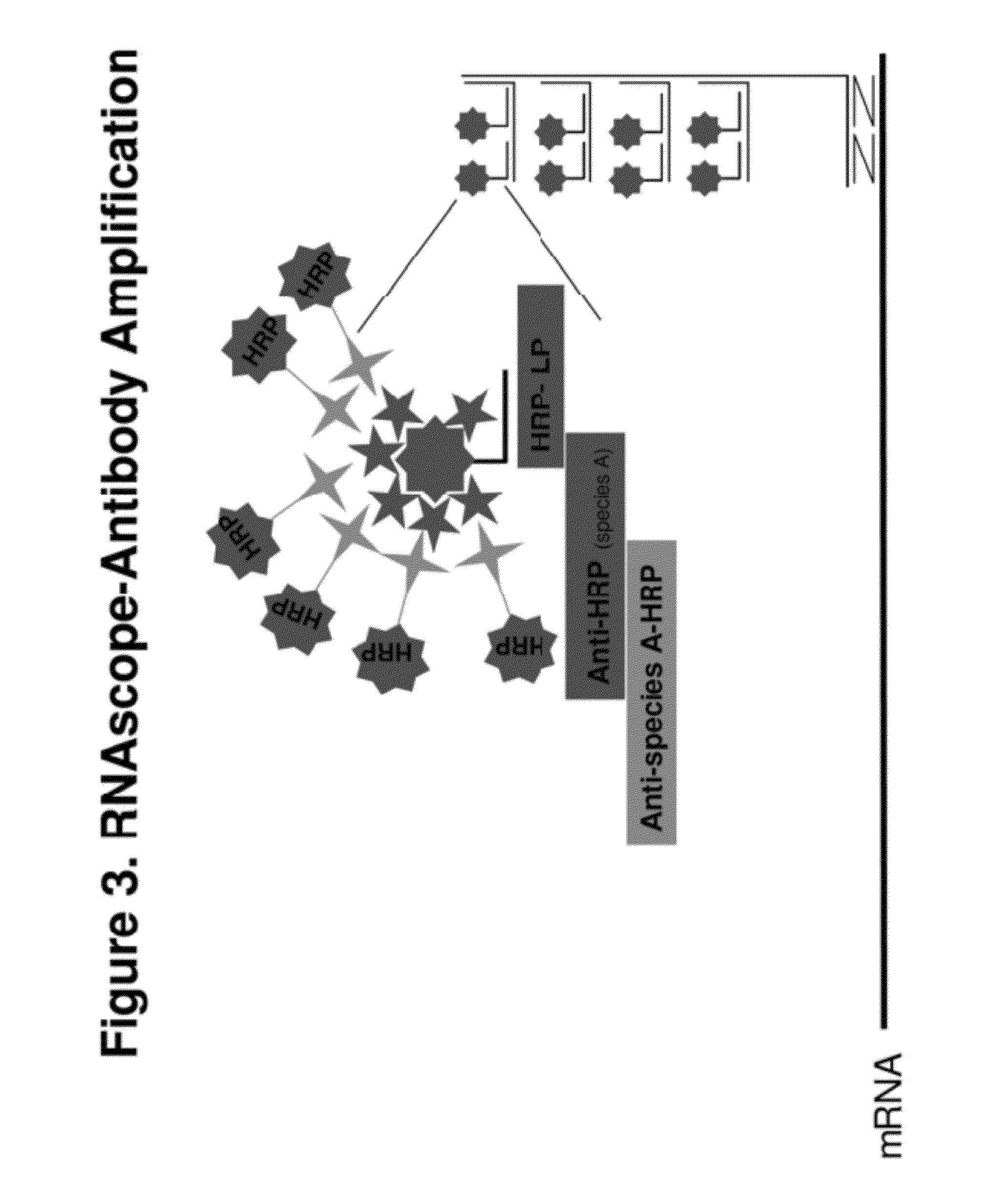
[0094]The signal intensity is enhanced further by TSA-based ISH signal amplification. An HRP is conjugated to each label probe. Reagent containing ty...

example 2

Multiplex RNAscope®+General ISH Signal Amplification Assay

[0099]The following prophetic example of a multiplexed amplification showing how the method disclosed in the present invention can detect two target nucleic acids with high sensitivity. To explore the potential of the disclosed invention for in situ detection of low copy RNA transcripts and its capability for multiplex detection, 18S and Her-2 are used as the model genes.

[0100]In the first amplification, 18S gene is captured on capture probes designed for 18S. The capture probe for 18S is designed as described in example 1. The capture-probe-hybridized-18S gene is then sequentially hybridized to preamplifiers, amplifiers and label probes. The label probe is then conjugated to HRP. The conjugated LP-HRP complexes are then detected with sequential incubations of tyramide-biotin, streptavidin-HRP and DAB. The product of the hybridization produces brown color at locations where target gene 18Ss are detected.

[0101]Before starting ...


PUM

PropertyMeasurementUnit
Fluorescent in situ hybridizationaaaaaaaaaa
tissue morphologyaaaaaaaaaa
molecular structureaaaaaaaaaa

Abstract

Disclosed is a method for in situ detection of one or more target nucleic acids based on a combination of RNAscope® method and a general ISH signal amplification method. This new method produces high signal intensity and while keeps low background noise of signal amplification. The result can be consistently reproduced and the method can be easily adopted for routine clinic diagnostic use. Further, the invention relates to a kit, comprising the components of RNAscope® assay and a general ISH signal amplification assay, for sensitive detection of one or more target nucleic acids.

Description

CROSS-REFERENCE TO RELATED APPLICATIONS[0001]This application claims priority to and benefit of U.S. provisional patent application No. 61 / 405,503, filed on Oct. 21, 2010, with attorney docket no. 12790-021-888, entitled “ULTRA SENSITIVE METHOD FOR IN SITU DETECTION OF NUCLEIC ACIDS” by Wu et al., and U.S. provisional patent application No. 61 / 412,276, filed on Nov. 10, 2010, with attorney docket no. 12790-022-888, entitled “ULTRA SENSITIVE METHOD FOR IN SITU DETECTION OF NUCLEIC ACIDS” by Wu et al. Each of these applications is incorporated herein by reference in its entirety for all purposes.FIELD OF THE INVENTION[0002]The invention relates generally to nucleic acid chemistry and biochemical assays. More particularly, the invention relates to methods for in situ detection of nucleic acid analytes in a sample.BACKGROUND OF THE INVENTION[0003]In situ hybridization (ISH) is a technique that allows detection and localization of specific nucleic acid molecules in morphologically preser...

Claims


Application Information

Patent Timeline
Patent Type & Authority Applications(United States)
IPC IPC(8): C12Q1/68
CPCC12Q1/6818C12Q1/6841C12Q1/682C12Q2563/131C12Q2537/143C12Q2537/125G01N33/6803
Inventor WU, XINGYONGWANG, HUEI-YU FAYSU, NANWANG, LI-CHONGLUO, YULING
Owner ADVANCED CELL DIAGNOSTICS INC
Why Eureka
Try Eureka