

Materials and methods for determining subtelomere DNA sequence
a technology of telomeres and dna sequences, applied in the field of materials and methods for determining subtelomere dna sequences, can solve the problems of mapping and sequencing efforts, incomplete subtelomere coverage, and loss of important gene functions, and achieve the effect of rapid and accurate determination of subtelomere dna sequences
- Summary
- Abstract
- Description
- Claims
- Application Information
AI Technical Summary
Benefits of technology
Problems solved by technology
Method used
Image

Examples
example 1
Determination of Telomeric Site of HHV-6 Integration
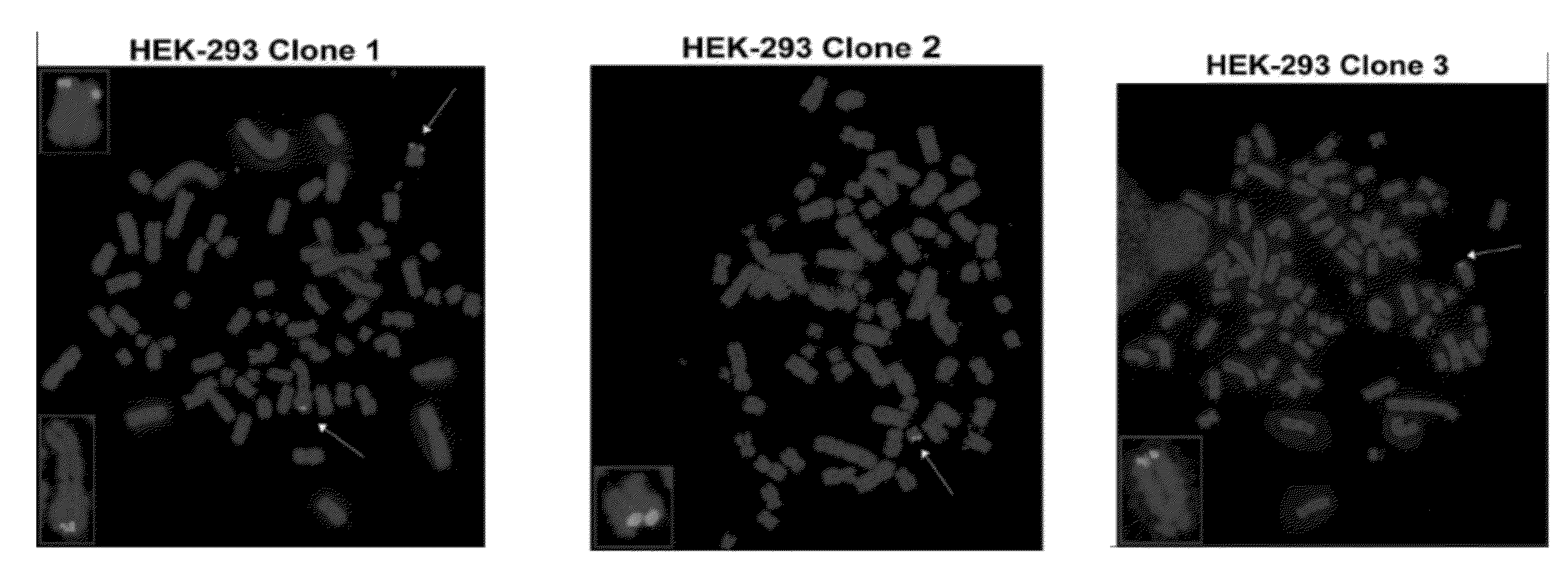
[0135]This Example illustrates methods for determining telomeric sites in which HHV-6 genomic DNA is inserted. Briefly, HEK-293 cells at 50% confluency were infected with HHV-6A (U1102) at 0.1 multiplicity of infection (MOI). After incubation for five days, the infected cells were washed to remove extracellular virus. Single cells were introduced into a 96-well plate and expanded. The results of ORF U94-specific PCR showed that 10 out of 22 clones have viral genome (FIG. 10a). Three PCR-positive clones and one PCR-negative clone were examined by FISH to determine the integration of HHV-6 genomic DNA. In two PCR-positive clones, FISH analysis identified integrated HHV-6A in one chromosome. In the third PCR-positive clone, the virus had integrated into two chromosomes (FIG. 10b).
[0136]The specific chromosomes into which HHV-6 had integrated in the HEK-293 cells was not identified using standard cytogenetic methods, due to the aneuplo...
example 2
Determination of Chromosomal Integration of HHV-6 DNA Using Fish
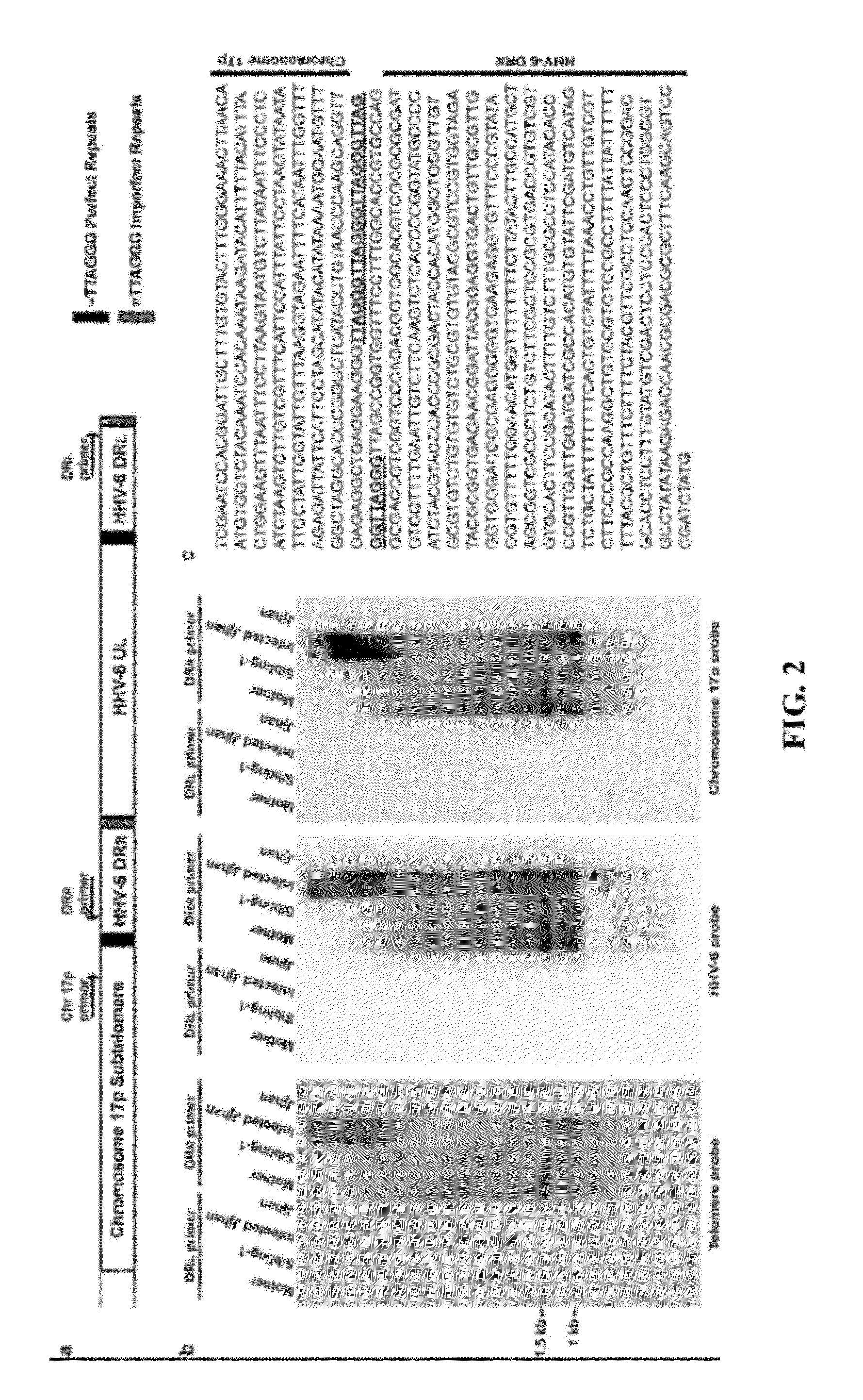
[0138]This Example illustrates methods for determining whether viral genomic DNA is integrated into the host chromosomes. Briefly, fluorescence in situ hybridization (FISH) was performed in T-cell cultures derived from peripheral blood samples. PBMCs were stimulated for 72 hours with 20 ng / ml PHA, and then cultured in RPMI 1640 medium containing 50 U / ml IL-2 and 10% FCS. Metaphase chromosomes were generated according to standard cytogenic protocol (Kowalska et al., Chromosome Research, 2007), stained with DAPI and hybridized with various probes as follows: FITC-conjugated HHV-6A (U1102 strain) cosmid probes pMF311-2, pMF335-6 (green) (40), and cy5-conjugated telomere peptide nucleic acid (PNA) probe (red) (DakoCytomation) (FIG. 7). The experiments were performed in two independent laboratories (University of Minnesota, Minneapolis, Minn. and Children's Cancer Research Institute, Vienna, Austria). Each laboratory was bli...
example 3
Confirmation of Chromosomal Integration of HHV-6 Genomic DNA by PCR
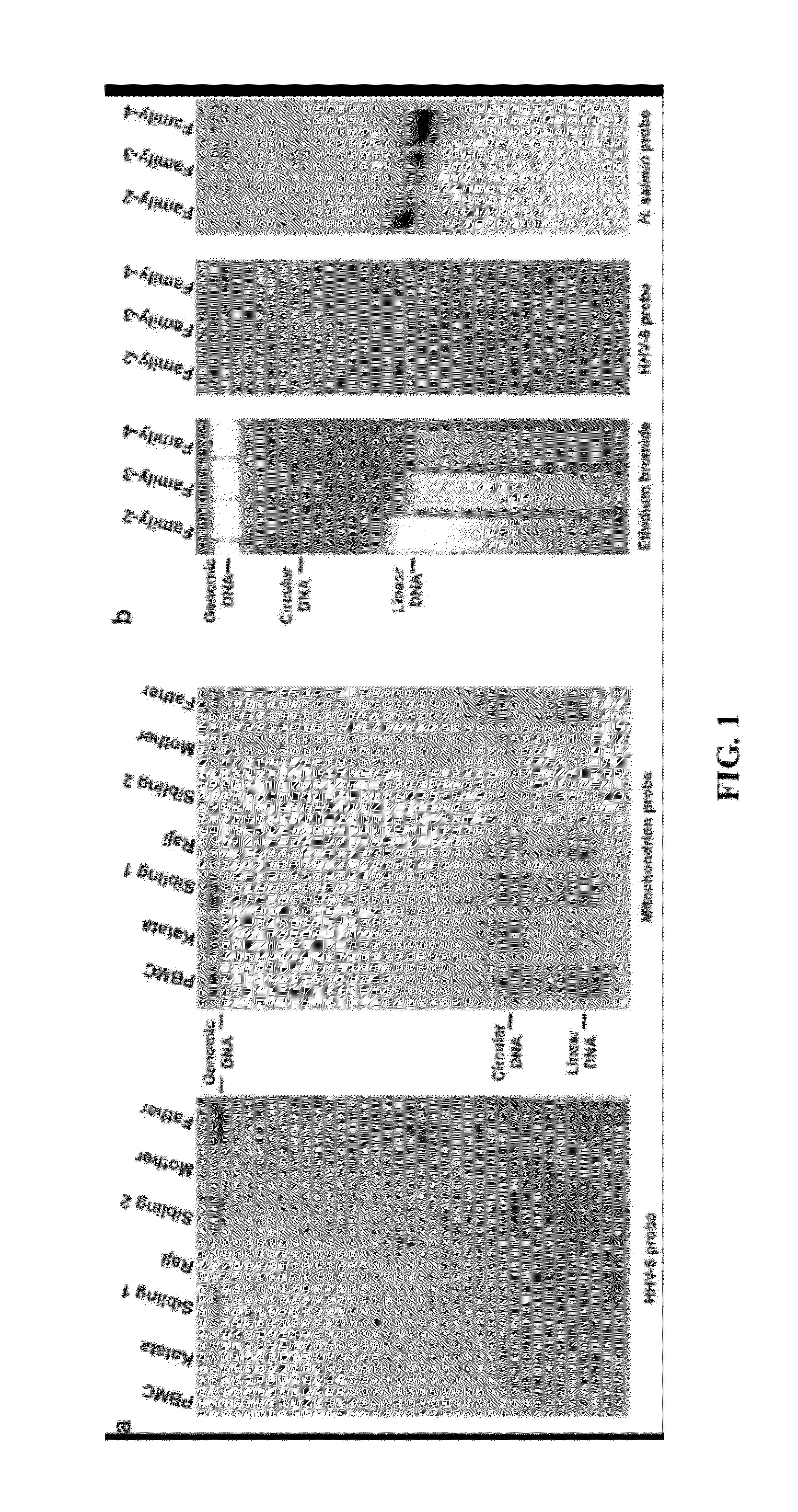
[0143]This Example confirms that HHV-6 genomic DNA integrates into chromosomes of host cells during viral infection. The majority of herpesviruses establish latency via circular nuclear episomes. However, as shown in Example 2, no HHV-6 episome is detected in host cells using the Gardella assay. To confirm that HHV-6 DNA only integrates into host chromosomes, a highly sensitive PCR assay was employed to detect the presence of small numbers of episomes, which might not have been detectable by the Gardella assay. Briefly, DNA was isolated from HHV-6A integrated HEK-293, T-cells from a member of Family-2, and T-cells from a member of Family-1 immortalized using HVS strain C484. The isolated DNA was subjected to CsCl / ethidium bromide gradient ultracentrifugation (FIG. 3). HHV-6 DNA was detected only in the linear fraction, and no episomal (ccc) DNA fraction was detected. Since the present PCR assay can detect as few as 1...
PUM
| Property | Measurement | Unit |
|---|---|---|
| Temperature | aaaaa | aaaaa |
| Temperature | aaaaa | aaaaa |
| Temperature | aaaaa | aaaaa |
Abstract
Description
Claims
Application Information