Compounds and related methods for manipulating parp-1-dependent cell death

Inactive Publication Date: 2012-05-17
VALTED
0 Cites 1 Cited by

AI-Extracted Technical Summary

Problems solved by technology

While Applicants have previously suggested after performing a proteomic screen for PAR-binding proteins that AIF could be a candidate protein for PAR...

Method used

[0054]With reference to FIG. 1A, it will be apparent to one skilled in the art that there are various ways in which this parthanatos mechanism can be interfered with. For example, reducing PAR abundance with PARG expressed in the cytosol or interfering with PAR through neutralizing antibodies can reduce mitochondrial AIF release and subsequent cell death. Simply reducing the cellular concentration of PAR in this manner, however, it is an unfavored approach because, as noted above, PAR has a significant role in the creation of a variety of nuclear proteins such as histones. Similarly, directly reducing the amount of AIF in the cells is also a non-optimal approach for inhibiting parthanatos because of AIF's roles in respiration. However, Applicants have discovered that PAR binds specifically to a certain portion of AIF (which portion Applicants refer to herein as a “PAR binding motif” or “PBM”) that acts as the transducer that mediates AIF release from the mitochondria, which in turn allows AIF to translocate to the nucleus, and further discovered that this PBM is separate and apart from the portions of AIF involved in respiratory functions. Thus, as shown in FIG. 1A, natural PAR on the outer membrane of the mitochondria (which is labeled in FIG. 1 as “WT-PAR” for “wild-type PAR”) which bears a PBM will bind with PAR and release from the mitochondrial membrane. However, AIF which has been genetically modified to “delete” the PBM (of preferred embodiment of which is labeled in FIG. 1 as “Pbm-PAR” and defined below) remains on the membrane of the mitochondria and thus does not trigger cell death.
[0055]As detailed in the experiments set forth below, PAR bound to AIF saturably and with high affinity. However, Applicants have confirmed through experiment that if PAR fails to bind to AIF, such as by genetically modifying AIF or otherwise chemically inhibiting this binding, then AIF is not released from mitochondria subsequent to PARP-1 activation. Further, Applicants have found that the cells survive the toxic stimuli if AIF release is inhibited in this manner. Applicants thus identified PBMs in the various forms of AIF found in different species, and have identified similarities to the various PBMs to facilitate genetic modification.
[0058]In particular, identification of AIF as a PAR-binding protein opens up opportunities for the development of compounds that inhibit the interaction of PAR with AIF, thus potentially protecting against parthanatos. Alternatively, agents could be identified that enhance the release of AIF, thereby promoting parthanatos and serving as potential cancer chemotherapeutic agents.
[0099]To determine whether Pbm-AIF fails to bind to PAR in cultured cortical neurons, neuronal cultures were transduced by lentivirus with WT-AIF-Flag or Flag-tagged Pbm-AIF (“Pbm-AIF-Flag”). Neurons from Hq mice, which have an 80% reduction in WT-AIF due to a pr...

Benefits of technology

[0010]Additionally, it is an object of one or more embodiments of the present invention to provide target sequences for genetic manipulation to alleviate unwanted PARP-1-induced cell death in target cells of a patient.
[0011]While Applicants have previously suggested after performing a proteomic screen for PAR-binding proteins that AIF could be a candidate protein for PAR binding (see J. P. Gagne et al., Nucleic Acids Res., 2008), the actual interaction between PAR and AIF heretofore has been unproven and unexplained. Applicants have discovered that AIF contains a PAR-binding motif (“PBM”), that PAR binding to AIF is required for AIF release from the mitochondria to occur, and that th...

Abstract

Apoptosis inducing factor (“AIF”) contains a PAR-binding motif (“PBM”) that binds to Poly(ADP-ribose) (“PAR”). Binding of PAR to AIF via the PBM is required for AIF release from the mitochondria to occur, and that this PAR-related release is a key step in the programmed cell death process known as parthanatos, both in vitro and in vivo. Preventing or disrupting this release can inhibit parthanatos and thus be the basis for treatments for patients suffering from diseases or medical conditions during which parthanatos commonly occurs, including Parkinson's disease or diabetes, or patients who have had and are recovering from heart attack, stroke and other ischemia reperfusion-related injuries. Alternatively, agents could be identified that enhance the release of AIF, thereby promoting parthanatos and serving as potential anti-tumor chemotherapeutic agents.

Application Domain

FungiSenses disorder +16

Technology Topic

DiseaseHeart disease +13

Image

  • Compounds and related methods for manipulating parp-1-dependent cell death
  • Compounds and related methods for manipulating parp-1-dependent cell death
  • Compounds and related methods for manipulating parp-1-dependent cell death

Examples

  • Experimental program(20)

Example

Example 1
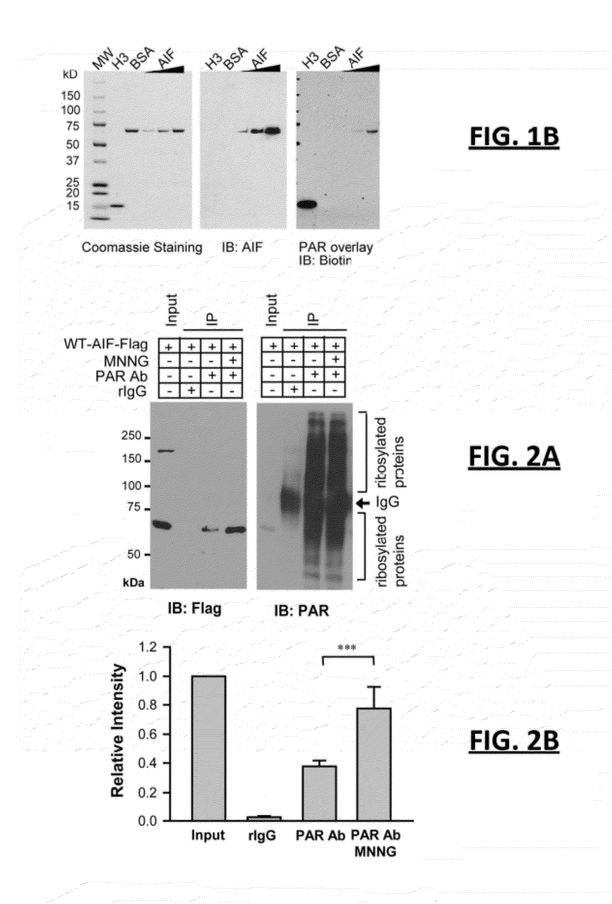
[0075]The following experiment was performed to confirm whether AIF in fact binds to PAR. Applicants performed 3 trials of an overlay assay on recombinant AIF with affinity-purified biotin-labeled PAR. Histone H3, which binds to PAR with high affinity, was included as a positive control and bovine serum albumin (“BSA”) was included as a negative control, as described by P. Chang et al. (Nat Cell Bio, 2005). FIG. 1B depicts three photos side by side for gels representative of all trials for these overlay assays. As shown in FIG. 1B, AIF bound to biotin-labeled PAR in a concentration-dependent manner (see the increasingly darker bands from left to right in each gel at approximately 70 kDa under AIF). It can also be seen that PAR polymer bound with AIF in a similar pattern to histone H3, while BSA failed to bind with PAR polymer. Furthermore, to confirm this result, an electrophoretic mobility shift assay (“EMSA”) was performed for AIFm using 32P-labeled PAR and histone H1 as a positive control. AIFm was found to cause a shift of PAR polymer similar to that cause by histone H1, a known PAR-binding protein. Thus, Applicants took the results of these two tests as confirming that AIF binds to PAR in vitro.

Example

Example 2
[0076]The following experiment was performed to confirm whether AIF in fact binds to PAR in intact cells. Applicants exposed HeLa cells stably transduced with lentivirus C-terminal Flag-tagged mouse wild type AIF (WT-AIF-Flag) to MNNG, a DNA alkylating agent that activates PARP-1 and kills cells primarily through parthanatos. PAR immunoprecipitation was performed from postnuclear fractions, which is the fraction prepared from whole cell lysates after removing nuclear proteins. FIG. 2A comprises two side by side black and white photos of representative gels obtained by Applicants, showing the impact of MNNG upon these HeLa cells (left photo being HeLa cells without MNNG, and right photo being HeLa cells 2 hours after MNNG treatment at 50 μM for 15 min). In the left gel of FIG. 2A, it can be seen that WT-AIF-Flag co-immunoprecipitated with PAR in resting cells. However, following MNNG treatment (the right photo of FIG. 2A), the interaction between AIF and PAR was significantly increased. Notably, WT-AIF-Flag did not co-immunoprecipitate with IgC, confirming the specificity of the interaction. FIG. 2B is a chart reporting the data regarding the relative intensity of the interaction pre and post MNNG for these experiments (*** indicates p<0.001).

Example

Example 3
[0077]Next, to determine whether endogenous AIF interacts with PAR polymer, the interaction between endogenous PAR and AIF was explored in primary cortical neurons under both resting conditions and after NMDA glutamate receptor stimulation. This stimulation is known to activate PARP-1 potently and kill neurons through parthanatos. For this test, cortical neurons were homogenized and fractionated in CSS as described above, and FIG. 3A comprises a notated black and white photograph of a representative gel obtained from co-immunoprecipitation of endogenous AIF with PAR polymer is post nuclear fractions isolated from the cortical neurons 2 hours after NMDA treatment (500 μM for 5 minutes). Applicants found that endogenous AIF interacted with PAR in non-stimulated cortical neurons (see the first three columns of the gel photograph of FIG. 3A), but that interaction that was significantly increased following NMDA treatment (compare against the right three columns of the gel of FIG. 3A). FIG. 3B is a chart reporting the data regarding the relative intensity of the interaction in neurons resting (in CSS) and 2 hours post-MNNG treatment at 500 μM for 5 min (*** indicates p<0.001 by Student's t-test, n=6). Together, the data from the experiments of Examples 1-3 suggest that AIF is a PAR-binding protein and that PARP-1 activation increases the AIF-PAR interaction.

PUM

PropertyMeasurementUnit
Cell death

Description & Claims & Application Information

We can also present the details of the Description, Claims and Application information to help users get a comprehensive understanding of the technical details of the patent, such as background art, summary of invention, brief description of drawings, description of embodiments, and other original content. On the other hand, users can also determine the specific scope of protection of the technology through the list of claims; as well as understand the changes in the life cycle of the technology with the presentation of the patent timeline. Login to view more.
Why Eureka
Try Eureka