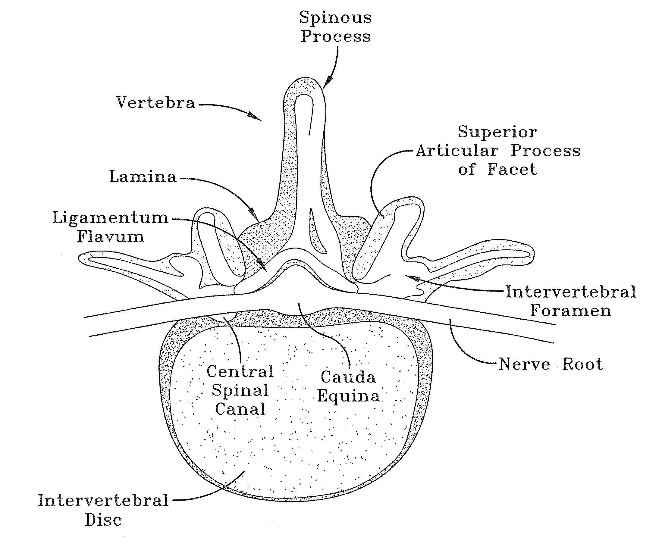
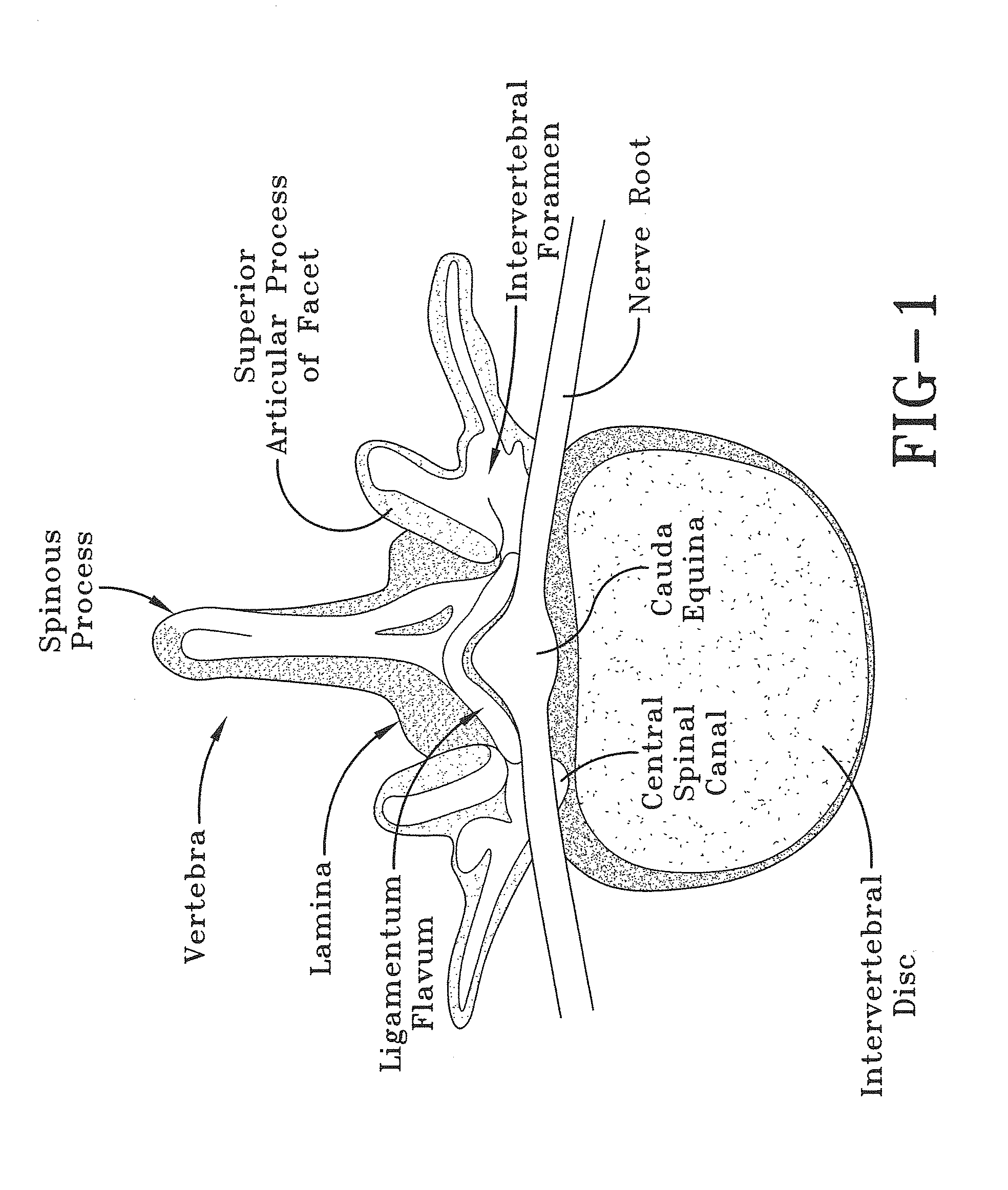
However, the hallmark of cauda equina syndrome is severe neural compression below the conus causing frank loss of bowel and bladder control.
This can manifest itself either with
urinary retention and inability to urinate with
severe pain from bladder expansion, or frank loss of bowel and bladder control causing the patient to either urinate or defecate on himself or herself without realizing that this has occurred.
The fact that
saddle anesthesia is often present would also cause the patient to not realize that such an accident had happened.
When
stenosis, or nerve compression, is present however the patient will often experience
severe pain radiating into the lower extremities following the distribution of the nerves being compressed.
The patient may also experience
weakness, numbness, and / or tingling with difficulty walking or standing.
Despite this, however, even cases of
stenosis, or nerve compression, are not true surgical emergencies.
As such, patients are typically advised that
surgery for
stenosis is a “
quality of life” issue that the patient considers when he or she is ready and wishes to proceed.
However, when present, this is a true surgical emergency, and
surgery that is not performed on an emergent basis has been found to lead to a dramatic decrease in success rates and postoperative function.
However, regardless of the cause, the common thread is that there is severe nerve compression below the conus causing the sacral roots to
shut down with loss of bowel and bladder control.
The results of cauda equina syndrome are often very serious, being both painful and having many debilitating effects.
However these are subjective measurements and there is no objective measure as to what normal tone and volition would be.
Furthermore, because cauda equina syndrome is relatively rare, many clinicians may not have experience with a truly abnormal rectal exam in which the tone is diminished or absent.
Perianal
sensation is often difficult to test.
Patients are in pain and are usually very uncooperative.
Without the cooperation of the patient, it is difficult to position the patient properly.
This is problematic because the hallmark of determining if
neurologic function is present includes both motor and a sensory examination.
Further, the conventional perianal sensory pinprick tests also suffer from a variety of deficiencies.
Such deficiencies include, but are not limited to, difficulty in accessing the perianal area with a safety pin and applying the appropriate force when pricking the perianal area with the safety pin.
Thus, cauda equina syndrome is considered a condition in which every physician should be familiar and also one in which is it considered unacceptable practice if it is missed.
However, cauda equina syndrome is rare and such procedures are expensive and cumbersome, particularly when ordered after hours in an emergency room situation.
Due to the perceived rarity of cauda equina syndrome, there has been no practical probe developed for routine examination for cauda equina syndrome, resulting in missing the occurrence of this syndrome.
When a missed cauda equina syndrome does occur, and surgical management is delayed, the success rates for
recovery of bowel and bladder dysfunction are significantly diminished.
This in turn may result in unfortunate and expensive treatments, as well as huge awards from
medical malpractice lawsuits.
A study of these cases demonstrated that
time to diagnosis and
surgery ranged from under 24 hours to over one month, and did not predict which cases would rule for the plaintiff.
Severity of initial symptoms also did not predict which cases would rule for the plaintiff.
This was consistently considered a significant factor in determining failure to provide the
standard of care.
Nevertheless, time to surgery weighed very little on the verdicts which were handed down.
It was the lack of an adequate rectal examination which appeared to carry the most weight in determining guilt in these cases.
Despite the fact that the claimant appeared to have received adequate care, the fact that a rectal examination was not performed on initial presentation, even though the symptoms were atypical for cauda equina syndrome, made a huge
impact on the overall outcome of the case with an enormous penalty attributed to the treating providers for not having checked rectal tone.
However, cauda equina syndrome is also very rare, and even the busy spinal surgeon will only see one or two cases per year.
This is a primary impediment to performing an adequate rectal examination that was frequently cited in these cases; that the treating provider was not a specialist and would not know if the rectal exam was truly abnormal or not.
That said, this proved to be an inadequate defense in these cases.
It is also not reasonable from a cost perspective to order an MRI on every patient with
lumbar complaints.
A simple inexpensive device to rule out this potentially devastating condition is thus necessary and not currently available.
The most common symptom is pain, particularly pain in the back, on one or both hind legs, or
tail; however, because an animal cannot report symptoms it is frequently difficult to determine if the stenosis has become severe.
However, it is difficult to perform a neurologic examination in a dog that cannot follow commands, and reflexes have poor sensitivity and specificity in determining if severe stenosis is present.
Furthermore, myelography is painful, has associated risks, and is expensive and invasive and as such is a poor tool for routine screening.
Although it would alert the owner and veterinarian that impending or significant neurologic compromise is occurring, an anal exam is not typically performed on a routine basis in these dogs, even when they have reached an age where severe stenosis and loss of
neurologic function is common.
A health problem which many physicians must be aware of is the enlargement and changes in the
normal shape of a man's
prostate gland which may be an indication of urinary symptoms and
prostate cancer.
Although it is usually just an initial test to determine whether further tests should be made, the skipping of the test or a misjudgment of the test results could lead to dire consequences.
None of the foregoing patents provide a
system for determining cauda equina syndrome or apparatus for quickly and accurately determining if the foregoing syndrome is present or there is a risk that it is present.
However, no single device is known which specifically determines if a patient has cauda equina syndrome.

