
Monopolar RF-Surgical Snares

a monopolar and rf-surgical technology, applied in the field of monopolar rf-surgical snares, can solve the problems of inability to securely ensnare or ensnare at all with a polypectomy snare, inability to ectomize, and inability to treat or treat easily

Active Publication Date: 2012-07-05
ENDOX FEINWERKTECHN
View PDF5 Cites 55 Cited by
  • Summary
  • Abstract
  • Description
  • Claims
  • Application Information

AI Technical Summary

Benefits of technology

"The invention is about improving monopolar and asymmetrical snares used for the removal of pathological tissue, particularly tumors, from the gastrointestinal tract. The goal is to develop RF-surgical snares that can remove large tumors in one piece from the organ wall without causing damage to the wall. The snares should be designed to be pressed against the wall of a hollow organ before and during RF-surgical incision, without cutting the organ wall. The invention also includes a method for insulating the snare portions to prevent current leakage and improve the overall insulation of the snare. The asymmetrical snare has a shorter snare portion that is electrically insulated and connected to the manipulation wire, which requires less electric power and reduces the risk of unintended RF-surgical effects."

Problems solved by technology

If a malignant tumor has grown into the blood or lymph vessels, malignant tumor cells may find their way through these vessels into other, and particularly vital, organs and there form metastases which cannot be easily treated or be treated at all.
If a pathological mucosal-submucosal area projects too little or not at all above the normal mucosal-submucosal area, it cannot be securely ensnared or ensnared at all with a polypectomy snare and cannot be ectomised.
However, as the size of the polyps ectomised or mucosal-submucosal areas resected in toto and en bloc has increased, the resulting problems and complications set out below have increased with the use of the snares available hitherto for EPE and EMR and the RF generators available for operating these snares.
One of these problems consists in the fact that particularly with en-bloc separation of large polyps or large-area mucosal-submucosal areas, particularly if through submucosal injection these become even greater than they are already before injection, is the electrical power required for this.
If this tissue is heated too slowly or with too long a time lag, which it is feared may result in a delay in initial incision, the heat created here may diffuse from the tissue into the adjacent tissue close to the snare and damage the adjacent tissue thermally.
If for the abovementioned reason the snare is applied close to the organ wall, the muscularis propria may be thermally damaged by it.
Thermal damage to the muscularis propria or even the serosa of a hollow organ of the gastrointestinal tract may result in perforation of this organ and will not infrequently make an open surgical operation necessary.
Bipolar snares have, however, not proved satisfactory in clinical applications.
For this reason bipolar snares have not proved satisfactory in practice.
Although snares of this kind require less RF current during the initial-incision phase and during the incision phase than snares of the same size without insulation, they have the disadvantage that the RF-surgically effective part of the snare viewed from an endoscope is always behind the polyp, that is, is out of sight, and there is a risk that the distal tip of the snare can perforate the organ wall unmonitored.
This form of electrode may, however, hinder accurate incision.
Both with electrosurgical instruments in accordance with U.S. Pat. No. 5,078,716 and DE 100 28 413 A1 and coagulating instruments in accordance with DE 25 14 501 the RF-surgically effective electrode surfaces can be disposed only at the distal end of the snare, with the disadvantage that these, seen through an endoscope, are always behind the tissue to be removed and the incision therefore has to be made without visual monitoring, which is a risky process, particularly with large polyps or mucosal-submucosal areas.
A further problem is the placing of the snare around in particular large or oddly shaped polyps or around polyps or mucosal-submucosal areas which have become enlarged through the injection of physiological NaCl solution or other injectables into the submucosa, that is, by submucosal injection, enlarged or oddly shaped polyps or mucosal-submucosal areas.
A further problem is the placing and guidance of the snare as close to the organ wall as possible in order to separate the tumor close to the muscularis propria for the above-mentioned reasons.
With multi-strand or braided standard snares this is normally not practicable, because these snares are too soft or too flexible for this.
The use of these snares is, however, hazardous in so far as because of the pressure against the organ wall they can also cut into or even through the organ wall.
A further problem, particularly with large polyps or mucosal-submucosal areas with an abnormally slippery surface may be that the snare slips out of the intended position while being applied or manipulated.
Since wide open snares, especially when being placed around flat polyps or mucosal-submucosal areas, cannot or must not be pressed against the organ wall concerned sufficiently firmly, they may, when the snare is closed, slip over the slippery mucous membrane and out of the intended position.
On asymmetrical snares claws, hooks or teeth of this kind interfere with the function of the above-described driving device.
Although endoscopic polypectomy and endoscopic mucosaresection are regarded today as clinically established methods, these methods are presumably also because of the above-described problems attended with various complications.
Both because of the above-described problems and because of the complication rate rising due to the mean diameter of the tumor the hitherto available abovementioned methods and snares only can only be used to remove polyps or mucosal-submucosal areas in toto and en bloc with a maximum mean diameter of up to approx.
With these methods larger polyps or mucosal-submucosal areas can be removed only by a piecemeal technique and often not radically or completely, a circumstance which may result in relapses.
These methods, however, call for a high degree of manual dexterity, experience, constant training, a willingness to take risks and long operating times. So far there have been only a very few experts who practise these methods.

Method used


Image

  • Monopolar RF-Surgical Snares
  • Monopolar RF-Surgical Snares
  • Monopolar RF-Surgical Snares

Examples


Embodiment Construction

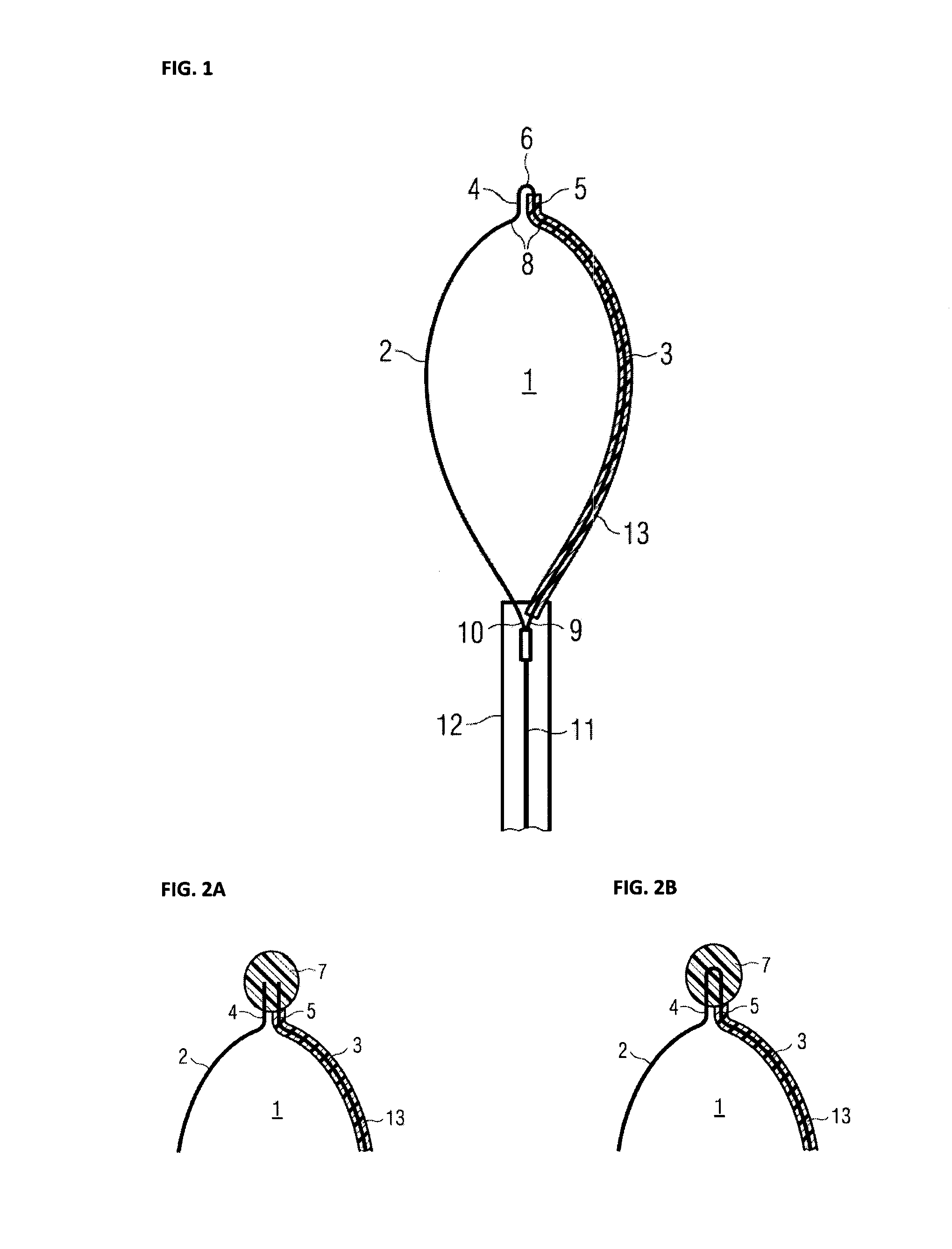
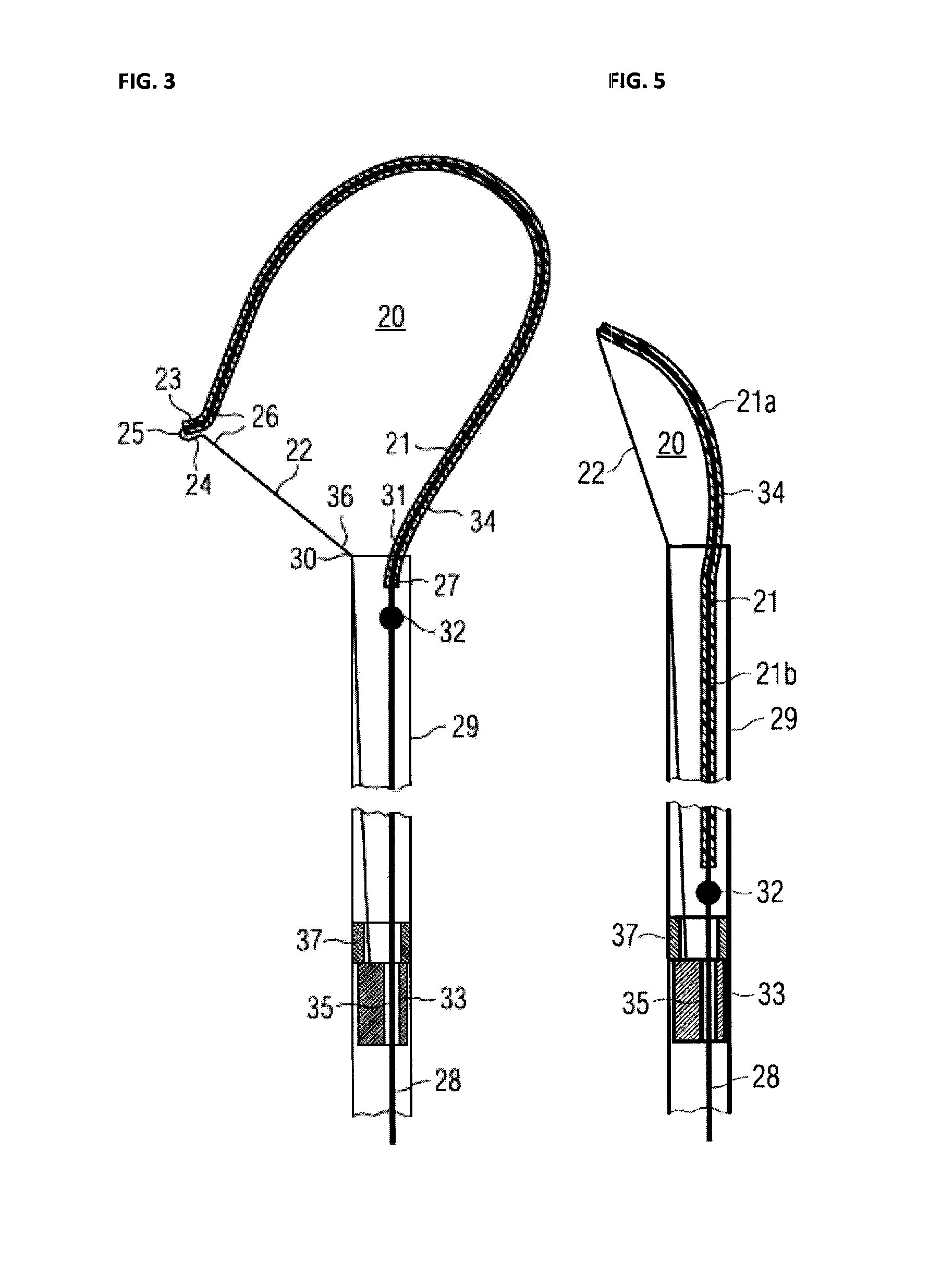
[0078]FIG. 1 shows an exemplary embodiment of a monopolar symmetrically opening and closing polypectomy snare, with which tissue to be removed can be ensnared and RF-surgically removed. This snare includes two snare portions 2, 3 essentially of equal length made from elastic metallic round wire, flat wire, wire strand and / or the like which are connected together at their distal ends 4, 5 directly 6 or by means of a connecting element 7 shown schematically in FIG. 2 and which form here a resilient bending element 8 and which are or can be connected at their proximal end 9,10 to a manipulation wire 11 which is guided inside a tube or catheter 12 and is used to pull the snare 1 into and push the snare 1 out of the catheter 12 and which can also be used as a conductor for the RF current required for the RF-surgical removal of biological tissue. One of the two snare portions, here, for example, snare portion 3, is completely insulated with an electrical insulation means 13 and the other ...


PUM


Abstract

Symmetrical and asymmetrical snares are used for endoscopically controlled methods by which tumors in hollow organs of the gastrointestinal tract can be ensnared and RF-surgically removed from the organ wall for diagnostic and / or therapeutic purposes. These snares are characterised in that one of the two snare portions, in the case of asymmetrical snares the longer of the two snare portions, is completely electrically insulated or is made from electrically non-conductive material and that the other of the two snare portions is made from electrically conductive material and its surface is not electrically insulated. In one embodiment, the asymmetrical snare is characterised in that the long snare portion is so much flexurally stiffer than the short snare portion that it tensions the short snare portion like the string of a bow.

Description

PRIORITY CLAIM[0001]This application is a continuation of pending International Application No. PCT / EP2010 / 060877 filed on Jul. 27, 2010, which designates the United States and claims priority from German Patent Application No. 10 2009 036 158.8 filed on Jul. 28, 2009.BACKGROUND OF THE INVENTION[0002]1. Field of the Invention[0003]The invention relates to radio-frequency-surgical (RF-surgical) snares for endoscopically monitored methods by which tissue in hollow organs, particularly pathological mucosal-submucosal areas or tumors of the gastrointestinal tract, can be ensnared and removed from the wall of the hollow organ by RF-surgical means for diagnostic and / or therapeutic purposes.[0004]2. Description of Relevant Art[0005]It is known that malignant tumors, also known as carcinomas, of the gastrointestinal tract, that is, the oesophagus, the stomach and the colon, cause metastases into other organs and are one of the four most common causes of death worldwide. 1 million new cases ...

Claims


Application Information

Patent Timeline
Patent Type & Authority Applications(United States)
IPC IPC(8): A61B18/18
CPCA61B18/14A61B2017/00867A61B2018/00083A61B18/1492A61B2018/1407A61B2018/141C08L2201/12A61B2018/00601
Inventor FARIN, GUNTHERHERNIK, MATTHIAS
Owner ENDOX FEINWERKTECHN
Learn More