Systems and Methods for Analyzing Body Fluids

a body fluid and system technology, applied in the field of body fluid composition and components determination, can solve the problems of large proportion of blood specimens that require further testing, large calibration and control of cbc instruments, maintenance and skilled operators,

Active Publication Date: 2013-03-21
ROCHE DIAGNOSTICS HEMATOLOGY INC
View PDF4 Cites 18 Cited by
  • Summary
  • Abstract
  • Description
  • Claims
  • Application Information

AI Technical Summary

Benefits of technology

[0016]By way of example, in the case of analyzing blood or bone marrow, aspects of the present invention may used process a slide and optionally capture an image of the slide. The image may be later used for performing various tests that provide for a count of various cell types, or an assessment of the morphology of the cells. One example is a complete blood count including image-based counting and assessment of the morphology of the formed elements of blood, including RBCs, WBCs, and platelets. Embodiments of the present invention may improve the accuracy of the CBC as a result of direct visualization of the formed elements of blood. The use of the disclosed systems and processes for applying a monolayer of cells onto a slide enables assessment of certain cell types, particularly of abnormal and immature WBCs that are found in cases of abnormal bone marrow function including hematological malignancies. Further, the present invention may decrease costs associated with instrumentation; decrease cost of consumables and reagents; and require less operator time and reagents, fewer repeated tests, and fewer moving parts. It may also reduce the turnaround time for many of the CBC tests that currently require visualization of blood cells after the instrumental portion of the test is completed, by allowing cells to be visualized on a monitor instead of under a microscope.
[0017]Aspects of the present invention are effective at preserving cell morphology. This may be important for patients with hematological malignancies such as chronic lymphocytic leukemia (CLL) or acute myeloid leukemia (AML). The systems and processes for creating a monolayer of cells from body fluid may enable detection of a larger number of morphologically well preserved blast cells and other immature or fragile cells. This would allow their more accurate recognition at an earlier stage of the leukemic or other disease process. Certain aspects of the present invention provide for preparing a substantially uniform distribution of cells across a test area of a slide.
[0018]Aspects of this invention may relate to the application of cells from body fluids to a slide and include possibly mixing the cells contained in the body fluid with a diluent, collecting a sub-sample (aliquot) of a known volume from the solution, and then depositing the aliquot onto a substratum such as a slide using a dispensing device or applicator. The cells may be allowed to air dry or may be fixed (using a fixative solution) or both, depending on the examination that is anticipated. The cells may also be stained. The stained cells on the substratum may be counted and examined by an automated imaging system utilizing a computer or viewed by manual microscopic examination. Digital images may be shown on a computer display to reduce the need for manual microscopic review.
[0020]Systems and methods of the present invention provide a number of improvements over prior art techniques. For example, an embodiment of the present invention may be used to determine the number of cells in a sample of the cervix that are infected by the Human Papilloma Virus (this may indicate the viral burden, which is a prognostic factor to assess if an abnormality may progress, remain stable, or regress). Embodiments of the present invention may be able to determine how many viral or infected cells are in the sample. Additionally, certain embodiments of the present invention may be able to determine the differential cell count in an non-gynecologic sample collected from a body cavity effusion. In further embodiments, the system or method could determine that there is a large number of acute inflammatory cells in a sample (which the system or method may use to determine the presence of a bacterial infection). Similarly, if an embodiment of the present invention determined there were a high number of lymphocytes in a particular sample this may suggest a viral infection, autoimmune disease, or tuberculosis.

Problems solved by technology

Flow based CBC instruments generally require extensive calibration and control, maintenance, and skilled operators, and they have substantial costs associated with acquisition, service, reagents, consumables and disposables.
One significant problem with these systems in routine use is that a large proportion of blood specimens require further testing to complete the assessment of the morphologic components of the CBC.
This process is often done manually by skilled medical technologists, which increases the cost and time to receive results from the tests.
In addition to additional work involved in performing manual differential counts, this process has a number of additional technical limitations.
These include distortions of cell morphology because of mechanical forces involved in smearing the cells onto the slide, and cells overlapping one another, which makes visualization of individual cell morphology difficult.
There are several limitations to current cytologic preparation methods.
These include distortions of cell morphology because of mechanical forces involved in smearing or sedimenting the cells onto the slide.
The deposition of cells onto the slide may result in cells overlapping one another, so that individual cell morphology cannot be visualized.
Additional limitations associated with current cytologic preparation methods include, although are not limited to, the following: inhomogeneous sampling of a specimen due to differential rates of sedimenting cells onto a slide; the loss of cell clusters during certain types of preparation methods; the loss of small cells in methods that depend on density gradient methods of preparation; damage or nonspecific loss of inflammatory cells in centrifugation methods; and the inability to determine the absolute number of cell types in a sample, which may be important in determining the number of abnormal cells in a sample or in counting the number of inflammatory cells to determine the predominant type of inflammation that is in a sample.
Examples of these limitations are found with the Pap testing techniques that may not adequately display certain cell clusters because of smearing or filtration processes used to prepare the slide.
The density gradient preparation method employed by the SurePath method may not capture small, buoyant cells that float atop the gradient.
Centrifugation methods may result in the inconsistent loss of small lymphocytes, which can be problematic when an accurate differential count of inflammatory cells is required.

Method used


Image

  • Systems and Methods for Analyzing Body Fluids
  • Systems and Methods for Analyzing Body Fluids
  • Systems and Methods for Analyzing Body Fluids

Examples


Embodiment Construction

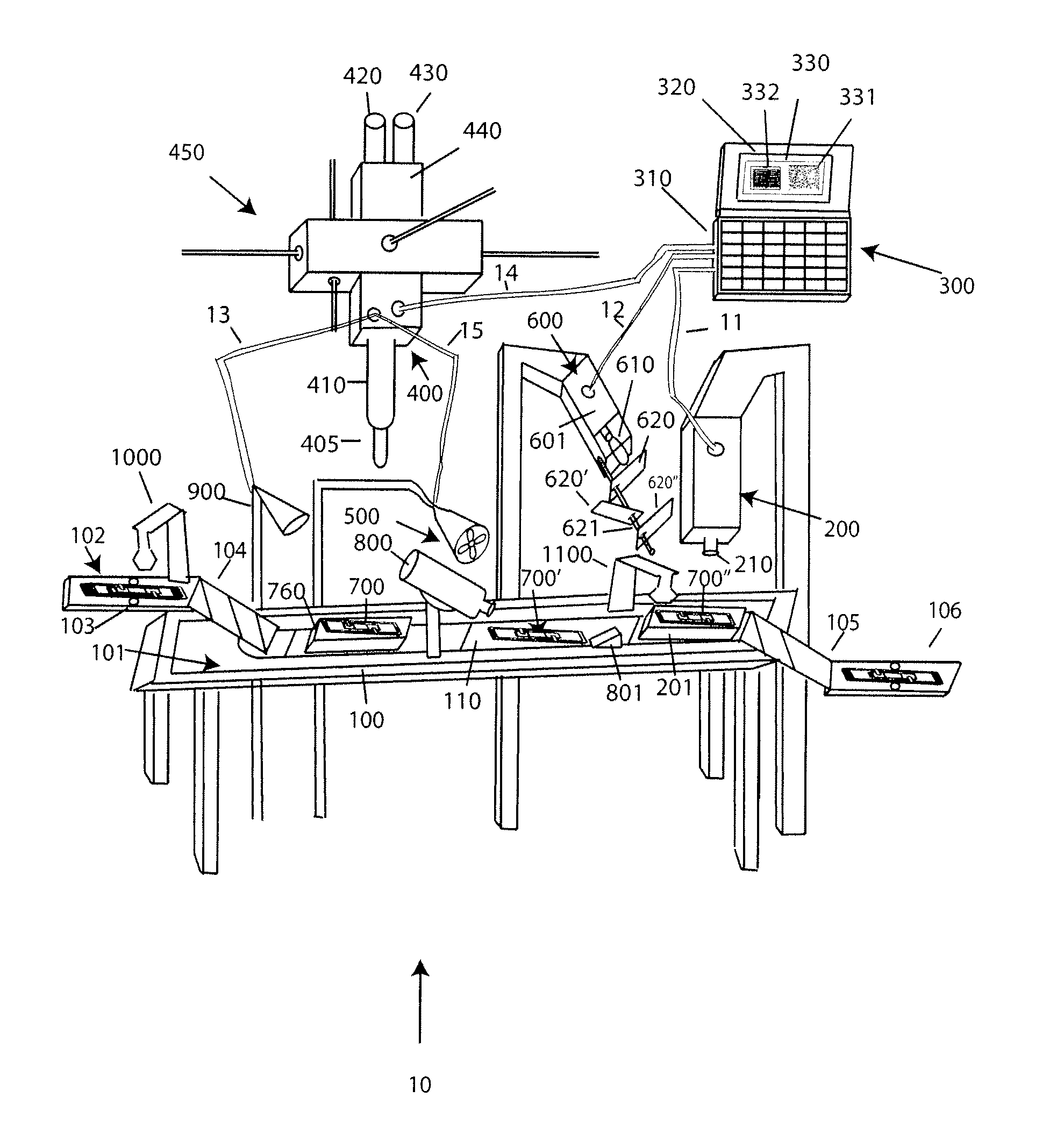
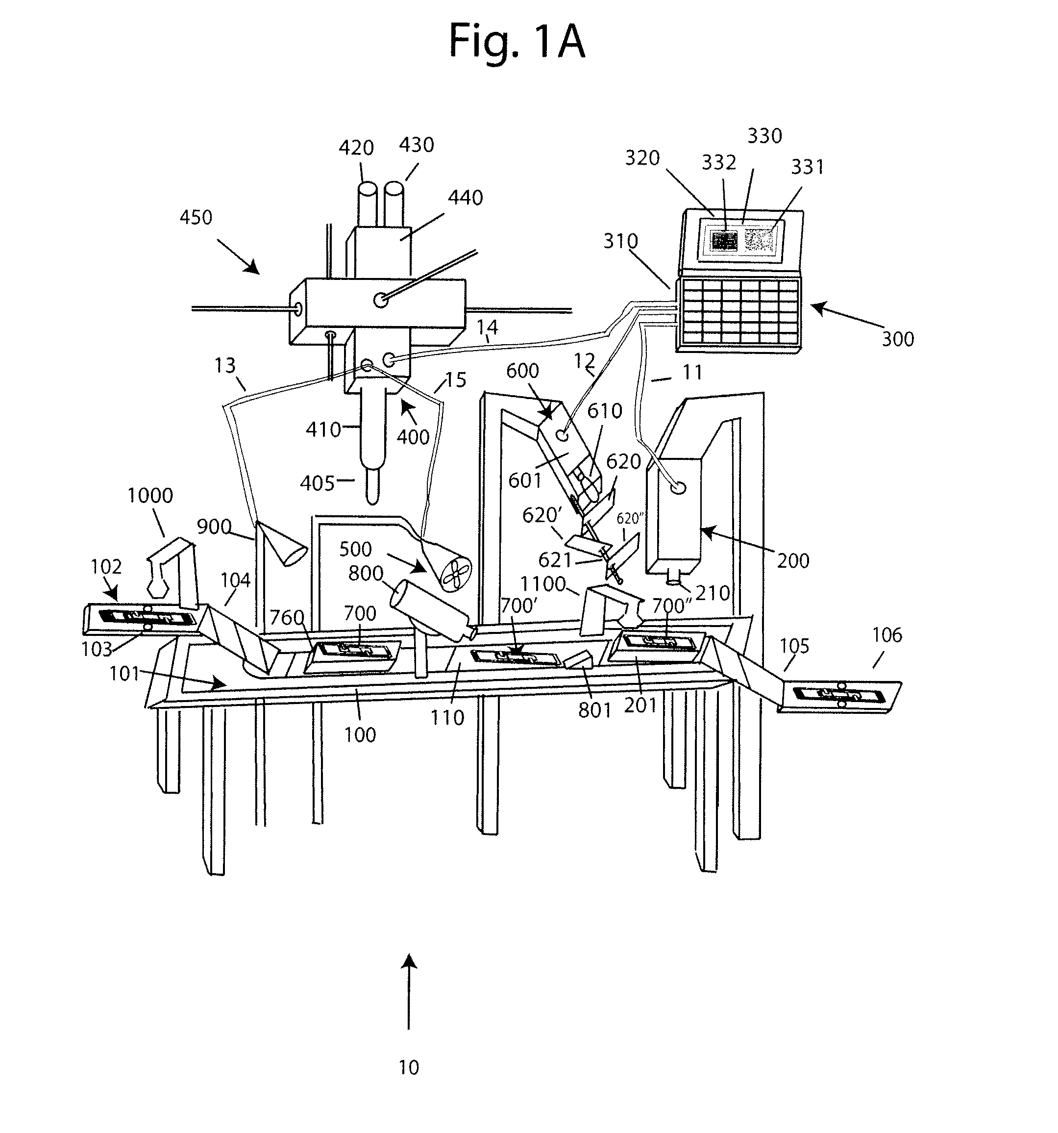
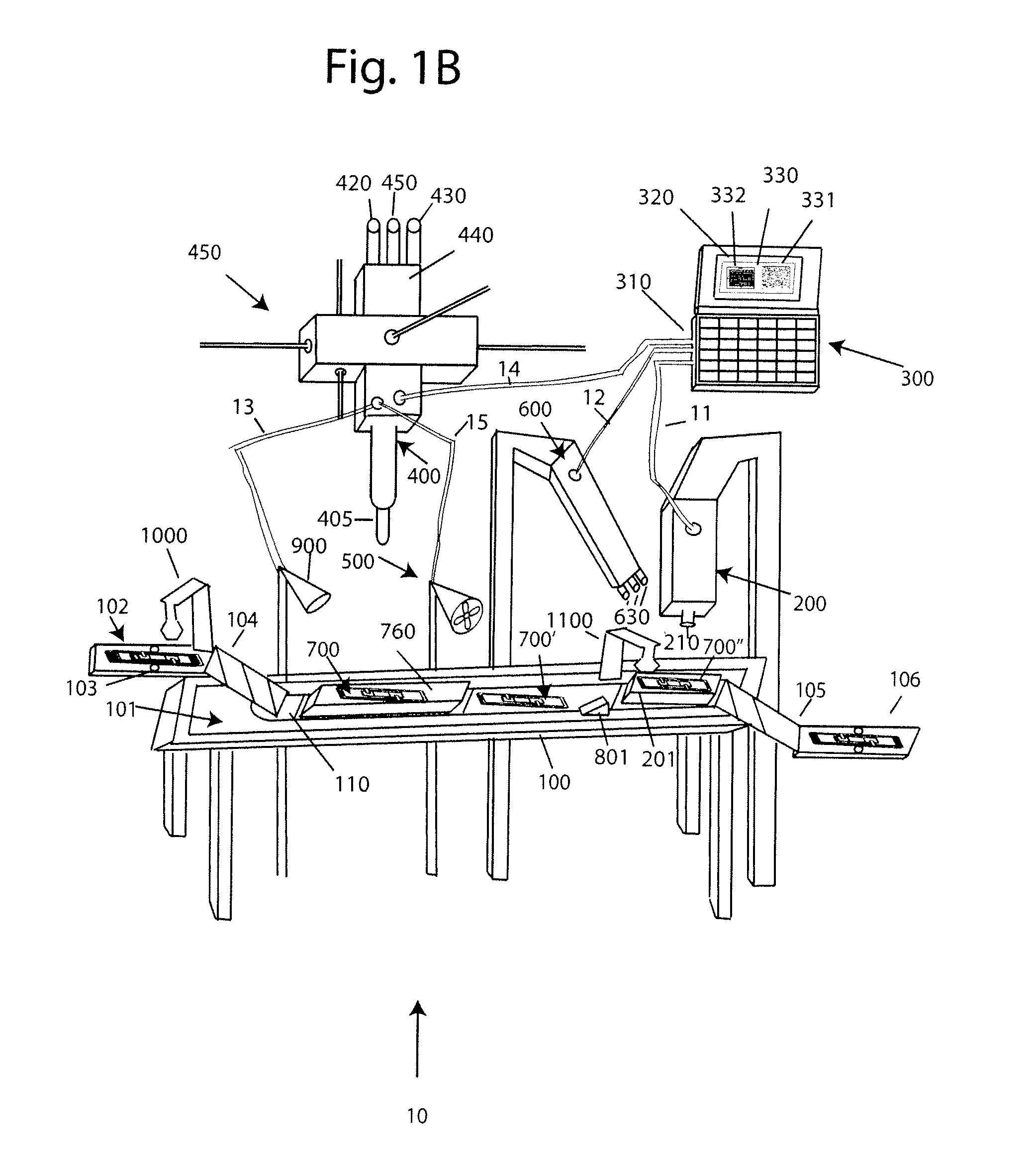
[0030]With reference to FIG. 1A, a system 10 for analyzing body fluids is disclosed. The system may comprise a platform 100, a light receiving device 200, a computer 300, an applicator 400, a gas circulation device 500, a light source 600, a dispenser 800, a discharge device 900, a slide labeler 1000, and slide label reader 1100. The following sections below include capitalized headings intended to facilitate navigation through the specification, which are not intended to be limiting of the invention in any manner.

The Platform 100

[0031]In embodiments that feature a platform 100, an advancer 110 may be configured to receive one or more slide apparatuses 700-700″. The advancer 110 may be attached to a surface, such as the top surface 101, of the platform. The advancer 110 may take the form of a belt as shown in FIG. 1A, the system may use a mechanical arm, gravity, magnetism, hydraulics, gears, or other locomotion techniques to move the slide apparatus along the surface 101 of the pla...


PUM


Abstract

Systems and methods analyzing body fluids contain cells including blood, bone marrow, urine, vaginal tissue, epithelial tissue, tumors, semen, and spittle are disclosed. The systems and methods utilize an improved technique for applying a monolayer of cells to a slide and generating a substantially uniform distribution of cells on the slide. Additionally aspects of the invention also relate to systems and method for utilizing multi-color microscopy for improving the quality of images captured by a light receiving device.

Description

PRIORITY CLAIM[0001]This application claims the benefit of priority to U.S. Provisional Application 61 / 173,186; filed Apr. 27, 2009. This application is a continuation-in-part of U.S. Ser. No. 12 / 430,885; filed Apr. 27, 2009; which claims the benefit of priority to U.S. Provisional Application 61 / 047,920; filed Apr. 25, 2008.FIELD OF THE INVENTION[0002]This invention relates to a system and process for determining composition and components of fluids. More specifically the present invention provides improved techniques for viewing cellular morphology, and determining the number of a particular type of cell in a portion of a body fluid.BACKGROUND OF THE INVENTION[0003]Pathology is a field of medicine where medical professionals determine the presence, or absence of disease by methods that include the morphologic examination of individual cells that have been collected, fixed or air-dried, and then visualized by a stain that highlights features of both the nucleus and the cytoplasm. T...

Claims


Application Information

Patent Timeline
Patent Type & Authority Applications(United States)
IPC IPC(8): H04N7/18G06K9/00
CPCG01N1/2813G01N1/312G01N15/1475H04N7/18G01N35/00029G06K9/00G01N2015/0073G01N2015/008G01N2015/0084G01N2015/1006G06F18/00
Inventor WINKELMAN, JAMESTANASIJEVIC, MILENKOZAHNISER, DAVIDLINDER, JAMES
Owner ROCHE DIAGNOSTICS HEMATOLOGY INC
Why Eureka
Try Eureka