Rapid Diffusion of Large Polymeric Nanoparticles in the Mammalian Brain

a polymer nanoparticle and brain technology, applied in the field of drug delivery, can solve the problems of virus carrying therapeutic genes, complex ecs, and inability to efficiently penetrate many particulate drug delivery systems, and achieve the effects of improving drug and gene delivery, improving drug loading efficiency, and increasing drug payload

Active Publication Date: 2013-07-18
THE JOHN HOPKINS UNIV SCHOOL OF MEDICINE
View PDF10 Cites 253 Cited by
  • Summary
  • Abstract
  • Description
  • Claims
  • Application Information

AI Technical Summary

Benefits of technology

[0018]The ability to achieve brain penetration with larger particles will significantly improve drug and gene delivery within the CNS since larger particles offer higher drug payload, improved drug loading efficiency, and significantly longer drug release durations.

Problems solved by technology

While the blood brain barrier has long been considered the crucial interface for therapeutic efficacy within the CNS, more recently poor distribution of agents within the brain itself has emerged as a major delivery challenge.
Importantly, the ECS may be more complex in certain pathogenic states, such as intrinsic invasive brain tumors.
For substances with few adhesive interactions, this size is large enough to allow diffusion of nanoparticles, but too small to allow efficient penetration of many particulate drug delivery systems and viruses carrying therapeutic genes.
Thus, the brain ECS poses a formidable barrier to therapy with some of the most advanced new treatment modalities.
A key factor and limitation to diffusion is the ‘mesh spacing’ or ‘pore size’ within the brain ECS.
This data has been criticized for poor preservation of the tissue architecture and therefore, artifactual results (Cragg 1980; Van Harreveld A & Trubatch J (1979) J Microsc 115(3):243-256).
While these models reduce the problems inherent to EM measurements, the modeling calculations based on gross particle spread and Fick's Law cannot resolve or analyze trajectories of individual particles and, therefore, are unable to assess micro-rheology, anisotropy, and small-scale Brownian motion.
In addition, these methods do not account for bulk flow phenomenon or convective forces introduced by the injection process.
Measurement of the brain ECS pore size has proven to be challenging.
Artifacts introduced with tissue preservation and processing as well as the structural heterogeneity of the tissue including anisotropic, electro-statically charged regions and dead-space micro-domains, make many measurements unreliable.
While this represents a significant improvement from the earlier EM-based methods, these calculations did not account for convective and bulk flow forces, anesthetic-induced changes, tissue anisotropy, or potential adhesive interactions with the experimental probes.

Method used


Image

  • Rapid Diffusion of Large Polymeric Nanoparticles in the Mammalian Brain
  • Rapid Diffusion of Large Polymeric Nanoparticles in the Mammalian Brain
  • Rapid Diffusion of Large Polymeric Nanoparticles in the Mammalian Brain

Examples


example 1

[0120]Materials and Methods

[0121]Brain Tissue Preparation

[0122]All experiments were carried out at Johns Hopkins University School of Medicine in accordance with National Institutes of Health guidelines and local Institutional Animal Care and Use Committee regulations. Brain tissue slices were prepared from 130-160 g female Sprague-Dawley rats. Animals were anesthetized with ketamine-xylazine and then administered an intracardiac injection of Euthanosol. After euthanasia, the brain was rapidly removed and immersed in chilled artificial cerebrospinal fluid (ACSF, Harvard Apparatus) supplemented with 10% glucose. Coronal slices were prepared using a rodent brain slice matrix kit (Zivic Instruments, Pittsburgh, Pa.). The matrix and razor blades were washed with 0.9% normal saline and placed on ice prior to inserting the excised rat brain. Placement of the brain and sectioning of the brain was carried out based on instrument instructions such that 1 mm thick slices were obtained. Slices...

example 2

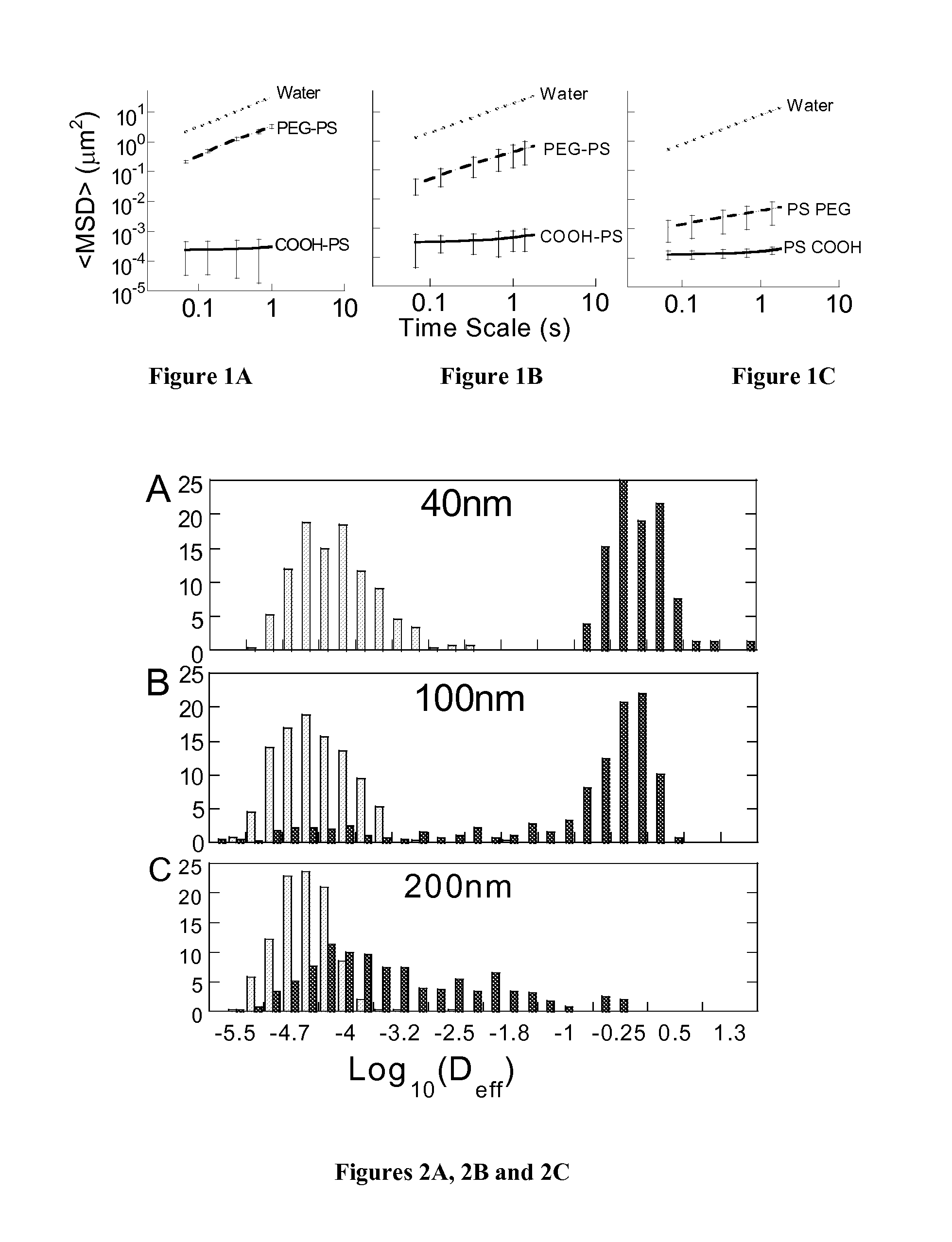
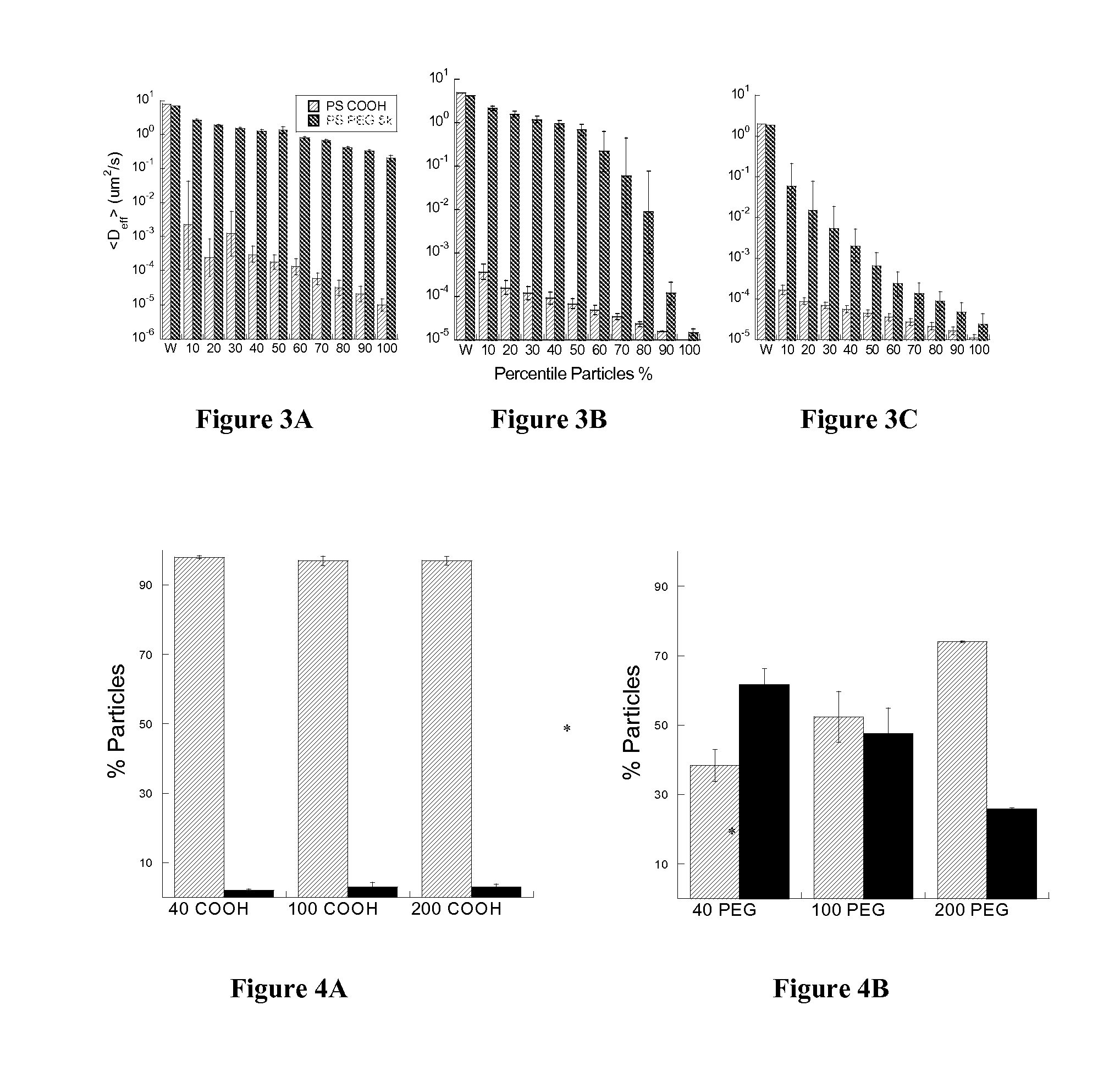
Determination of Size and Effect of Charge on Transport of Nanoparticles through Brain Tissue

[0152]Materials and Methods

[0153]Nanoparticle Preparation and Characterization

[0154]Forty- to 200-nm red fluorescent carboxyl modified polystyrene particles (Molecular Probes, Eugene OR) were covalently modified with methoxy-PEG-amine (molecular mass 5kDa; Creative PEG Works, Winston-Salem, N.C.) by carboxyl amine reaction. Briefly, 100 μL of 2% polystyrene particle suspension were washed and resuspended to 4-fold dilution in ultrapure water. An excess of MeO-PEG5000-NH2 was added to the particle suspension in a 1.5 mL Eppendorf tube and mixed to dissolve the PEG. Sulfo-NHS (Sigma) was added to each tube, and 200 mM borate buffer, pH 8.2, was added to a 4-fold dilution of the starting volume. The pH of each reaction tube was adjusted to pH 7.80, and then EDC was added to a concentration of 6.4 mM to each tube. Particle suspensions were placed on a rotary incubator for 4 hours and then ultrac...


PUM

PropertyMeasurementUnit
diameteraaaaaaaaaa
diameteraaaaaaaaaa
particle sizeaaaaaaaaaa

Abstract

Non-adhesive particles as large as 110 nm can diffuse rapidly in the brain ECS, if coated with hydrophilic coatings such as PEG coatings and preferably having neutral surface charge. The ability to achieve brain penetration with larger particles will significantly improve drug and gene delivery within the CNS since larger particles offer higher drug payload, improved drug loading efficiency, and significantly longer drug release durations.

Description

CROSS-REFERENCE TO RELATED APPLICATIONS[0001]This application claims benefit of and priority to U.S. Provisional Patent Application No. 61 / 381,754 filed on Sep. 10, 2010.FIELD OF THE INVENTION[0002]This invention is generally in the field of drug delivery, and in particular, a method of delivering drugs to the brain using coated particles to penetrate brain tissue.BACKGROUND OF THE INVENTION[0003]While the blood brain barrier has long been considered the crucial interface for therapeutic efficacy within the CNS, more recently poor distribution of agents within the brain itself has emerged as a major delivery challenge. See Arifin, et al. Pharm Res 26, 2289 (2009), Bobo et al., Proc Natl Acad Sci USA 91, 2076 (1994), Kunwar et al., Neuro Oncol 12, 871 (2010), Kunwar et al., J Clin Oncol 25, 837 (2007), Rosso et al., Cancer Res 69, 120 (2009), and Sampson et al., J Neurosurg 113, 301 (2010).[0004]The extracellular space (ECS) in the brain represents the major pathway for movement of m...

Claims


Application Information

Patent Timeline
Patent Type & Authority Applications(United States)
IPC IPC(8): A61K9/00
CPCA61K9/0085A61K49/0093A61K49/0065A61K9/5138A61P25/00
Inventor HANES, JUSTINWOODWORTH, GRAEME F.NANCE, ELIZABETH A.
Owner THE JOHN HOPKINS UNIV SCHOOL OF MEDICINE
Why Eureka
Try Eureka