Echogenic medical device

a medical device and echogenic technology, applied in the field of medical devices, can solve the problems of increasing the cost and complexity of the procedure, exposing the patient to potentially harmful radiation, and the image obtained via fluoroscopy may not achieve sufficient clarity, so as to enhance the echogenicity of the geometric configuration

Inactive Publication Date: 2013-07-25
COOK MEDICAL TECH LLC
View PDF8 Cites 27 Cited by
  • Summary
  • Abstract
  • Description
  • Claims
  • Application Information

AI Technical Summary

Benefits of technology

[0011]In another form thereof, a medical device configured for insertion into the body of a patient and ultrasound-guided movement therein to a target site is disclosed. A shaft has a proximal portion and a distal portion, wherein the distal portion extends to a distal end. An echogenic region at the distal portion comprises a plurality of geometric configurations disposed along the distal portion. At least some of the geometric configurations extend into a matrix of the shaft and define at least two wall angles1, 2. Each of the wall angles is configured and positioned to enhance an echogenicity of the geometric configuration under ultrasound visualization.

Problems solved by technology

However, the use of x-ray exposes the patient to potentially harmful radiation.
Additionally, health care workers must typically transport the patient to a specially-equipped radiology facility to obtain the x-ray, thereby increasing the cost and complexity of the procedure.
Further, the images obtained via fluoroscopy may not achieve sufficient clarity to provide the desired level of detail to the medical professional.
However, the video camera provides a field of view limited to only the immediate region.
Surgical instrumentation within the immediate region that is obstructed by body features or by other instrumentation cannot be visualized.
Similarly, instrumentation outside of the immediate region, such as outside the lumen in which the endoscope has been positioned, cannot be visualized with the endoscopic video camera.
For example, the device to be observed under ultrasound may not be easily visible at certain angles relative to the ultrasound probe.
In addition, the ultrasound image may not provide sufficient detail to enable the medical professional to determine with a high degree of confidence the particular orientation of the instrument in the viewing region, such as the degree of rotation of the device in the region.

Method used


Image

  • Echogenic medical device
  • Echogenic medical device
  • Echogenic medical device

Examples


Embodiment Construction

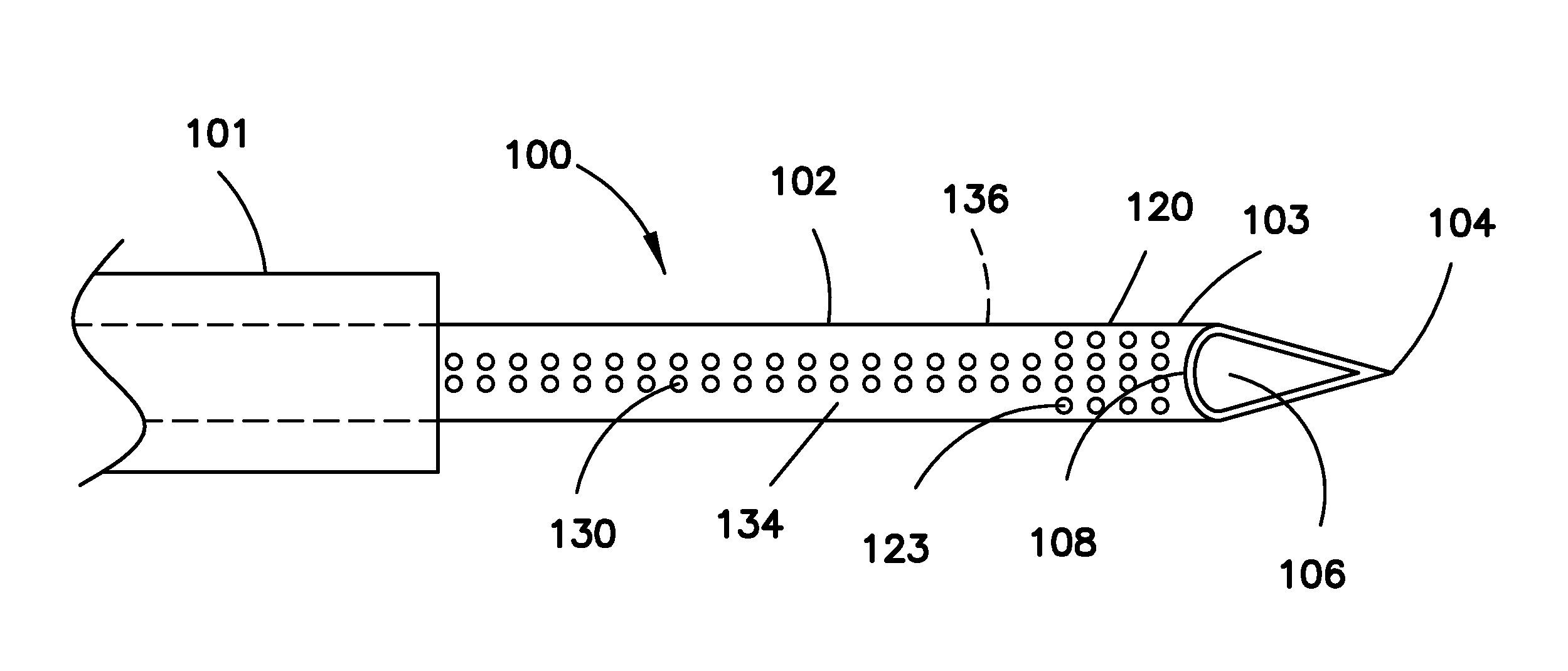
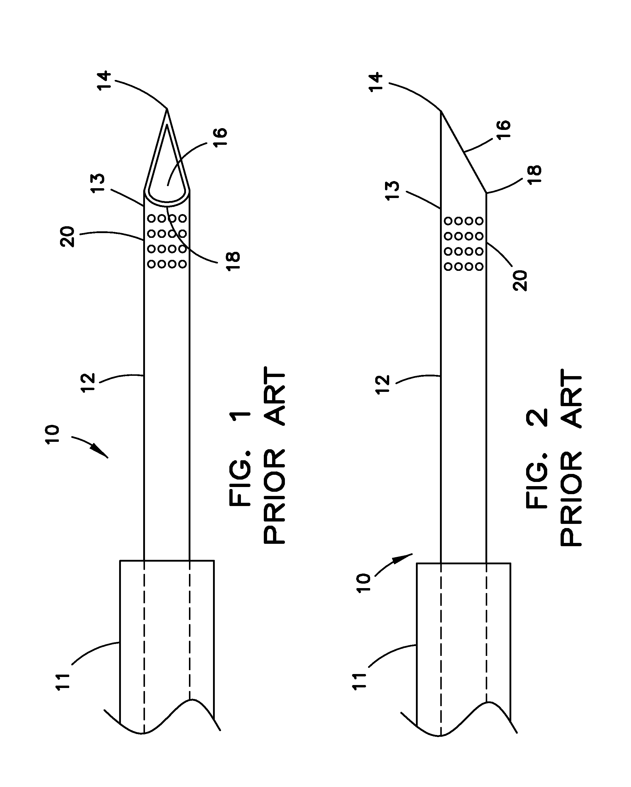
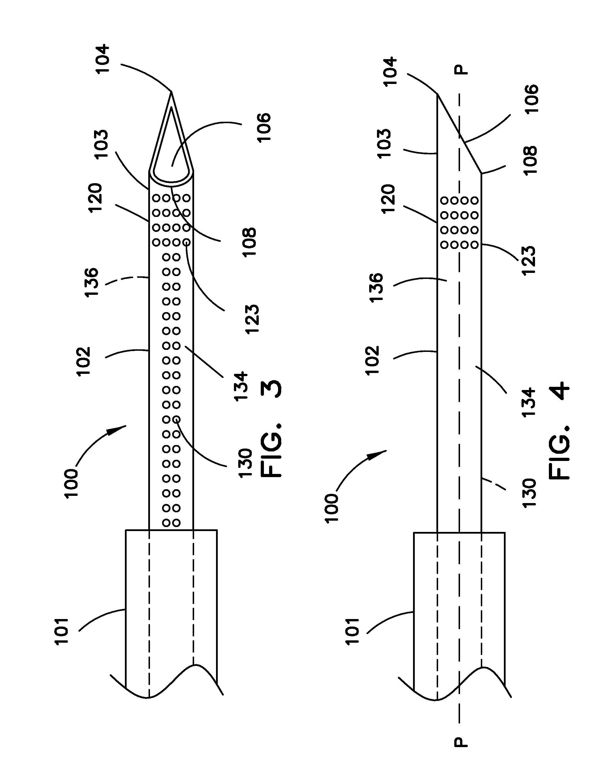
[0028]For purposes of promoting an understanding of the present invention, reference will now be made to the embodiments illustrated in the drawings, and specific language will be used to describe the same. It should nevertheless be understood that no limitation of the scope of the invention is thereby intended, such alterations and further modifications in the illustrated device, and such further applications of the principles of the invention as illustrated therein being contemplated as would normally occur to one skilled in the art to which the invention relates.

[0029]In the following discussion, the terms “proximal” and “distal” will be used to describe the opposing axial ends of the device, as well as the axial ends of various component features of the device. The term “proximal” is used in its conventional sense to refer to the end of the device (or component thereof) that is closest to the operator during use of the device. The term “distal” is used in its conventional sense ...


PUM


Abstract

An echogenic medical device includes a shaft having a proximal portion, a distal portion, first and second opposing longitudinal sides, and a passageway extending therethrough. The distal end includes a beveled opening communicating with the passageway, and extending between a distal tip portion disposed along the first longitudinal side and a heel portion disposed along the second longitudinal side. A first echogenic region extends circumferentially around the shaft at the distal portion. The first echogenic region is structured for providing a signal visible along the circumference of the shaft under ultrasound visualization. A second echogenic region extends along a length of the second longitudinal side and is substantially aligned with the heel portion. The second echogenic region is structured and arranged for providing a generally linear signal visible under ultrasound examination along the second longitudinal side, and substantially not visible along the first longitudinal side.

Description

RELATED APPLICATION[0001]The present patent document claims the benefit of the filing date under 35 U.S.C. §119(e) of Provisional U.S. Patent Application Ser. No. 61 / 590,495, filed Jan. 25, 2012, which is hereby incorporated by reference in its entirety.BACKGROUND OF THE INVENTION[0002]1. Technical Field[0003]The present invention relates to a medical device constructed for visualization within the body of a patient under medical imaging. More particularly, the invention relates to an echogenic medical device configured such that the location and rotational orientation of the device in the body of a patient may be observed in real time under ultrasound visualization.[0004]2. Background Information[0005]The ability to monitor the location and orientation of surgical instrumentation within intraluminal and extraluminal regions of the body of a patient has attained increased importance in recent years. Instruments formed of fluoroscopic and radiopaque materials are widely used to creat...

Claims


Application Information

Patent Timeline
Patent Type & Authority Applications(United States)
IPC IPC(8): A61B8/08
CPCA61B8/0841A61B17/3403A61B2019/5425A61B2017/3413A61B2017/00455A61B2090/3925
Inventor FISCHER, JR., FRANK J.
Owner COOK MEDICAL TECH LLC
Why Eureka
Try Eureka