
Robust Calibration and Self-Correction for Tissue Oximetry Probe

a tissue oximetry and self-correction technology, applied in the field of optical systems, can solve the problems of inaccurate saturation measurement, unstable tissue oxygenation state of patients, and fluctuation of existing oximeters, so as to reduce redundancy of reflectance data, improve accuracy, and improve the probability of derived optical properties.

Active Publication Date: 2013-12-05
VIOPTIX
View PDF2 Cites 36 Cited by
  • Summary
  • Abstract
  • Description
  • Claims
  • Application Information

AI Technical Summary

Benefits of technology

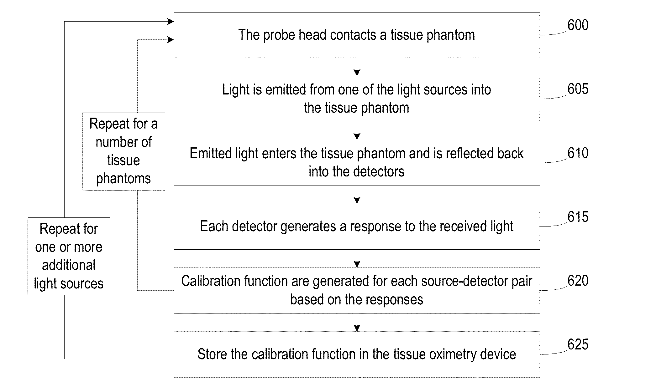
[0016]The above described embodiments of the tissue oximetry device and the method of use enable robust detector calibration and provides for identification of local inhomogeneity in real tissue and for discarding reflectance data from moles or other tissue aberrations. The positioning of the detectors with respect to the light sources provides a relatively large number of unique source-to-detector distances that increase the probability that derived optical properties are accurate by decreasing reflectance data redundancy and also enables a fast, reliable correction for dissimilarities in the data caused by power differences between sources.

Problems solved by technology

Thus, oximeters are often used in clinical settings, such as during surgery and recovery, where it may be suspected that the patient's tissue oxygenation state is unstable.
While existing oximeters have been sufficient for post-operative tissue monitoring where speed of measurement is less critical, existing oximeters fluctuate substantially and give inaccurate saturation measurements when used during surgery where various elements can interfere with accurate reading, such as if the oximeter comes in contact with blood.

Method used


Image

  • Robust Calibration and Self-Correction for Tissue Oximetry Probe
  • Robust Calibration and Self-Correction for Tissue Oximetry Probe
  • Robust Calibration and Self-Correction for Tissue Oximetry Probe

Examples


Embodiment Construction

[0030]Spectroscopy has been used for noninvasive measurement of various physiological properties in animal and human subjects. Visible light (e.g., red) and near-infrared spectroscopy is often utilized because physiological tissues have relatively low scattering in this spectral range. Human tissues, for example, include numerous chromophores such as oxygenated hemoglobin, deoxygenated hemoglobin, melanin, water, lipid, and cytochrome. The hemoglobins are the dominant chromophores in tissue for much of the visible and near-infrared spectral range. In the 700-900 nanometer range, oxygenated and deoxygenated hemoglobin have very different absorption features. Accordingly, visible and near-infrared spectroscopy has been applied to measure oxygen levels in physiological media, such as tissue hemoglobin oxygen saturation and total hemoglobin concentrations.

[0031]Various techniques have been developed for visible and near-infrared spectroscopy, such as time-resolved spectroscopy (TRS), fr...


PUM


Abstract

A method for calibrating detectors of a self-contained, tissue oximetry device includes emitting light from a light source into a tissue phantom, detecting in a plurality of detectors the light emitted from the light source, subsequent to reflection from the tissue phantom, and generating a set of detector responses by the plurality of detectors based on detecting the light emitted from the light source. The method further includes determining a set of differences between the set of detector responses and a reflectance curve for the tissue phantom, and generating a set of calibration functions based on the set of differences. Each calibration function in the set of calibration functions is associated with a unique, light source-detector pair. The method further includes storing the set of calibration function in a memory of the self-contained, tissue oximetry device.

Description

CROSS-REFERENCE TO RELATED APPLICATIONS[0001]This patent application claims the benefit of U.S. provisional patent applications 61 / 642,389, 61 / 642,393, 61 / 642,395, and 61 / 642,399, filed May 3, 2012, and 61 / 682,146, filed Aug. 10, 2012, which are incorporated by reference along with all other references cited in this application.BACKGROUND OF THE INVENTION[0002]The present invention relates generally to optical systems that monitor oxygen levels in tissue. More specifically, the present invention relates to oximeters that include sources and detectors on sensor heads for emitting and detecting light.[0003]Oximeters are medical devices used to measure oxygen saturation of tissue in humans and living things for various purposes. For example, oximeters are used for medical and diagnostic purposes in hospitals and other medical facilities (e.g., surgery, patient monitoring, or ambulance or other mobile monitoring for, e.g., hypoxia); sports and athletics purposes at a sports arena (e.g.,...

Claims


Application Information

Patent Timeline
Patent Type & Authority Applications(United States)
IPC IPC(8): A61B5/1495A61B5/1455
CPCA61B5/1495A61B5/14551A61B5/0075A61B5/14552A61B2560/0431A61B5/74A61B5/1455A61B90/39A61B5/7405A61B5/742A61B5/7475A61B5/7282A61M35/003A61B5/72A61B5/14546A61B90/11A61B5/0059A61B5/7246A61B2090/395A61B2090/065A61B2090/306A61B5/1459A61B2560/0475A61B2562/0271A61B2562/166
Inventor BECHTEL, KATE LEEANNWILFLEY, BRIAN
Owner VIOPTIX
Learn More