Vitro capture and analysis of circulating tumor cells

a technology of tumor cells and in vitro capture, applied in the field of cancer biology, can solve the problems of not capturing cells with low or no, poor sensitivity and specificity of metastatic cases, and a large number of cancer deaths

Active Publication Date: 2014-05-15
ROCHE MOLECULAR SYST INC +1
View PDF2 Cites 10 Cited by
  • Summary
  • Abstract
  • Description
  • Claims
  • Application Information

AI Technical Summary

Benefits of technology

[0007]In yet another embodiment, the invention is a method of assessing a risk of developing a metastatic tumor in a patient carrying or having carried a tumor by capturing circulating tumor cells from a patient's sample using a mammalian seprase-targeting affinity reagent. The patient's sample may comprise whole blood, body fluid, any cell-containing blood fraction, a fragmented tumor, a tumor cell suspension, or a cell culture established from a patient's sample, or the culture supernatant or a xenograft established from a patient's tumor. In variations of this embodiment, the method further comprises a step of detecting one or more of the following biomarkers: EpCAM, CD146, CK5, CK7, CK18, CK19, CD44, Cd44v6, EphB4, IGF-1R, BCL2, HER2, HER3, CA19-9, CEA, CD133, MUC1, N-cadherin, Survivin, EGFR, KRAS, BRAF, p53, Pi3KCA, PTEN, KRT19, CD34, CD24, ACT2, VIM, NANOG, CXCR4 and TWIST1 in the captured cel

Problems solved by technology

Metastasis to distal organs causes the majority of cancer deaths.
However, reports have suggested that this approach does not capture cells with low or no expression of EpCAM; and the assay shows poor sensitivity and specificity for metastatic cases.
Therefore, attempts to capture all CTC sub-populations (especial

Method used


Image

  • Vitro capture and analysis of circulating tumor cells
  • Vitro capture and analysis of circulating tumor cells

Examples


example 1

[0054]Selection of Seprase(+) and EpCAM(+) Circulating Tumor Cells (CTCs) from the Peripheral Blood of Pancreatic Cancer Patients

[0055]The following clinical measurements were secured from an approved IRB (University of North Carolina, Chapel Hill, Lineberger Cancer Center). These measurements are intended to illustrate embodiments of the present invention and not intended to limit the scope of the invention. The current invention is intended to include all adenocarcinomas or solid tumors.

Materials & Methods

[0056]The CTC microchips used for selection of seprase and EpCAM positive cells were fabricated in a thermoplastic (cyclic olefin co-polymer, COC or poly(methylmethacrylate), PMMA). The CTC microchips consisted of an architecture comprised of 50 sinusoidally-shaped channels that emanated from a common input and converged into a common output (see Adams et al., Highly Efficient Circulating Tumor Cell Isolation form Whole Blood and Label-Free Enumeration Using Polymer-based Microfl...

example 2

Determining the Ratio of Seprase(+) and EpCAM(+) CTCs from the Peripheral Blood of Patients with Various Cancers

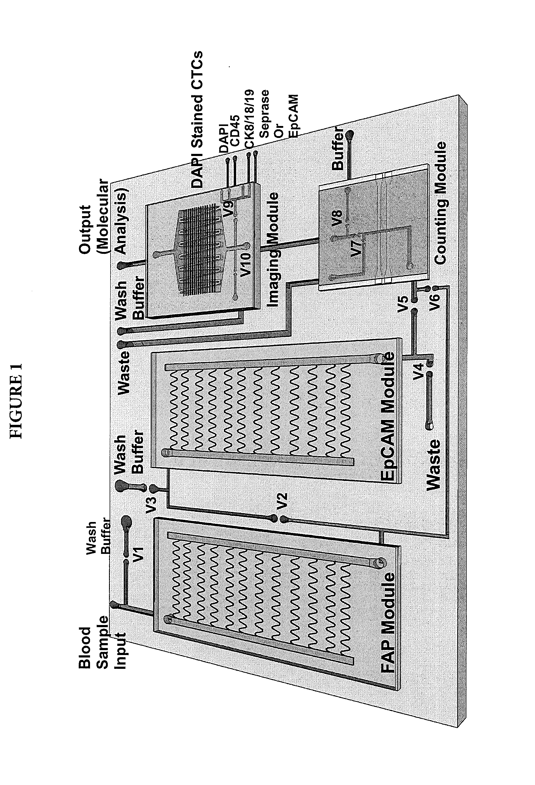
[0066]CTC microfluidic chips were fabricated in COC substrates. The chip design consisted of a 26.3×20.5 mm footprint with inlet and outlet leading channels (20.5 mm long, 400 μm wide, and 150 μm deep) connecting a series of 50-curvilinear channels that in concert formed the cell selection bed. FIG. 1 shows the CTC selection chip design. Each curvilinear selection channel was 30.6 mm long, 150 μm deep, and 25 μm wide. The surface area of the CTC selection bed was 596 mm2 (11 mm2 / channel) with 45.1 mm2 of that surface area in the lead channels. The chip's total volume was 9.4 μL (138 nL / channel) with 2.5 μL volume for the lead channels.

[0067]The depth of these channels increased the throughput as well as provided reduced pressure drop throughout the selection channels, especially when occupied by captured CTCs. On average, the widths of microchannels are only slightly large...

example 3

Expression Analysis of CTCs Captured with Seprase-Targeting Affinity Reagent

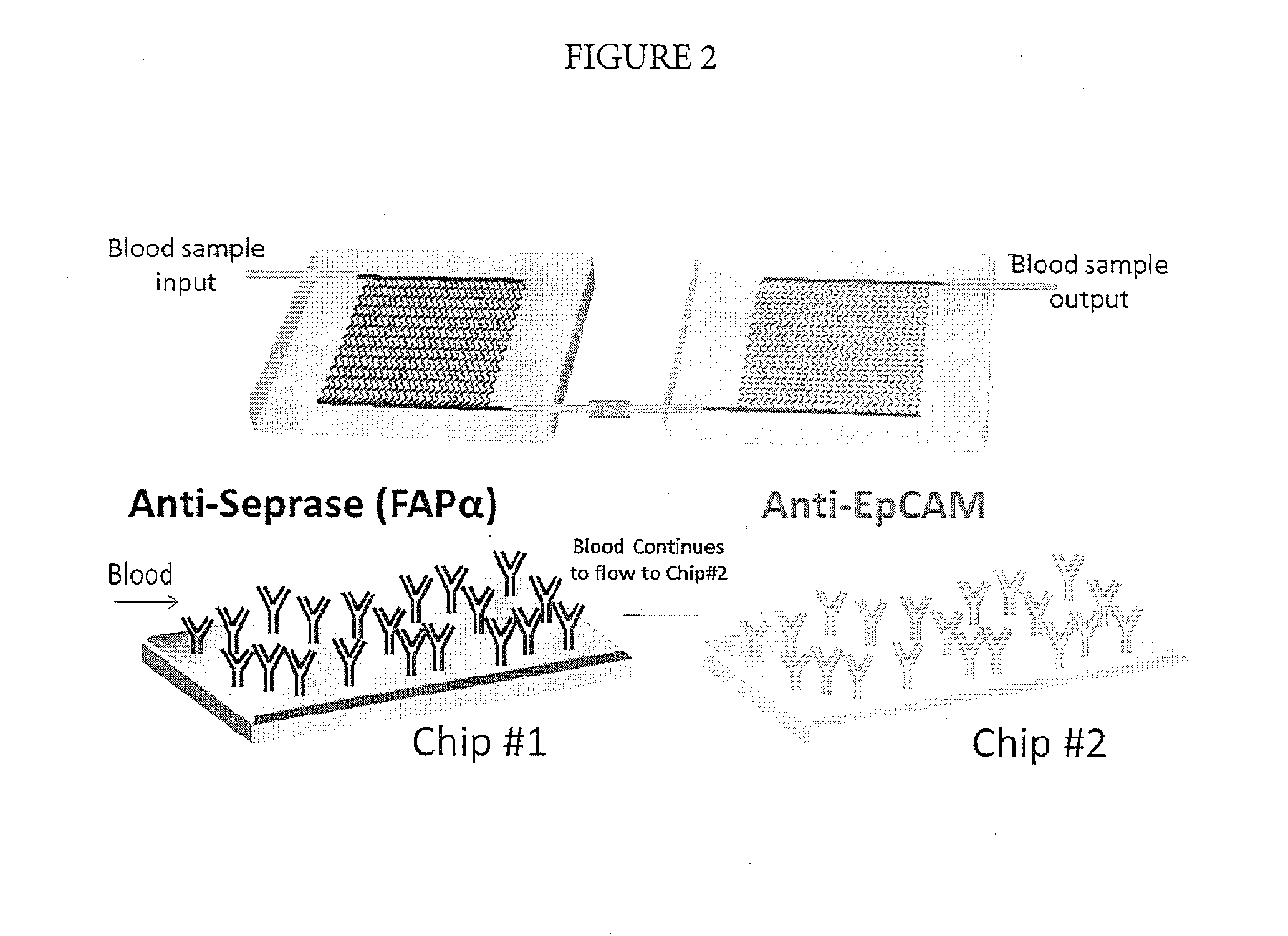
[0075]Using the methods described herein, CTCs were captured, released, enumerated via impedance detection (no staining performed), and collected in a microtube. The collected CTCs were spun spin down and lysed using ˜20 μL of lysing solution from a commercially available Cell-to-CT™ kit (Life Technologies, Grand Island, N.Y.). Cells-to-CT™ technology enables reverse transcription of lysates from 10-105 cells without isolating or purifying RNA. Eliminating the RNA isolation step substantially expedites and simplifies gene expression analysis of cells. As an alternative, CTCs lysis and isolation of RNA was performed directly using the CTC capture bed. This can be performed when another set of serially connected chips, modified with FAPα and EpCAM, is used to enumerate cells and characterize their phenotype via immunostaining.

[0076]Samples after lysis were treated with DNase to remove residual gDNA from the to...


PUM


Abstract

This invention provides methods and compositions for capturing circulating tumor cells (CTCs) as well as various divergent CTC phenotypes using seprase-specific affinity reagents. Methods of analyzing CTCs and assessing their metastatic potential in vivo and in vitro are also disclosed.

Description

FIELD OF THE INVENTION[0001]This invention relates to the field of cancer biology and more specifically, to methods and compositions for in vitro capture and analysis of circulating tumor cells, the source of cancer metastasis.BACKGROUND OF THE INVENTION[0002]Metastasis to distal organs causes the majority of cancer deaths. Circulating tumor cells (CTCs) originate from a primary tumor and initiate a metastasis cascade that ultimately results in metastatic tumors appearing at distal sites. See Sun et al. (2011) Circulating tumor cells: advances in detection methods, biological issues, and clinical relevance, J. Cancer Res. Clin. Oncol. 137:1151-1173. Several genetic, morphological, immunological and physio-logical tests may be used to identify CTCs. See Id., and Man, et al. (2011) Currently used markers for CTC isolation—advantages, limitations and impact on cancer prognosis, J. Clin. Exper. Pathol. 1:1. Because the number of CTCs in peripheral blood even in patients with advanced me...

Claims


Application Information

Patent Timeline
IPC IPC(8): G01N33/569
CPCG01N33/56966B01L3/5027G01N33/57492G01N2333/96433G01N33/54386G01N33/577G01N2333/96466
Inventor MARTIN, GEORGESOPER, STEVENWITEK, MAKGORZATAYEH, JEN JEN
Owner ROCHE MOLECULAR SYST INC
Why Eureka
Try Eureka