
Intra-fraction motion management system

a motion management system and motion management technology, applied in the field of radiation therapy, can solve the problems of radiation reaching tissue-destructive levels, destroying cancer cells, and normal structure included in the target volume being subject to damage, so as to avoid potential damage to surrounding tissue

Inactive Publication Date: 2014-07-31
ELEKTA AB
View PDF5 Cites 15 Cited by
  • Summary
  • Abstract
  • Description
  • Claims
  • Application Information

AI Technical Summary

Benefits of technology

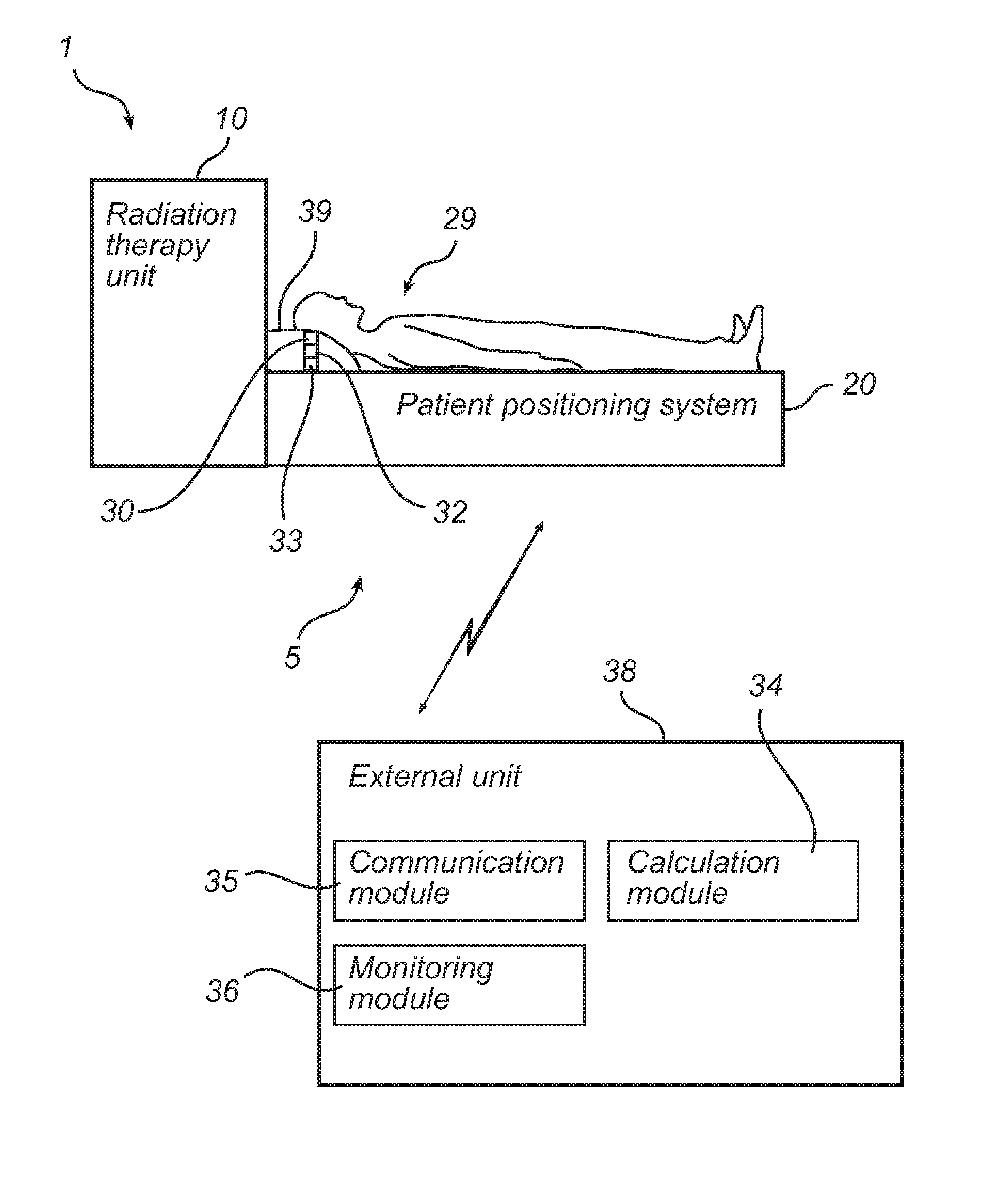
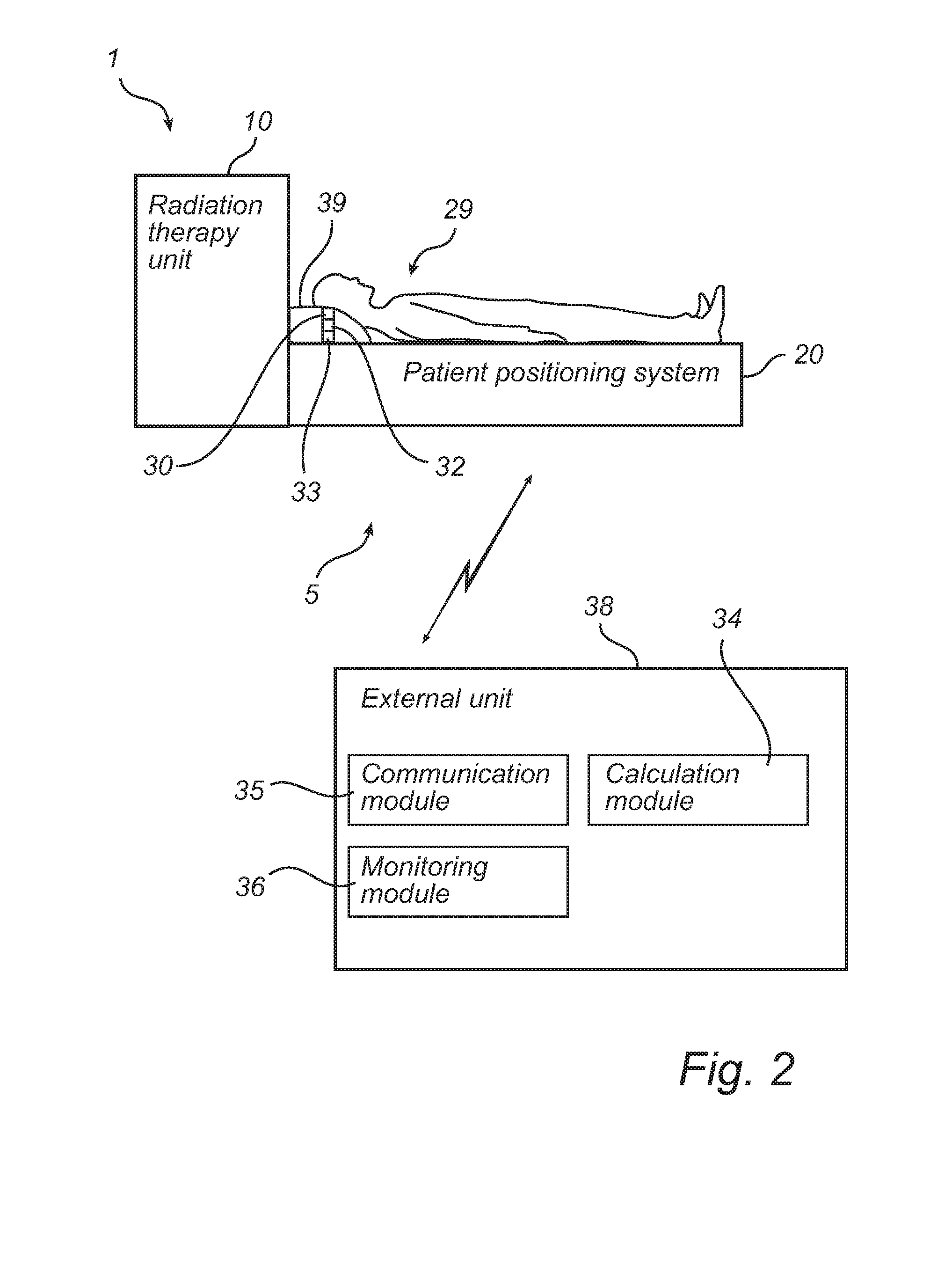
The present invention is a non-invasive motion detection system for monitoring changes in the position of bones or parts of bones in the human body, particularly for patients undergoing radiation therapy. The system uses ultrasonic transducers to measure the time it takes for signals to travel between the transducers and the bone. By measuring these changes, the system can accurately detect and track changes in the position of the bone or part of bone. This allows for real-time monitoring of patient motion, which can help to prevent damage to surrounding tissue and improve the effectiveness of the therapy. The system is easy to use, low-cost, and comfortable for patients. It can also be integrated with existing radiation therapy systems. The technical effects of the invention include improved accuracy and efficiency in motion detection, reduced risk of injury, and improved patient comfort.

Problems solved by technology

However, tissue destruction occurs where the radiation beams from all radiation sources intersect or converge, causing the radiation to reach tissue-destructive levels.
These x-rays can destroy the cancer cells while sparing the surrounding normal tissue.
Because destructive doses are used, however, any normal structure included in the target volume is subject to damage.
For example, damage to the neural tissue caused by the radiation may lead to irreparable damages such as partial paralysis.
In the different fixation devices used today it is not possible to immobilize that patient to such an extent that motions of body parts of the patient is completely eliminated or prevented.
If a spinal tumor in the cervical region is treated, very small movements of a cervical vertebra may result in that the radiation is mainly or partly delivered to the surrounding tissue instead of to the tumor, which may result in severe damages to the surrounding tissue.
However, these prior art methods are associated with drawbacks.
For example, imaging methods such as X-ray imaging or optical imaging require extensive image processing which may lead to complex and expensive solutions.
X-ray imaging also exposes the patient for radiation, which may be injurious.
Invasive solutions may be uncomfortable for the patient and may also be injurious for the patient.
Furthermore, the prior art systems may have problems in withstanding the gamma radiation generated in, for example, a Perfexion® system (a radiation therapy system provided by the applicant).
Further, the prior art systems are often bulky which makes it difficult to use them together with, for example, the Perfexion® system.

Method used


Image

  • Intra-fraction motion management system
  • Intra-fraction motion management system
  • Intra-fraction motion management system

Examples


Embodiment Construction

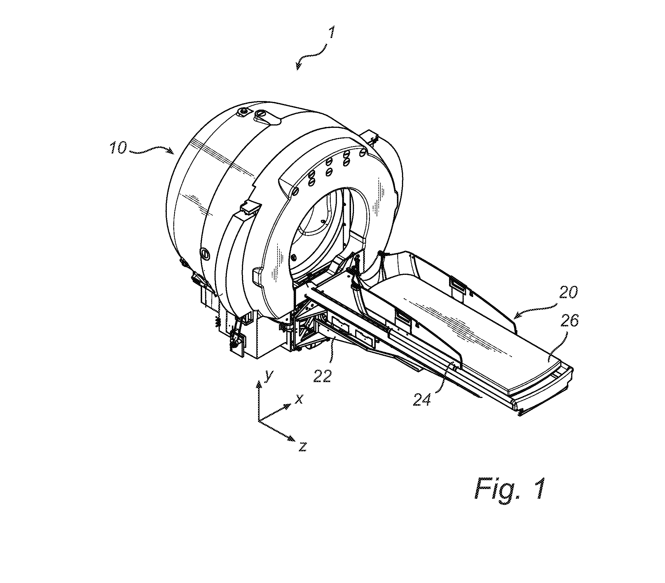
[0051]With reference to FIG. 1, a radiation therapy system 1 for which the present invention is applicable comprising a radiation unit 10 and a patient positioning unit 20 will be discussed. In the radiation unit 10, there are provided radioactive sources, radioactive source holders, a collimator body, and external shielding elements. The collimator body comprises a large number of collimator channels directed towards a common focus point, in a manner as is commonly known in the art.

[0052]The collimator body also acts as a radiation shield preventing radiation from reaching the patient other than through the collimator channels. Examples of collimator arrangements in radiation therapy systems applicable to the present invention can be found in WO 2004 / 06269 A1, which is hereby incorporated by reference. However, the present invention is also applicable to radiation therapy systems using other arrangements for collimating radiation into a fixed focus point, such as is disclosed in U....


PUM


Abstract

The present invention relates to the field of radiation therapy. In particular, the invention concerns systems and methods for monitoring intra-fraction motions of patients in connection with treatment cancer in radiation therapy system. Ultrasound waves are generated in a direction of a bone or a part of a bone of the patient being in a fixed relation to a cancer tumor to be treated by radiation therapy such that the generated ultrasound waves are reflected by the bone or part of bone using an ultrasonic transducer and sensing the reflected ultrasound waves at least one ultrasonic transducer. Time intervals between generated ultrasound waves and corresponding sensed reflected ultrasound signals are determined for the at least one ultrasonic transducer. Based on the time intervals, motions of the bone or part of bone are monitored using changes of the time intervals, wherein the motions indicate that the patient or a part of the patient has moved.

Description

FIELD OF THE INVENTION[0001]The present invention relates to the field of radiation therapy. In particular, the invention concerns systems and methods for monitoring intra-fraction motions of patients in connection with treatment cancer in radiation therapy system.BACKGROUND OF THE INVENTION[0002]The development of surgical techniques has made great progress over the years. For instance, for patients suffering from cancer tumors requiring surgery, non-invasive surgery is now available which is afflicted with very little trauma to the patient.[0003]One system for non-invasive surgery is sold under the name of Leksell Gamma Knife®, which provides such surgery by means of gamma radiation. The radiation is emitted from a large number of fixed radioactive sources and is focused by means of collimators, i.e. passages or channels for obtaining a beam of limited cross section, towards a defined target or treatment volume. Each of the sources provides a dose of gamma radiation which is insuf...

Claims


Application Information

Patent Timeline
IPC IPC(8): A61B8/00A61N5/10A61G13/12A61B8/08
CPCA61N5/1049A61B8/0875A61B8/4483A61N2005/1097A61G13/121A61N2005/1058A61N5/1067A61N5/1069
Inventor CHEN, RUIWILLIAMS, MALCOLM
Owner ELEKTA AB
Learn More