Antibody for detecting epithelial ovarian cancer marker and method for diagnosing epithelial ovarian cancer

an epithelial ovarian cancer and marker technology, applied in the field of anti1, can solve the problems of poor prognosis of the disease, the highest mortality among gynecologic cancers, and the difficulty of detection of epithelial ovarian cancer, and achieve the effect of high accuracy ra

Active Publication Date: 2014-08-28
NAT INST OF ADVANCED IND SCI & TECH
View PDF0 Cites 8 Cited by
  • Summary
  • Abstract
  • Description
  • Claims
  • Application Information

AI Technical Summary

Benefits of technology

[0041]The antibody of the present invention and / or the fragment thereof is capable of specifically recognizing and detecting a glycosyltransferase MGAT5B polypeptide fragment as an epithelial ovarian cancer marker.
[0042]The hybridoma of the present invention can stably supply an anti-MGAT5B monoclonal antibody capable of specifically recognizing and detecting a glycosyltransferase MGAT5B as an epithelial ovarian cancer marker.
[0043]The method for diagnosis of epithelial ovarian cancer according to the present invention can detect epithelial ovarian cancer conveniently and relatively low invasively with high accuracy rate. As a result, whether or not a test subject has epithelial ovarian cancer can be determined early.

Problems solved by technology

Hence, this disease has poor prognosis and exhibits the highest mortality among gynecologic cancers.
In addition, epithelial ovarian cancer is generally difficult to detect by palpation before the ovary is enlarged at the advanced stage of symptoms.
Hence, epithelial ovarian cancer is often undetected in ordinary examination or diagnosis methods.
Although echography, MRI, CT, or the like is relatively effective for the early detection of epithelial ovarian cancer, this examination itself is extensive work with a high cost.
In addition, these approaches, unfortunately, do not always have high diagnostic accuracy to distinguish between benign and malignant tumors.
These proteins, however, present specificity problems in such a way that CA125 exhibits positivity to a non-cancer benign gynecologic disease such as endometriosis or CA72-4, CA19-9, and STN exhibit positivity to various cancers of the digestive system including the stomach and the large intestine in addition to ovarian cancer.
The markers differ in reactivity among these histological types and therefore, do not correctly reflect the progression of cancer in some cases.
Unfortunately, this histological type is therefore rarely detected even at the advanced stage.
Thus, these enzymes are not secreted to the outside of the cells and are therefore rarely detected in the body fluids of normal individuals.
In ovarian cancer, however, it is known that glycosyltransferases are abnormally cleaved due to the increased expression level of a certain kind of protease and released to the outside of the cells.
Hence, this glycosyltransferase exhibits a serum concentration of approximately 200 ng / mL even in normal individuals and thus fails to distinguish between benign disease and cancer by its enzymatic activity alone.
This may lead to specificity problems as ovarian cancer markers.

Method used


Image

  • Antibody for detecting epithelial ovarian cancer marker and method for diagnosing epithelial ovarian cancer
  • Antibody for detecting epithelial ovarian cancer marker and method for diagnosing epithelial ovarian cancer
  • Antibody for detecting epithelial ovarian cancer marker and method for diagnosing epithelial ovarian cancer

Examples


example 1

Preparation of Anti-MGAT5B Antibody and Hybridoma Producing the Antibody

1. Selection and Preparation of Immunogen

[0142]An immunogen MGAT5B gene was selected by real-time PCR analysis on transcripts of a total of 186 glycosyltransferases known in the art in ovarian cancer cell lines RMG-I, RMG-II, and RMG-V (all derived from clear cell adenocarcinoma), RMUG-S (derived from mucinous adenocarcinoma), peripheral blood cells, and normal tissues of the large intestine, the liver, and the stomach. Specifically, their transcription levels in the ovarian cancer cell lines and the normal tissues were ranked by the comparison of mean values and the comparison of maximum values to select a top glycosyltransferase as an ovarian cancer marker candidate. MGAT5B, one of the selected candidates, was ranked in first in terms of both the mean value and the maximum value and had a much higher transcription level in ovarian cancer cells than in normal tissues. In addition, MGAT5B was placed in a higher ...

example 2

Confirmation of Antigen Specificity of Monoclonal Antibody

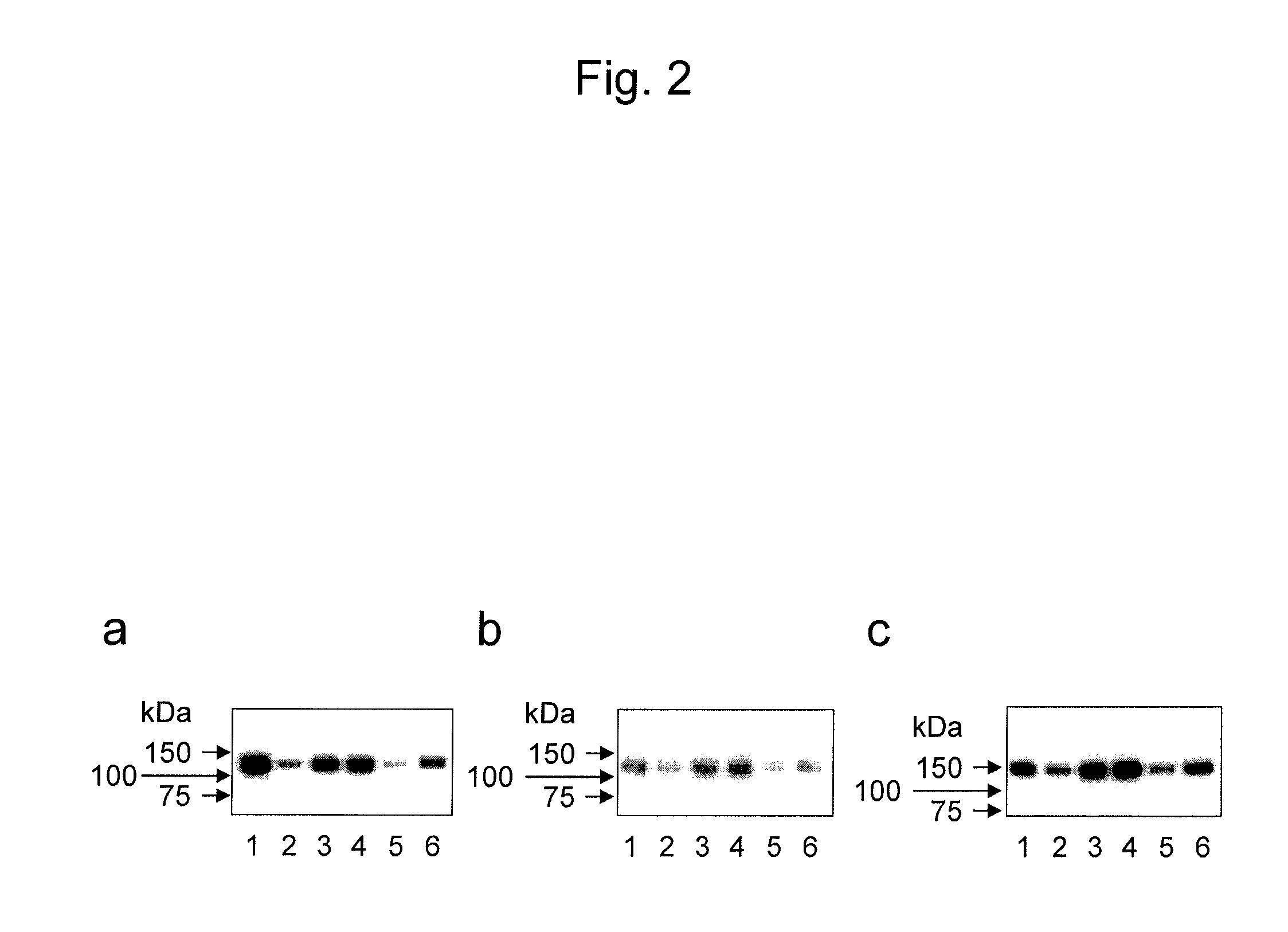
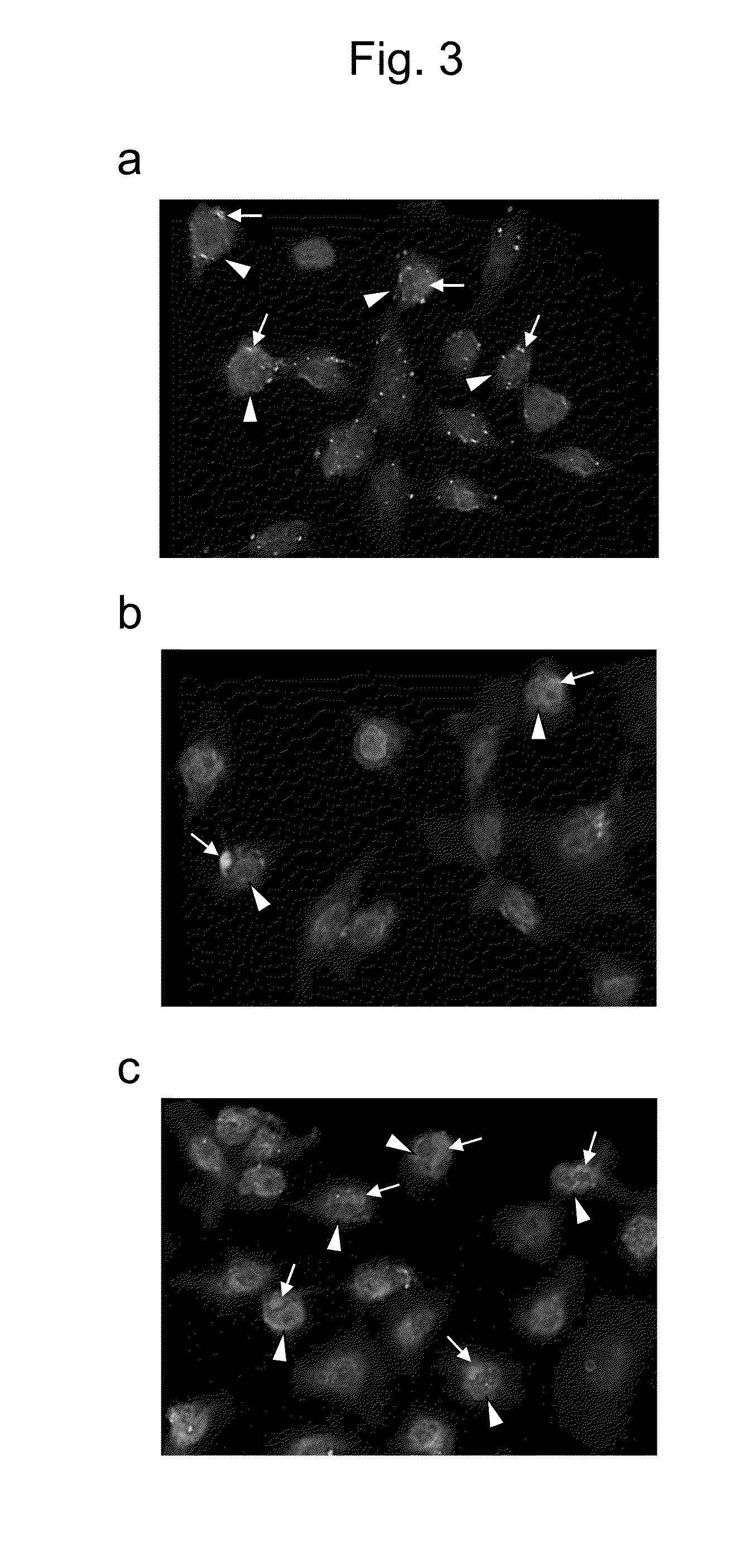
[0157]The hybridoma-derived anti-MGAT5B monoclonal antibodies (GT131-12 antibody and GT131-18 antibody) obtained in Example 1 were studied for their antigen specificity.

(Method)

[0158]10 ng of the FLAG-MGAT5B polypeptide fragment obtained in Example 1, 10 ng of the MGAT5B polypeptide fragment obtained by the cleavage of the FLAG tag from the fragment through enzymatic treatment, 1 μL of immunoglobulin- and albumin-free serum from a pool normal human serum (NHS) containing a serum mixture from a plurality of healthy adults, and 0.5 μL of the pool NHS were each electrophoresed using a 10% polyacrylamide gel under SDS-PAGE reduced conditions and transferred to a PVDF membrane. Each membrane was blocked with PBS containing 5% skimmed milk and then reacted with each purified MGAT5B monoclonal antibody (1 μg / mL) at room temperature for 90 minutes. After washing, the membrane was soaked with an HRP-labeled anti-mouse IgG antibody (GE...

example 3

Detection of MGAT5B Polypeptide Fragment by Immunoprecipitation Using Anti-MGAT5B Antibody

(Method)

[0160]The pool NHS was diluted 100-fold with a 50 mM tris buffer solution (pH 8.0) containing 0.15 M NaCl and 0.1% sodium azide. The FLAG-MGAT5B polypeptide fragment prepared in Example 1 was added thereto at a final concentration of 10 μg / mL. This mixture was used as an immunoprecipitation sample. 2 μg of each monoclonal antibody (GT131-2 antibody, GT131-7 antibody, GT131-12 antibody, or GT131-18 antibody) and 20 μL of a protein G gel (GE Healthcare Japan Corp.) were added to 0.5 mL of this sample and mixed at 4° C. for 4 hours using a rotator. The sample solution was discarded by centrifugation, and the protein G gel was washed with the above tris buffer solution. Then, 40 μL of an SDS-PAGE sample buffer was added thereto, and the mixture was heated at 98° C. for 5 minutes to obtain immunoprecipitated fractions.

[0161]Subsequently, the FLAG-MGAT5B polypeptide fragment, each fraction im...


PUM

PropertyMeasurementUnit
concentrationaaaaaaaaaa
dissociation constantaaaaaaaaaa

Abstract

It is intended to find a highly specific epithelial ovarian cancer marker and to provide an antibody capable of specifically recognizing and detecting the marker or a fragment of the antibody. The present invention provides an anti-β1,6-N-acetylglucosaminyltransferase 5B antibody for diagnosis of epithelial ovarian cancer, i.e., an antibody for detection of a glycosyltransferase β1,6-N-acetylglucosaminyltransferase 5B as an epithelial ovarian cancer marker. The antibody recognizes, as an epitope, a part of a polypeptide of the enzyme consisting of the amino acid sequence represented by SEQ ID NO: 1.

Description

TECHNICAL FIELD[0001]The present invention relates to an anti-β1,6-N-acetylglucosaminyltransferase 5B antibody for detection of an epithelial ovarian cancer marker, a hybridoma producing the antibody, and a method for diagnosis of epithelial ovarian cancer using the antibody.BACKGROUND ART[0002]Ovarian cancer is a cancer with a low incidence, compared with breast cancer or uterine cancer, among gynecologic cancers. Both incidence and mortality of this cancer, however, have been on the increase in recent years. In general, ovarian cancer is substantially asymptomatic early in the course of the disease and often found at the already advanced stage of symptoms. Hence, this disease has poor prognosis and exhibits the highest mortality among gynecologic cancers. Ovarian cancer is known to include, for example, surface epithelial-stromal tumors (hereinafter, referred to as “epithelial ovarian cancer”) developed from surface epithelial cells in the ovary and germ cell tumors developed from...

Claims


Application Information

Patent Timeline
Patent Type & Authority Applications(United States)
IPC IPC(8): G01N33/574C07K16/40
CPCG01N33/57449C07K16/40C07K2317/33C07K2317/34
Inventor SOGABE, MAKIKUBOTA, TOMOMITOGAYACHI, AKIRAIKEHARA, YUZURUNARIMATSU, HISASHISAWAKI, HIROMICHINAKANISHI, HAYAONAKANISHI, TORU
Owner NAT INST OF ADVANCED IND SCI & TECH
Why Eureka
Try Eureka