
Expandable cannula and method of use

a cannula and expansion technology, applied in the field of expandable cannula and method of use, can solve the problems of reducing the ability to achieve a current at the cutting electrode of sufficient density to initiate a cut, and reducing the size of the cutting electrode. , to achieve the effect of increasing the size of the working/hole, and expanding the hole in the tissu

Active Publication Date: 2014-09-18
KYPHON
View PDF6 Cites 3 Cited by
  • Summary
  • Abstract
  • Description
  • Claims
  • Application Information

AI Technical Summary

Benefits of technology

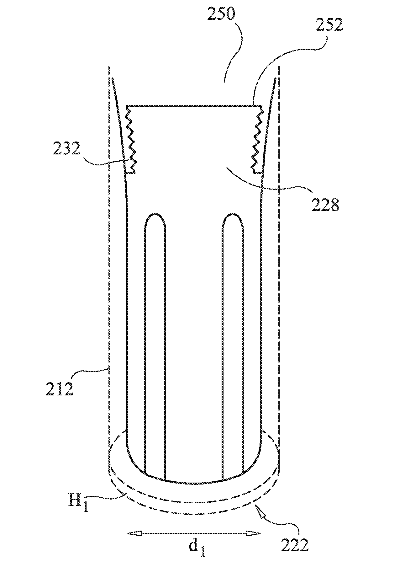
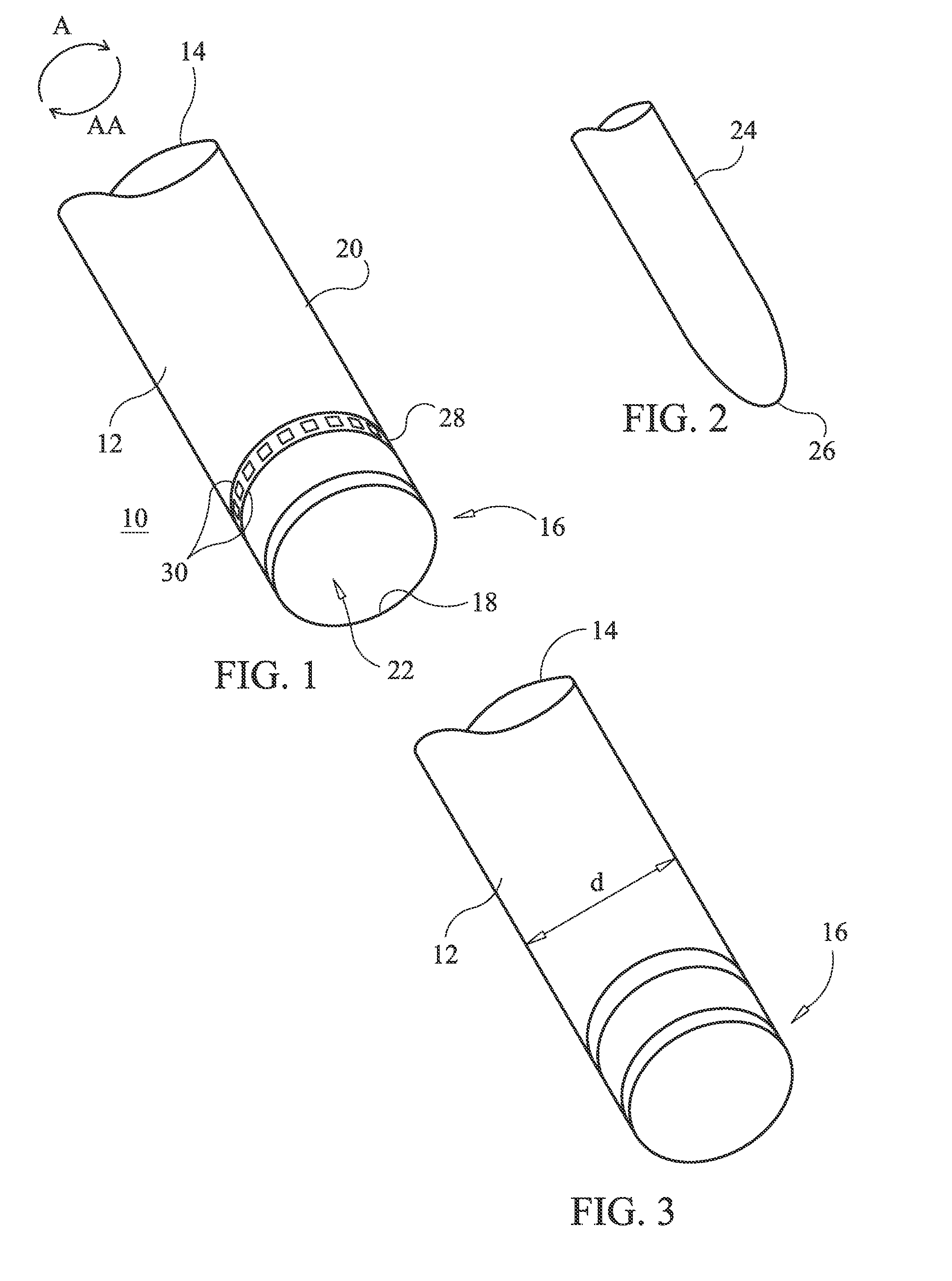
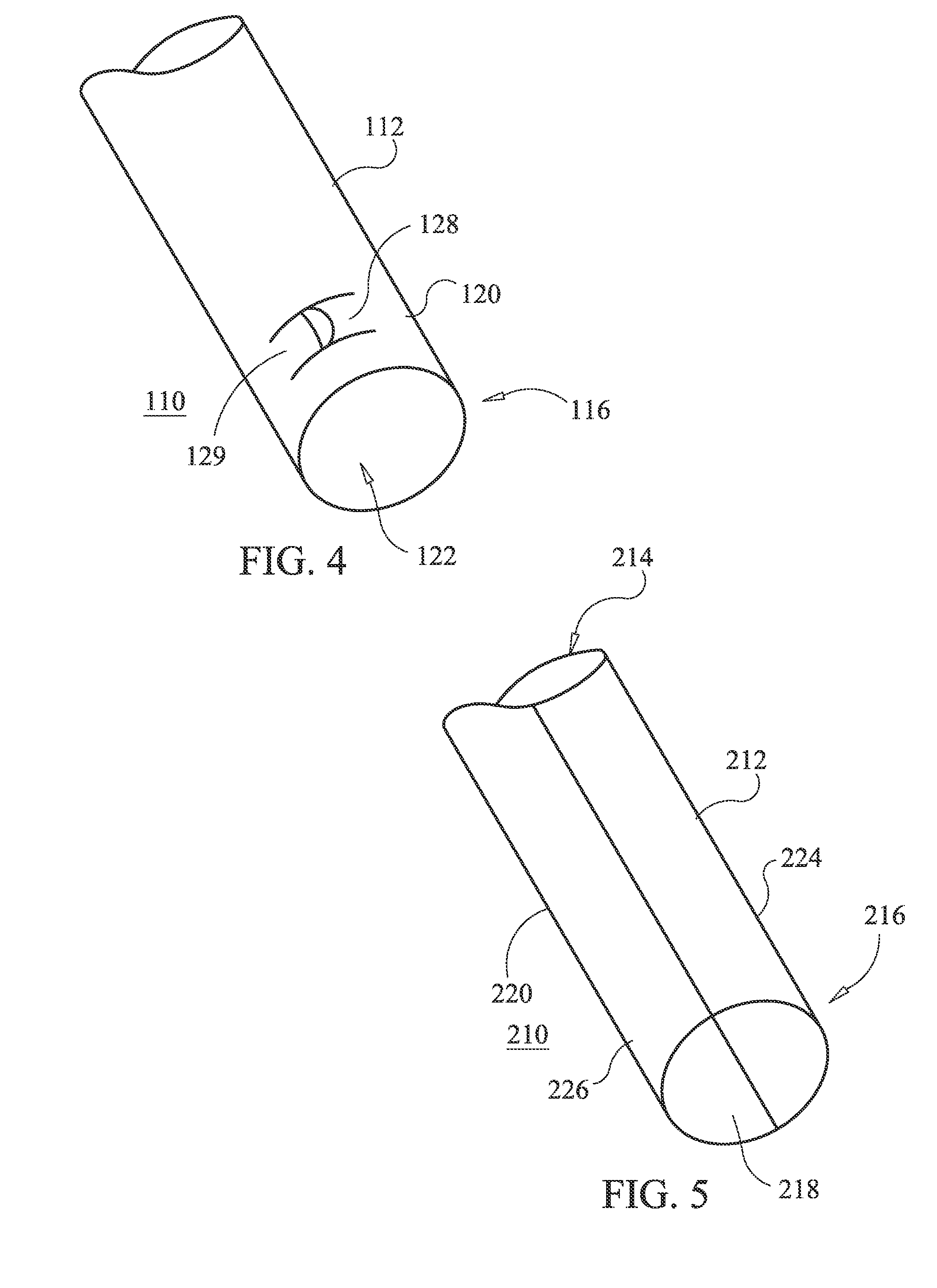
The patent describes a device that can be used to enlarge a hole in tissue. The device has a cannula with a retractable rod that can be expanded to increase the size of the hole. The device can be manipulated by a medical practitioner to widen the hole or sequential larger cannulas can be used to increase the size of the hole. The technical effect of the device is to provide a simple and effective way to enlarge a hole in tissue.

Problems solved by technology

Additionally, electrosurgical ablation procedures, where tissue surfaces and volume may be reshaped, cannot be duplicated through other treatment modalities.
Minimally invasive procedures in nerve and / or soft tissue such as the spine or the breast, however, are difficult to perform using standard scissors and scalpel.
Furthermore, in a closed environment, radio frequency current dissipates into the surrounding tissue causing a decreased ability to achieve a current at the cutting electrode of sufficiently high density to initiate a cut.
To overcome this problem, high power settings are often required to initiate the cut which often is painful and increases thermal damage to the tissue whether using a standard or a custom electrosurgical generator.
Another problem associated with cutting tissue is the control of bleeding.
However, while the bleeding is generally controlled, the cutting of tissue is often slower than with radio frequency energy and the knife edge readily dulls.
Other commercially available scalpels use ultrasonic energy generally at 50 kHz to heat the tissue so as to coagulate severed blood vessels but cut slower than a standard electrosurgical electrode and are costly as a custom ultrasonic generator is required.
A further disadvantage of using radio frequency energy is the generation of smoke.
Furthermore, the smoke often obscures visualization of the procedure.
This unnecessarily prolongs the operative time.
At the point of contact of the electric arcs with tissue, rapid tissue heating occurs due to high current density between the electrode and tissue.
Thus, the tissue is parted along the pathway of evaporated cellular fluid, inducing undesirable collateral tissue damage in regions surrounding the target tissue site.
This collateral tissue damage often causes indiscriminate destruction of tissue, resulting in the loss of the proper function of the tissue.
In addition, the device does not remove any tissue directly, but rather depends on destroying a zone of tissue and allowing the body to eventually remove the destroyed tissue.
Present electrosurgical techniques used for tissue ablation may suffer from an inability to provide the ability for fine dissection of soft tissue.
The distal end of electrosurgical devices are wide and flat, creating a relatively wide area of volumetric tissue removal and making fine dissections along tissue planes more difficult to achieve because of the lack of precision provided by the current tip geometries.
In addition, identification of the plane is more difficult because the large ablated area and overall size of the device tip obscures the physician's view of the surgical field.
The inability to provide for fine dissection of soft tissue is a significant disadvantage in using electrosurgical techniques for tissue ablation, particularly in arthroscopic, otolaryngological, and spinal procedures.
Traditional monopolar RF systems can provide fine dissection capabilities of soft tissue, but may also cause a high level of collateral thermal damage.
Further, these devices may suffer from an inability to control the depth of necrosis in the tissue being treated.
The high heat intensity generated by these systems causes burning and charring of the surrounding tissue, leading to increased pain and slower recovery of the remaining tissue.
Further, the desire for an electrosurgical device to provide for fine dissection of soft tissue may compromise the ability to provide consistent ablative cutting without significant collateral damage while allowing for concomitant hemostasis and good coagulation of the remaining tissue.
Another problem with currently available RF nerve ablation devices is that they attempt to destroy nerve tissue from a central location including the tip of the device and a 3-D spherical or cylindrical zone around it.
As a result, the further away the resecting ability is from the central zone the less effective the nerve destruction.
Consequently, often the nerve is not adequately ablated leading to continued pain symptoms.
Further, the health care practitioner may have difficulty positioning the tip of the device in the optimal location to get an optimal and consistent clinical result.
This may also result in unwanted necrosis of adjacent tissue, which can lead to clinical adverse events including subsequent repair of the necrotic tissue.
However, these devices require the insertion of relatively large cannulas that further complicate the surgical procedure and can cause nerve compression and pain with variable clinical efficacy.

Method used


Image

  • Expandable cannula and method of use
  • Expandable cannula and method of use
  • Expandable cannula and method of use

Examples


Embodiment Construction

[0026]Devices for efficient severing or cutting of a material or substance such as nerve, soft tissue and / or bone suitable for use in open surgical and / or minimally invasive procedures are disclosed. The following description is presented to enable any person skilled in the art to make and use the present disclosure. Descriptions of specific embodiments and applications are provided only as examples and various modifications will be readily apparent to those skilled in the art.

[0027]Lumbar spinal stenosis (LSS) may occur from hypertrophied bone or ligamentum flavum, or from a lax ligamentum flavum that collapses into the spinal canal. LSS can present clinical symptoms such as leg pain and reduced function. Conventional treatments include epidural steroid injections, laminotomy, and laminectomy. Surgical interventions which remove at least some portion of the lamina are usually performed through a relatively large incision, and may result in spinal instability from removal of a large...


PUM


Abstract

A cutting device includes an elongated shaft that extends between a proximal end and a distal end. A distal arm extends from the distal end of the elongated shaft. The distal arm includes an inner surface defining a cavity and an outer surface defining a blunt tip. At least one proximal arm extends from the distal end of the elongated shaft at a position proximal to the distal arm. The at least one proximal arm having an inner surface defines a cavity including a cutting portion configured to cut tissue.

Description

FIELD OF THE INVENTION[0001]The present invention relates generally to devices and methods for a surgical tool for creating access to and / or cutting a material or substance. More specifically, the devices and methods are useful for resecting nerve and / or soft tissue via a minimally invasive procedure to alleviate pain.BACKGROUND[0002]Standard methods of cutting tissue may include using a scalpel, scissors, and radio frequency energy. Electrosurgical procedures and techniques using radio frequency energy are currently used since they generally reduce patient bleeding and trauma associated with cutting operations. Additionally, electrosurgical ablation procedures, where tissue surfaces and volume may be reshaped, cannot be duplicated through other treatment modalities.[0003]Minimally invasive procedures in nerve and / or soft tissue such as the spine or the breast, however, are difficult to perform using standard scissors and scalpel. Furthermore, in a closed environment, radio frequenc...

Claims


Application Information

Patent Timeline
IPC IPC(8): A61B17/32A61B18/14A61B17/16A61B17/34A61B18/18A61B18/04
CPCA61B17/320016A61B17/1659A61B18/18A61B18/042A61B18/1487A61B17/3439A61B17/1615A61B17/3209A61B2017/0073A61B2018/00434A61B2018/00601A61B2018/1213
Inventor ARTHUR, AMY L.GOSHAYESHGAR, MOJAN
Owner KYPHON
Learn More