
Method and apparatus for electromagnetic treatment of cognition and neurological injury

a cognition and neurological injury technology, applied in the field of electromagnetic treatment devices, systems and methods, can solve the problems of inability to use electromagnetic devices with patients who are bedridden, metal-containing devices, toll of neurological deficits and mortality, etc., to accelerate or decelerate the production, enhance the cam-dependent no/cgmp signaling pathway, and increase the binding of ca2+

Inactive Publication Date: 2014-10-09
RIO GRANDE NEUROSCI
View PDF9 Cites 80 Cited by
  • Summary
  • Abstract
  • Description
  • Claims
  • Application Information

AI Technical Summary

Benefits of technology

The patent describes a protective helmet that can deliver electromagnetic treatment to the user's head. The helmet has a layer of padding to provide comfort and reduce impact forces on the head. The helmet also has a sensor that detects an impact parameter and activates the electromagnetic treatment device if the impact parameter exceeds a predetermined threshold. This invention can improve neuronal survival.

Problems solved by technology

Although PEMF / PRF therapy has been used for a variety of treatments, one challenge has been in providing a PEMF / PRF delivery device in a design configuration that accommodates the patient's injury and concurrent treatment.
For example, EMF devices are difficult to use with patients who are bed-ridden, bandaged, and engaged in ongoing treatment (or monitoring) by metal-containing devices.
The toll of neurological deficits and mortality from TBI continue in the military and private sectors and, to date, there are no widely successful medical or surgical interventions to prevent neuronal death.
Current medical practice has attempted to use pharmaceuticals to mitigate and prevent tissue damage and injury resulting from secondary physiological responses of traumatic brain injury with little success.
However, the data and safety monitoring board halted the trial after half of the patients were enrolled as it became apparent that MPSS significantly increased mortality of severe injuries from 17.9% to 21.1% (P=0.0001).
In comparison, for example, inflammation and swelling in the CNS can lead to secondary tissue damage and neuronal death.
Moderate to severe TBI can produce mechanical damage characterized by the disruption of cell membranes and blood vessels, resulting in direct and ischemic neuronal death.
Moreover, inflammation and swelling reduces blood flow to the brain and can cause damage and death of healthy brain tissue.
However, a significant challenge has been that current available EMF devices are difficult to use with patients who are bed-ridden, heavily bandaged, and / or wearing surgical, monitoring, or metal containing devices that can interfere with the delivery of therapeutic EMF.
For example, a TBI patient may be placed in an immobilizing body support article such as a head and neck brace during transport to a hospital, which limits access by EMF devices to the injured region.
After sustaining an injurious event such as a fall, patients are often left minimally assisted or completely unassisted for minutes to several hours.
Treatment for improving cognition has been limited to the use of pharmaceuticals (e.g. psychostimulants or cholinergic agents) that can target neurotransmitters or neuropathways in the central nervous system (CNS).
However, reliance on pharmaceutical treatments has several drawbacks including limited bioavailability of the drug and severe adverse side effects such as vomiting, convulsions, and bradycardia.
Furthermore, once administered, it is often difficult to completely limit the pharmacokinetics and effects of a psychopharmaceutical to a single target neuropathway.
For example, typical antipsychotic drugs (e.g. haloperidol) that target the brain's dopamine pathways have the unwanted side effect of blocking other dopamine pathways, which can cause extrapyramidal motor side effects that can persist long after the medication is discontinued.

Method used


Image

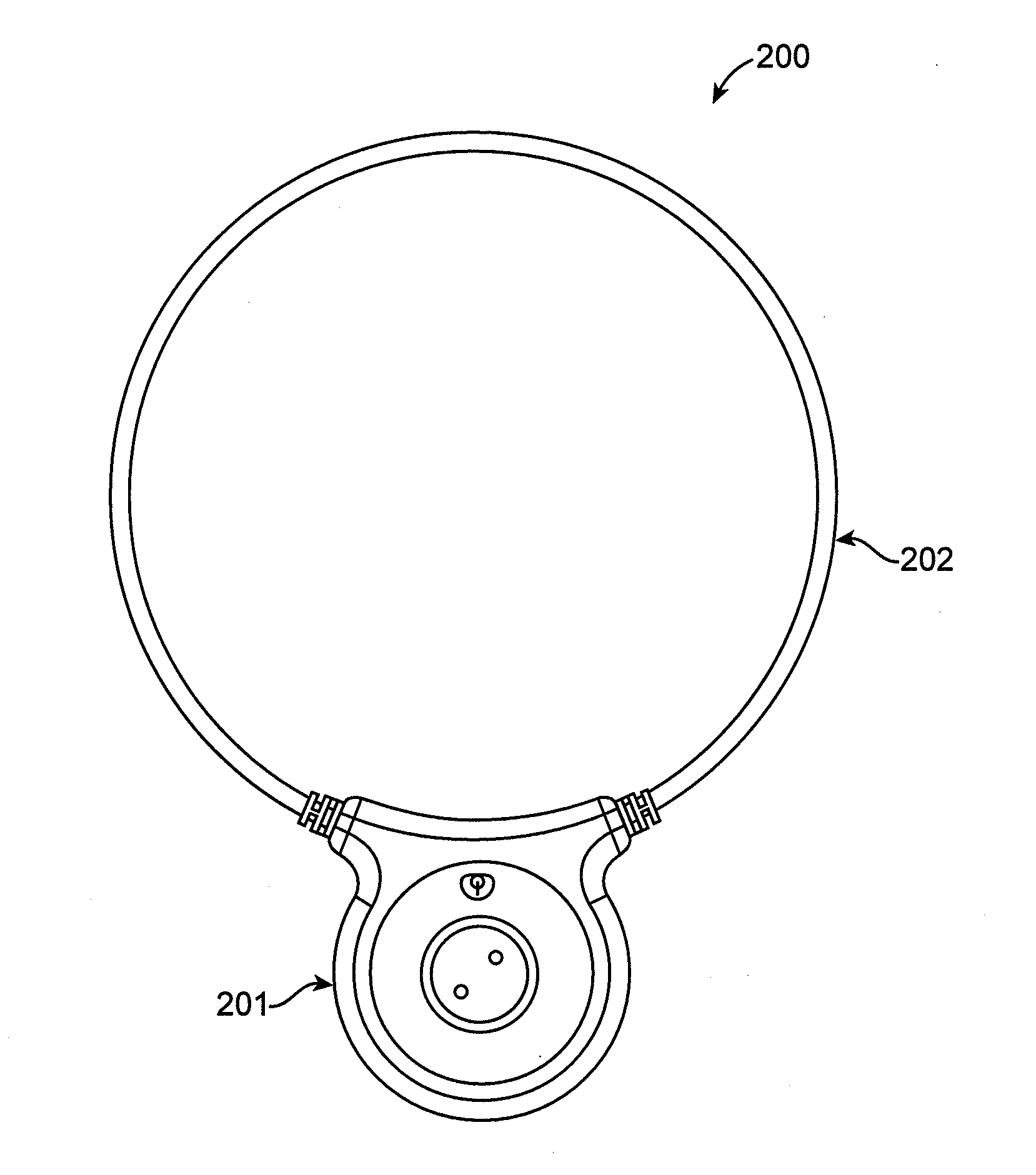
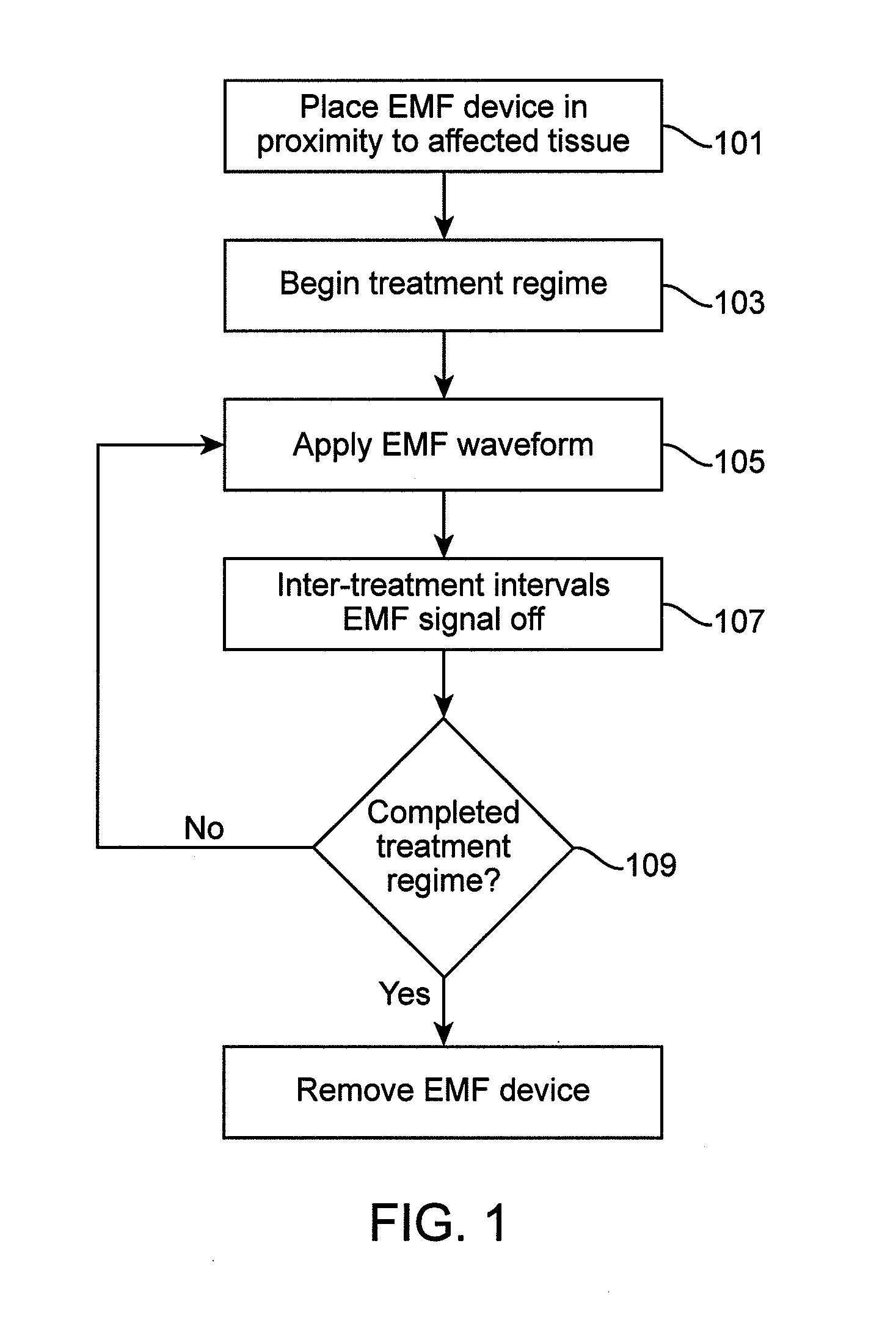
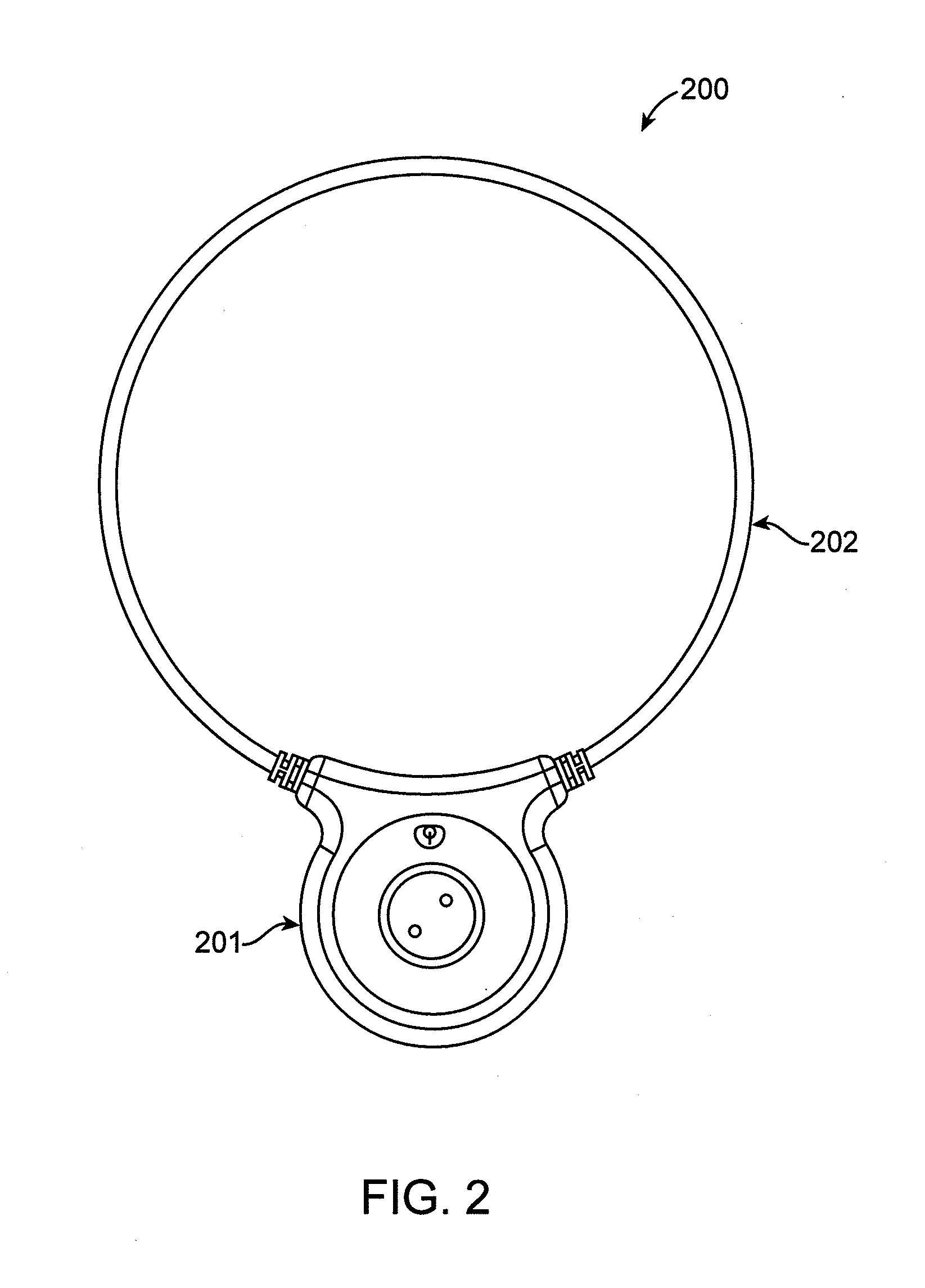
  • Method and apparatus for electromagnetic treatment of cognition and neurological injury
  • Method and apparatus for electromagnetic treatment of cognition and neurological injury
  • Method and apparatus for electromagnetic treatment of cognition and neurological injury

Examples


example 1

[0226]In this example experiments, designed to assess the EMF effect on NO release, were performed on a dopaminergic cell line (MN9D) in culture. Cells were plated at 100,000 cells / 35 mm dish in Dulbecco's Modified Eagle's medium (DMEM) containing 10% fetal calf serum and allowed to stabilize for 24 hours. Thereafter, serum was withdrawn and cells allowed to stabilize for 6 hours at 37° C. These cultures were placed at room temperature for 15 min to create a repeatable stress which caused cytosolic Ca2+ to rise, thereby activating CaM. Cells were then treated for 15 min with a non-thermal RF signal configured according to the teachings of this application, which consisted of a 27.12 MHz carrier pulse-modulated with a burst duration of 3 msec at 2 bursts / sec. In situ signal amplitude was 0.05 G which induced a mean electric field of approximately 18 V / m. The results in FIG. 17 show the EMF signal increased NO production by several-fold, and that this was inhibited by N-(6-Aminohexyl)...

example 2

[0227]In this example experiments, designed to assess the EMF effect on cAMP release, were performed on a dopaminergic cell line (MN9D) in culture. Cells were plated at 100,000 cells / 35 mm dish in Dulbecco's Modified Eagle's medium (DMEM) containing 10% fetal calf serum and allowed to stabilize for 24 hours. Thereafter, for the cAMP signaling experiments, serum was withdrawn and cells allowed to stabilize for 6 hours at 37° C. Cells were then treated for 15 min with a non-thermal RF signal configured according to the teachings of this application, which consisted of a 27.12 MHz carrier pulse-modulated with a burst duration of 3 msec at 2 bursts / sec. In situ signal amplitude was 0.05 G which induced a mean electric field of approximately 18 V / m. The results in FIG. 18 show the EMF signal increased cAMP production approximately 2-fold, and that this was inhibited by L-nitrosoarginine methyl ester (L-NAME), a cNOS inhibitor. These results demonstrate that an EMF signal configured accor...

example 3

[0228]In this example experiments, designed to assess the EMF effect on neurite outgrowth (differentiation), were performed on a dopaminergic cell line (MN9D) in culture. Cells were plated with or without fetal calf serum and 1 mM dibutyryl cyclic adenosine monophosphate (Bt2cAMP). At 1 day, immature cultures were divided into two groups and treated with a non-thermal RF signal configured according to the teachings of this application, which consisted of a 27.12 MHz carrier pulse-modulated with a burst duration of 3 msec at 2 bursts / sec. In situ signal amplitude was 0.05 G which induced a mean electric field of approximately 18 V / m. EMF treatment was 30 minutes a day for three days. Cultures assigned to control groups were exposed to the same conditions in the absence of EMF signals. After three days of treatment, cells were fixed and photographed for subsequent analysis with ImageJ. Measurements of neurite length, cell numbers, and number of cells with and without processes were qu...


PUM


Abstract

Methods and devices for providing therapeutic electromagnetic field treatment to a subject having a cognitive or neurological condition or injury. Treatment devices can include headwear incorporating electromagnetic treatment delivery devices providing electromagnetic treatment to a user's head area. Such devices include protective headwear such as helmets with electromagnetic delivery devices. Additionally, embodiments of the invention provide for wearable and adjustable electromagnetic treatment devices that can be used to provide electromagnetic treatment to multiple areas of the user's head. Embodiments of the invention provide for sequential electromagnetic treatment with a single or a plurality of treatment applicators which target a single or multiple cerebral regions as determined by imaging, non-imaging and physiological monitoring before, during and after electromagnetic treatment.

Description

CROSS REFERENCE TO RELATED APPLICATIONS[0001]This application claims priority to U.S. provisional patent application 61 / 556,068, filed Nov. 4, 2011, and titled “METHOD AND APPARATUS FOR ELECTROMAGNETIC TREATMENT OF COGNITION AND NEUROLOGICAL INJURY”.[0002]This application may also be related to any of the following patent applications, each of which is herein incorporated by reference in its entirety: U.S. patent application Ser. No. 11 / 003,108, filed Dec. 3, 2004, now U.S. Pat. No. 7,744,524 (“APPARATUS AND METHOD FOR ELECTROMAGNETIC TREATMENT OF PLANT, ANIMAL AND HUMAN TISSUE, ORGANS, CELLS AND MOLECULES”); U.S. patent application Ser. No. 12 / 771,954, filed Apr. 30, 2010, titled “APPARATUS AND METHOD FOR ELECTROMAGNETIC TREATMENT OF PLANT, ANIMAL AND HUMAN TISSUE, ORGANS, CELLS AND MOLECULES”; U.S. patent application Ser. No. 12 / 772,002, filed Apr. 30, 2010, titled “APPARATUS AND METHOD FOR ELECTROMAGNETIC TREATMENT OF PLANT, ANIMAL AND HUMAN TISSUE, ORGANS, CELLS AND MOLECULES”; ...

Claims


Application Information

Patent Timeline
Patent Type & Authority Applications(United States)
IPC IPC(8): A61N2/02A61B5/11A61B5/03A61B5/0488G01R33/48A61B6/03A61B5/0476A61B5/00A61N2/00
CPCA61N1/40A61N2/006A61N2/02A61N5/022A61N2/004A61B6/037A61B5/4836A61B5/6803G01R33/4806A61B5/11A61B5/0488A61B5/0476A61B5/031A61B5/746A61B5/0036A61B5/369A61B5/389
Inventor PILLA, ARTHUR A.DIMINO, ANDRE' A.HAGBERG, SEANGLUCKKSTERN, STEVEN M.
Owner RIO GRANDE NEUROSCI
Learn More