
Magnetic Resonance Based Method for Assessing Alzheimer's Disease and Related Pathologies

a magnetic resonance and pathology technology, applied in the direction of nmr measurement, instruments, applications, etc., can solve the problems of no cure, no definitive diagnostic currently available for longitudinal use, and neither of these modalities currently has the ability to see changes

Inactive Publication Date: 2014-10-09
OSTEOTRONIX MEDICAL PTE LTD
View PDF10 Cites 13 Cited by
  • Summary
  • Abstract
  • Description
  • Claims
  • Application Information

AI Technical Summary

Benefits of technology

The patent is about a non-invasive method to detect and monitor changes in brain structures and tissue in response to disease progression and treatment. The invention is particularly useful for diagnosing and monitoring Alzheimer's disease, but can also be used for other dementias and motor diseases. The technical effect is an accurate, non-invasive technique to detect and monitor changes in brain structures and tissue in response to disease and injury.

Problems solved by technology

The exact cause of AD is unknown and, although a variety of therapies that purport to lessen the effects of AD are available, there is at present no cure.
Though various in vivo diagnostics are available, there is no definitive diagnostic currently available for longitudinal use.
However, neither of these modalities currently has the ability to see changes on the structural size level of the columnar organization of neurons in-vivo.
As such, diagnosis is often not made until the disease has resulted in significant changes to a patient's behavior.
Four out of five of these biomarkers present serious drawbacks to use as a routine and a longitudinal diagnostic.
Use of CSF biomarkers involves painful and invasive sample withdrawal, and therefore is not able to be used routinely longitudinally.
The associated risk would preclude its use in clinical studies without clearly demonstrable patient benefit.
Further, these fluid sampling biomarkers cannot differentiate signal levels by anatomic position in the brain, as is possible with imaging biomarkers.
As the progress of various forms of dementia, as well as stages in pathology progression, are often distinguished by differential effects and rates of progression in different brain regions, this is a serious drawback to use of fluid biomarkers.
Along with being extremely costly, PET imaging requires use of radiotracers and positioning / calibration x-rays.
This makes implementation as a routine diagnostic, and especially as a longitudinal one, problematic.
Further, the role of Aβ plaques in disease etiology is not well understood; it is thought that these plaques may be incidental rather than causative in the disease process (see Mateen C. Moghbel et al., “Amyloid-β imaging with PET in Alzheimer's disease: is it feasible with current radiotracers and technologies?”, European Journal of Nuclear Medicine and Molecular Imaging, Oct. 19, 2011).
(Some PET agents, including PiB, also have half-lives too short to allow practical clinical use.)
Therapies that remove amyloid beta plaques from the brain have been shown to offer no improvement in, or slowing of loss of, cognition.
Use of PET radiotracers to determine Aβ load is problematic: there is often a striking discrepancy in the measured distribution of Aβ deposits in the brain from PET radiotracer images as compared to that measured by histopathological and immunohistochemical studies, which may be due in part to the low resolution of PET imaging (2-3 mm) causing partial volume effects when used to measure structures on the order of 100 microns (see Mateen C. Moghbel et al., “Amyloid-β imaging with PET in Alzheimer's disease: is it feasible with current radiotracers and technologies?”, European Journal of Nuclear Medicine and Molecular Imaging, Oct. 19, 2011).
It is difficult in many cases to differentiate dementia caused by a disease such as AD from that caused by CVD.
Another difficulty in assessing brain function in normal as well as diseased brains arises due to a lack of ability to, in vivo, determine the boundaries of the various control regions of the cerebral cortex or the different Brodmann's areas of which these are comprised.

Method used


Image

  • Magnetic Resonance Based Method for Assessing Alzheimer's Disease and Related Pathologies
  • Magnetic Resonance Based Method for Assessing Alzheimer's Disease and Related Pathologies
  • Magnetic Resonance Based Method for Assessing Alzheimer's Disease and Related Pathologies

Examples


Embodiment Construction

[0046]The current invention consists of adaptations / refinements to U.S. Pat. No. 7,932,720, to facilitate application of this prior art to brain pathology, specifically to the etiology attendant with onset and development of AD and other associated dementias, though the refinements also can be applied to probing regions of the brain to measure many other pathology and trauma-induced tissue effects.

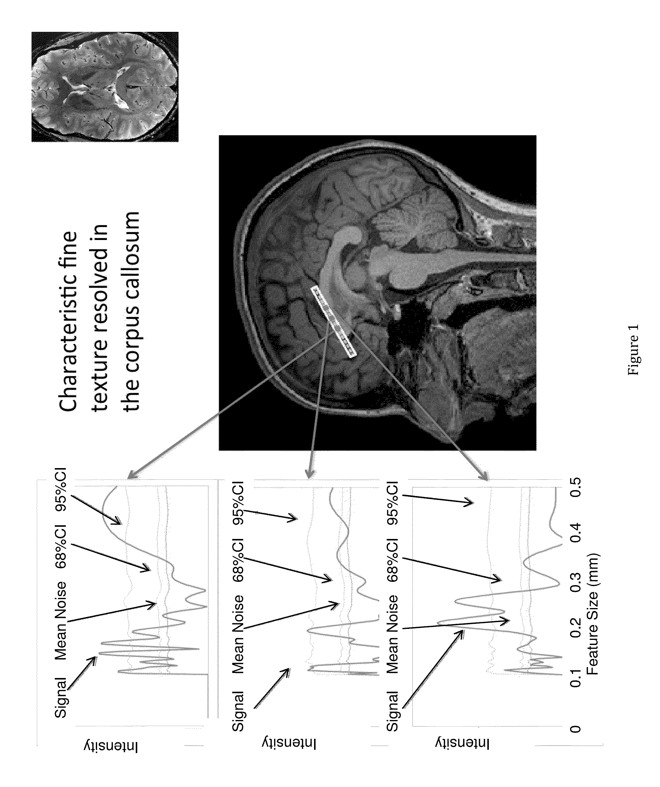
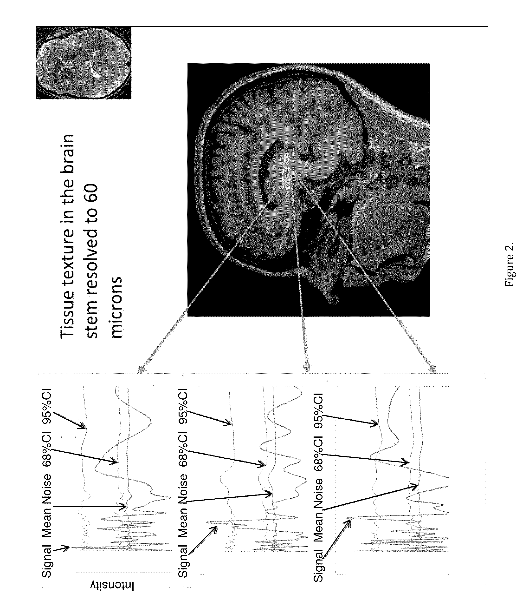
[0047]FIGS. 1 and 2 show the magnetic resonance fine texture measurement technique applied to brain. Structural wavelength spectra are generated from the indicated regions of interest along an axis of the selectively excited inner volume.

[0048]In order to define terminology for what follows, and with reference to the prior art magnetic resonance fine texture measurement technique, an internal volume in the anatomy of interest is excited by proper sequencing of magnetic field gradients and RF (Radio Frequency) pulses. Acquisition of the finely sampled 1D data is enabled by application of a ...


PUM


Abstract

The disclosed invention is a method for detecting indications of the presence of Alzheimer's disease (AD) and related dementia-inducing, motor-control-related pathologies, and other diseases in the human brain using a magnetic-resonance based technique for measuring fine tissue and bone textures. Specifically, the invention focuses on refinements / adaptations to a prior art magnetic resonance fine texture measurement technique that facilitates / enables pushing the detection limits closer to the cellular level, so as to be able to measure the fine scale structures and tissue changes that are known to be characteristic of the neurodegenerative processes involved in the development of these diseases.

Description

CROSS-REFERENCE TO RELATED APPLICATIONS[0001]This application is a continuation of International Application No. PCT / US2012 / 054934 filed Sep. 12, 2012 which claims the benefit of U.S. Provisional Patent Application No. 61 / 534,020 filed Sep. 13, 2011, U.S. Provisional Patent Application No. 61 / 596,424 filed Feb. 8, 2012 and U.S. Provisional Patent Application No. 61 / 639,002 filed Apr. 26, 2012.BACKGROUND OF THE INVENTION[0002]1. Field of the Invention[0003]The present invention relates to the field of diagnostic assessment of changes in brain structures and tissue in response to disease progression and treatment, specifically in response to Alzheimer's Disease (AD) and related forms of dementia such as Dementia with Lewy Bodies (DLB) and Frontotemporal Dementia (FTD), as well as motor diseases such as Amyotrophic Lateral Sclerosis (ALS), and Parkinson's disease, but also in all other pathologies that involve changes to normal brain structures such as CVD, (Cerebrovascular Disease), a...

Claims


Application Information

Patent Timeline
IPC IPC(8): A61B5/055G01R33/483G01R33/56
CPCA61B5/0555G01R33/483G01R33/5601A61B5/055A61B5/0042A61B5/4088G01R33/4833G01R33/5608A61B2576/026G16H30/40
Inventor JAMES, TIMOTHY W.JAMES, KRISTINFARR, LANCE W.CHASE, DAVID R.BRADY, J. MICHAELRAFFERTY, JAMESHEINRICH, JOHN P.
Owner OSTEOTRONIX MEDICAL PTE LTD
Learn More