
Method for detection of coagulation activity and biomarkers

a biomarker and coagulation activity technology, applied in the field of coagulation activity and biomarkers detection, can solve the problems of inconvenient clinical application, thrombosis in the venous system, acute death, etc., and achieve the effect of saving costs and time, facilitating correct conclusions regarding suitable medication, and sufficient and accurate results

Inactive Publication Date: 2014-10-16
KONINKLJIJKE PHILIPS NV
View PDF0 Cites 7 Cited by
  • Summary
  • Abstract
  • Description
  • Claims
  • Application Information

AI Technical Summary

Benefits of technology

The present invention is a method for carrying out various measurements on the same detection platform, even if the measurements have different biochemistry and physics. This method is convenient because it requires only a small amount of sample from an individual or patient, which can be used for multiple testing steps. The method can provide accurate results quickly, and can thus help clinicians make correct decisions regarding suitable medication. This method is also cost-effective and time-efficient. The application demonstrates that the method can be used to accurately measure the coagulation activity of a sample, and that the detection of clotting time can be done using the same optical detection system. The method is advantageous because it can measure the loss of mobility of magnetic particles near the bottom surface of the sample container, which minimizes the effects of unspecific binding or clustering of the beads and ensures accurate detection accuracy.

Problems solved by technology

Thrombosis in the venous system is also an important cause of acute death due to pulmonary embolism.
Typically, the assays for activation products of blood coagulation which also include markers of activated platelets such as β-thromboglobulin (β-TG) have been instruments of major interest in clinical research for studying the mechanisms of thrombosis, but have been proven to be unsuitable for clinical application, mainly because these tests are rather laborious and required expensive reagents and devices.
An important drawback of these assays, however, is the fact that the means and methods known in the art are specifically tailored and limited to the measurement of a single factor of coagulation activity, namely the blood clotting time of a blood sample.
Hence, the state of the art lacks means and methods capable of conducting an integrated evaluation combining the detection of one or more target molecules with the measurement of coagulation activity.

Method used


Image

  • Method for detection of coagulation activity and biomarkers
  • Method for detection of coagulation activity and biomarkers
  • Method for detection of coagulation activity and biomarkers

Examples


example 1

Measurement of Coagulation Activity by Determining Clotting Time of a Plasma Sample Using an Optomagnetic Assay

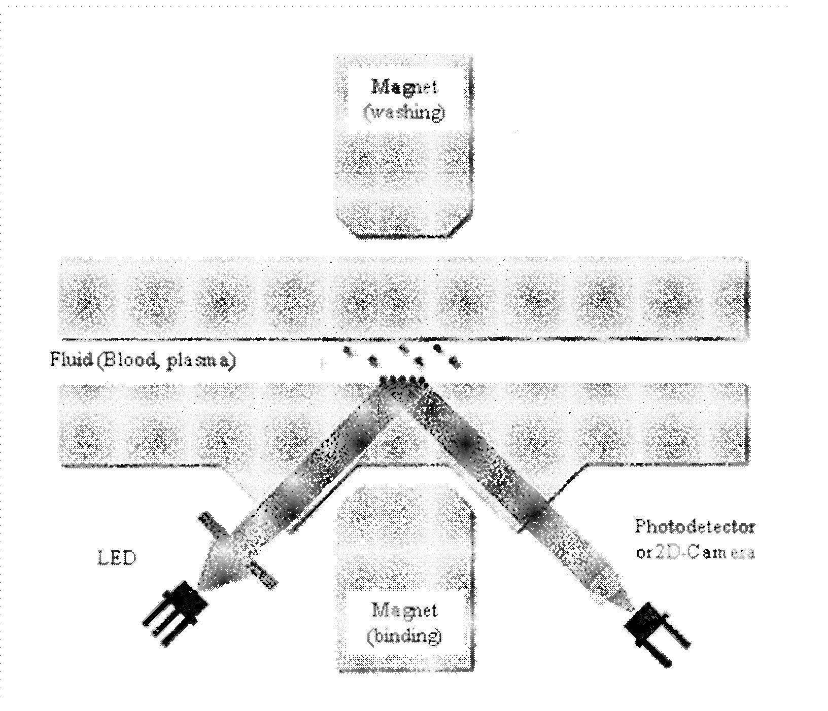
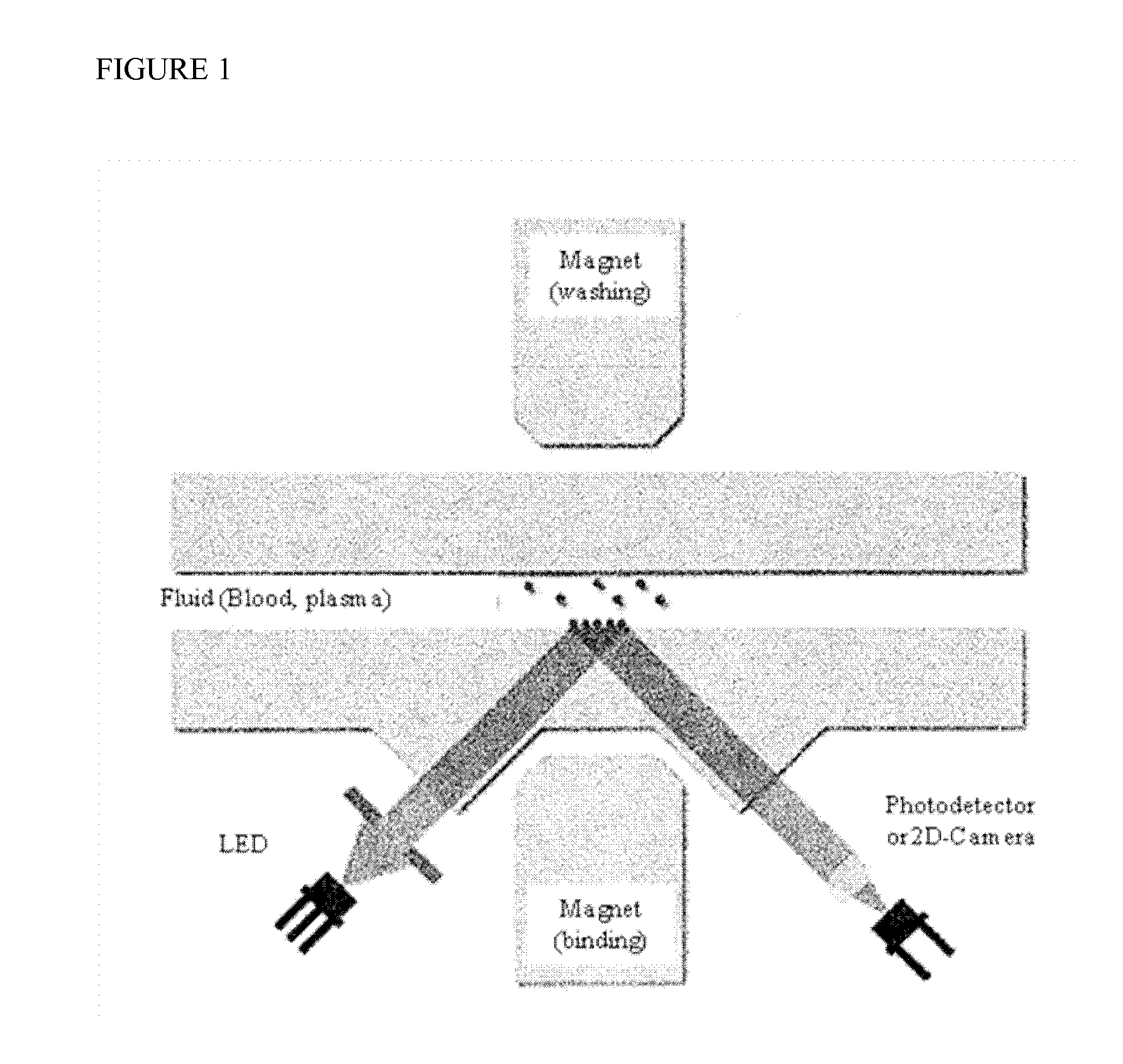
[0120]The coagulation cascade in plasma typically results in formation of fibrin networks which hamper the movement of magnetic nanoparticles in the liquid to be tested. An exemplary measurement of clotting time according to the invention is carried out on the Magnotech®-biosensor system using a detection based on frustrated total internal reflection (FTIR). The experimental setup is depicted in FIG. 1. Magnetic actuation of the magnetic particles occurs via the alternate action of two magnets, i.e. an upper magnet (washing magnet) and a lower magnet (binding magnet). The clotting time in this example is measured utilizing a standard Magnotech® biosensor cartridge for FTIR detection and 500 nm superparamagnetic beads. Likewise the beads may be already provided within a biosensor cartridge or added together with the blood sample mixture.

[0121]In this Example, a mixture of 5 ...

example 2

Measurement of Coagulation Activity by Determining Clotting Time of a Whole Blood Sample using an Optomagnetic Assay

[0125]The same measurement as exemplified in Example 1 can be carried out using a whole blood sample instead of plasma. Although whole blood sample comprises blood cells, preliminary tests have shown that the free movement of the magnetic particles are not compromised by the presence of hematocrit. Using the experimental setup as shown in FIG. 1, 5 μl of a whole blood sample is injected into the sample container already comprising the 500 nm superparamagnetic beads and the change in light signal is measured over time. The time period between injection (time point 0) and change in light signal corresponds to the clotting time and serves as measure of coagulation activity.

example 3

Simultaneous Measurement of Coagulation Activity by Determining Clotting Time of a Citrated Blood Sample and of the Presence / Amount of a Coagulation Unrelated Biomarker Troponin-I using an Optomagnetic Assay

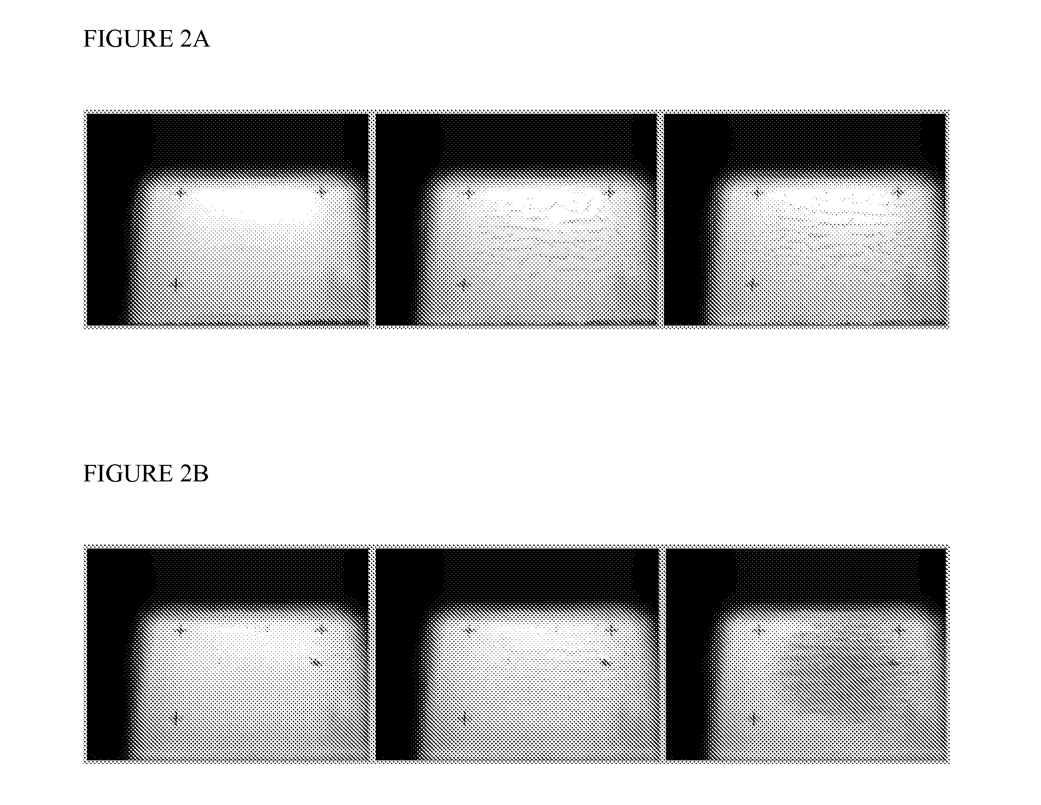
[0126]Example 1 and 2 demonstrate that coagulation activity can be successfully measured using an optomagnetic device as described in FIG. 1. In order to assess whether a simultaneous measurement using the same cartridge comprising superparamagnetic beads functionalized with a binding molecule directed against a coagulation unrelated biomarker within the blood sample is feasible, 5 μl citrated plasma is injected with or without 5 μl 0.125 M CaCl2 in PBS into a sample container containing 500 nm superparamagnetic beads which surface is covered with an antibody directed against the biomarker Troponin-I. Immediately after injection magnetic actuation is started and the biomarker is detected as a round spot on the cartridge surface within about 2 to 3 minutes after injection. The mea...


PUM


Abstract

The present invention relates to a method for simultaneous detection of the coagulation activity of a blood sample and of the presence and / or amount of at least one target molecule within said blood sample, comprising the steps of: i) introducing a blood sample into a sample container comprising magnetic particles and a sensor surface, wherein said magnetic particles are functionalized with a first binding molecule, wherein said first binding molecule is attached to said magnetic particles, wherein the first binding molecule is capable of specifically binding to the at least one target molecule within said blood sample, and wherein a second binding molecule is attached to a sensor surface at the bottom of said sample container, and wherein said second binding molecule of the sensor surface is capable of specifically binding to the at least one target molecule within said blood sample; ii) measuring the presence and / or amount of said target molecule by detecting the number of magnetic particles bound to said sensor surface; wherein the number of bound magnetic particles is directly or inversely related to the amount of said at least one target molecule present in the sample; iii) at the same time or at different times measuring the co-agulation activity of said blood sample, wherein said magnetic particles are magnetically actuated; and wherein the loss of mobility of said magnetic particles is detected by measuring the light reflected from said immobilized magnetic particles near or at said sensor surface; and wherein the change of detected light signal is indicative of an increase of viscosity and / or a clotting of the blood sample; and wherein the magnetic particles in steps i) to iii) are magnetically actuated using a magnetic field generator.

Description

FIELD OF THE INVENTION[0001]The present invention relates to a method for simultaneous detection of the coagulation activity of a blood sample and of the presence and / or amount of at least one target molecule within said blood sample, comprising the steps of:[0002]i) introducing a blood sample into a sample container comprising magnetic particles and a sensor surface, wherein said magnetic particles are functionalized with a first binding molecule, wherein said first binding molecule is attached to said magnetic particles, wherein the first binding molecule is capable of specifically binding to the at least one target molecule within said blood sample, and wherein a second binding molecule is attached to a sensor surface at the bottom of said sample container, and wherein said second binding molecule of the sensor surface is capable of specifically binding to the at least one target molecule within said blood sample;[0003]ii) measuring the presence and / or amount of said target molec...

Claims


Application Information

Patent Timeline
IPC IPC(8): G01N33/543
CPCG01N33/4905G01N33/54326G01N33/54306G01N33/86G01N33/68
Inventor VAN LIESHOUT, RON MARTINUS LAURENTIUS
Owner KONINKLJIJKE PHILIPS NV
Learn More