3-d holographic imaging flow cytometry

Active Publication Date: 2014-11-13
MASSACHUSETTS INST OF TECH
View PDF2 Cites 58 Cited by
  • Summary
  • Abstract
  • Description
  • Claims
  • Application Information

AI Technical Summary

Benefits of technology

[0009]In exemplary embodiments, the angular spectra, which are complex quantities, may be measured with phase shifting interferometry (PSI). A filtered back-projection algorithm may be applied for tomographic reconstruction. In PSI, however, the sample has to be stationary during the phase shifting step; thus, images are acquired in a discrete fashion, which limits imaging throughput. Moreover, the filtered back-projection algorithm is not ideal for thick cells. Thus, as taught herein, imaging throughput and applicability (particularly for thicker cells) may be increased by adopting off-axis digital holography for the angular spectra measurement and applying an algorithm based on the scalar diffraction theory for the tomographic reconstruction. In exemplary embodiments, the systems and methods taught herein also utilize a micro-fluidic channel specially designed for a high-numerical-aperture setup which may advantageously be utilized for 3-D refractive index imaging of cells continuously flowing in the micro-fluidic channel (as empirically demonstrated herein).
[

Problems solved by technology

In PSI, however, the sample has to be stationary during the phase shifting step; thus, images are acquired in a discrete

Method used


Image

  • 3-d holographic imaging flow cytometry
  • 3-d holographic imaging flow cytometry
  • 3-d holographic imaging flow cytometry

Examples


Embodiment Construction

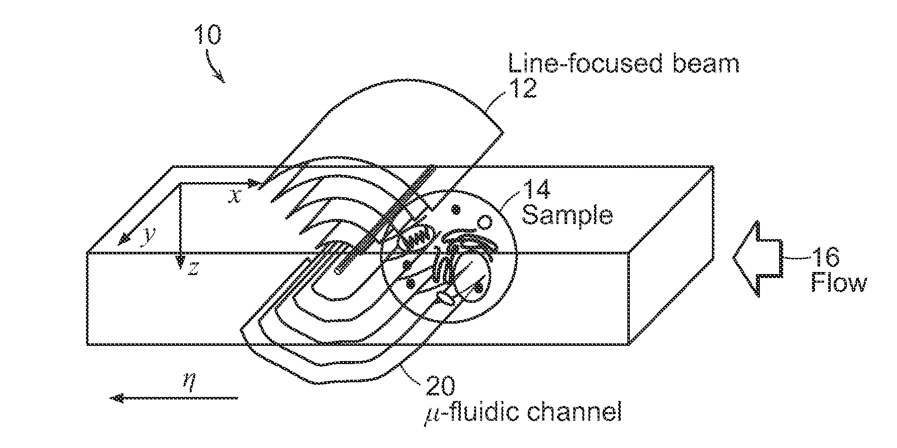
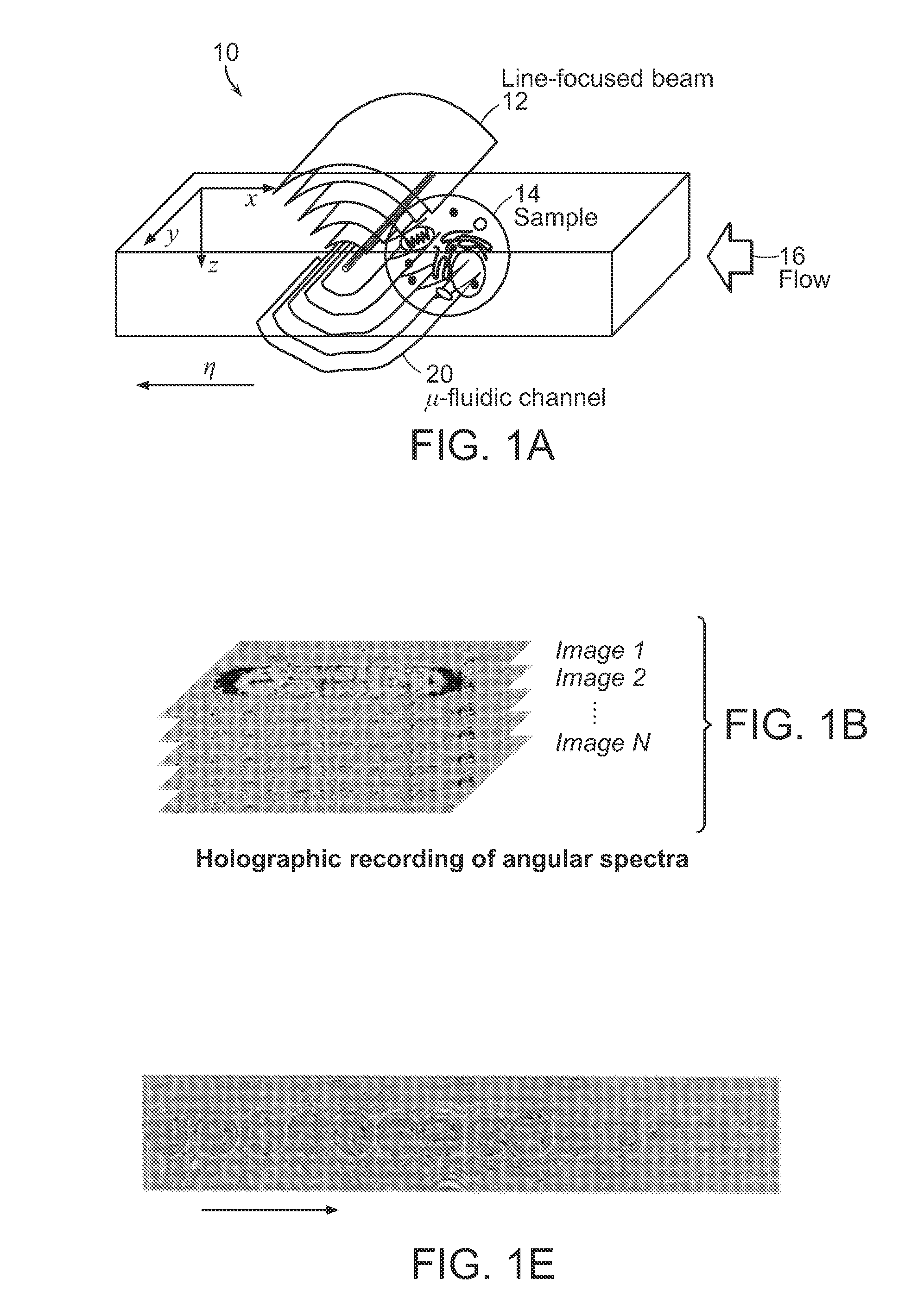
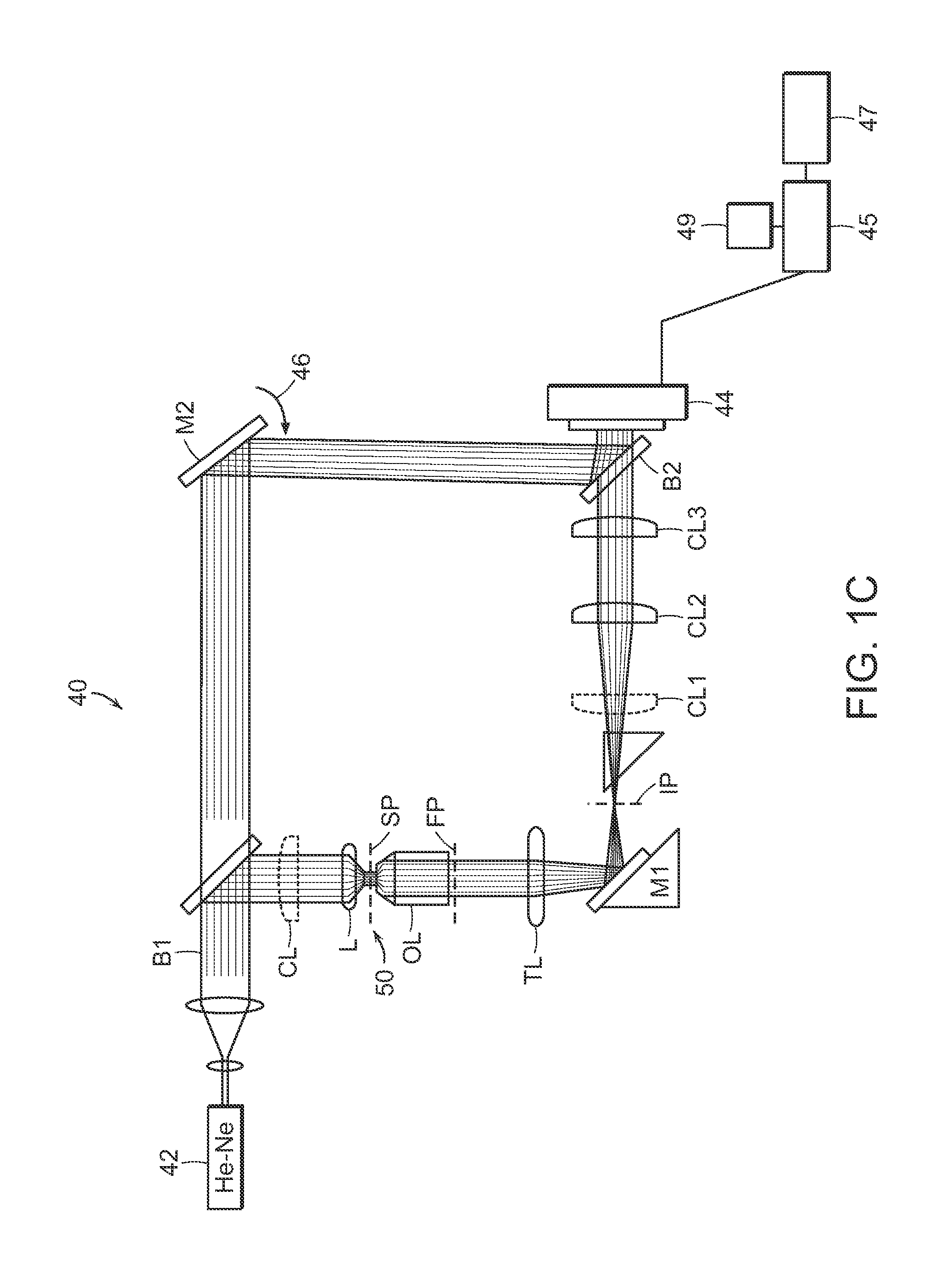
[0024]Refractive index (RI) of biological specimens is a source of intrinsic contrast that can be explored without any concerns of photobleaching or harmful effects caused by extra contrast agents. In addition, RI contains rich information related to the metabolism of cells at the cellular and subcellular levels. The subject application discloses a no-moving parts approach that provides three-dimensional (3-D) RI maps of biological samples continuously flowing in a microfluidic channel. Specifically, line illumination and off-axis digital holography is used to record the angular spectra of light scattered from flowing samples at high speed. In addition, an optical diffraction tomography algorithm is applied to obtain accurate RI maps of the samples from the measured spectra. As demonstrated in empirical studies described herein, the systems and methods of the present disclosure have proven effective in label-free 3-D imaging of live RKO human colon cancer cells and RPMI8226 multiple...


PUM


Abstract

Refractive index of biological specimens is a source of intrinsic contrast that can be explored without any concerns of photobleaching or harmful effects caused by extra contrast agents. This feature also contains rich information that can be related to the metabolism of cells at the cellular and subcellular levels. The present invention relates to systems and methods that can provide, without any moving parts, the 3-D refractive index map of continuously flowing biological samples in a micro-fluidic channel, for example.

Description

CROSS-REFERENCE TO RELATED APPLICATION(S)[0001]This application relates to and claims priority from U.S. Provisional Patent Application No. 61 / 761,079 filed on Feb. 5, 2013, the entire disclosure of which is incorporated herein by reference in its entirety.GOVERNMENT SUPPORT[0002]This invention was made with government support under Grant No. EB015871 awarded by the National Institutes of Health. The government has certain rights in the invention.BACKGROUND OF THE INVENTION[0003]Flow cytometry is a technology that interrogates a large number of cells flowing in a stream of liquid, typically in combination with optical spectroscopy. Flow cytometry has enabled cell characterization even at a genomic level. A contrast mechanism in flow cytometry is fluorescence, externally administered or generated by genetic manipulation. To observe biological specimens in their most native condition, however, stain-free methods utilizing intrinsic properties of a specimen are preferred. Furthermore, ...

Claims


Application Information

Patent Timeline
IPC IPC(8): G01N15/14G01N21/64
CPCG01N15/1434G01N21/64G01N2015/1445G01N15/1475G01N15/1484G01N21/53G03H1/0443G03H1/0866G03H2001/0033G03H2001/005G03H2001/0445G03H2210/12
Inventor SUNG, YONGJINLUE, NIYOMYAGOOB, ZAHIDDASARI, RAMACHANDRASO, PETER T.C.
Owner MASSACHUSETTS INST OF TECH
Why Eureka
Try Eureka