

System for Imaging Lesions Aligning Tissue Surfaces
a tissue surface and imaging technology, applied in the field of medical diagnostic devices and methods, can solve the problems of increasing the risk of developing melanoma, difficult treatment, death, etc., and achieve the effects of low cost, quick test performance, and convenient us
- Summary
- Abstract
- Description
- Claims
- Application Information
AI Technical Summary
Benefits of technology
Problems solved by technology
Method used
Image

Examples
Embodiment Construction
[0136]While various embodiments of the invention have been shown and described herein, it will be obvious to those skilled in the art that such embodiments are provided by way of example only. Numerous variations, changes, and substitutions may occur to those skilled in the art without departing from the invention. It should be understood that various alternatives to the embodiments of the invention described herein may be employed.
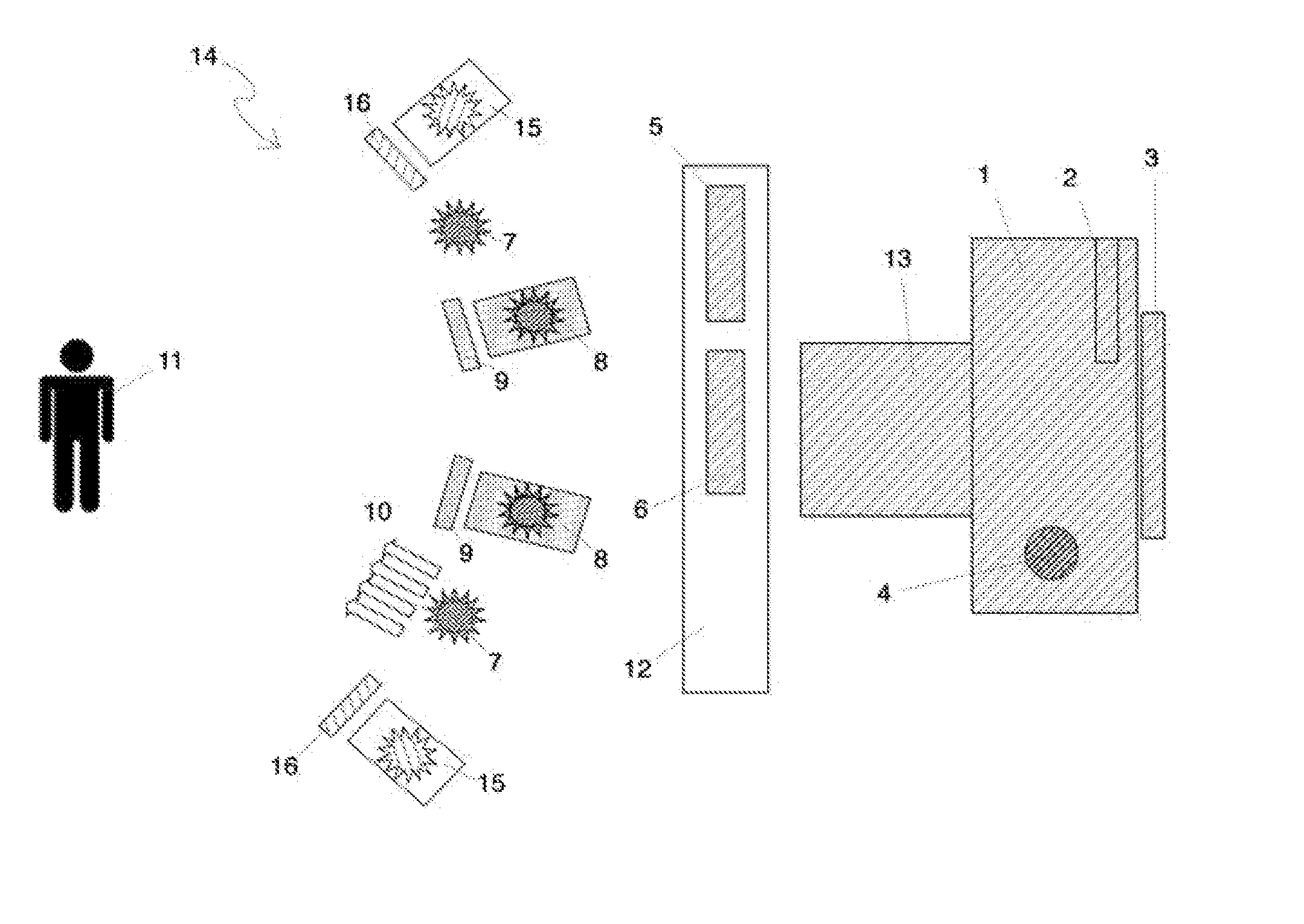
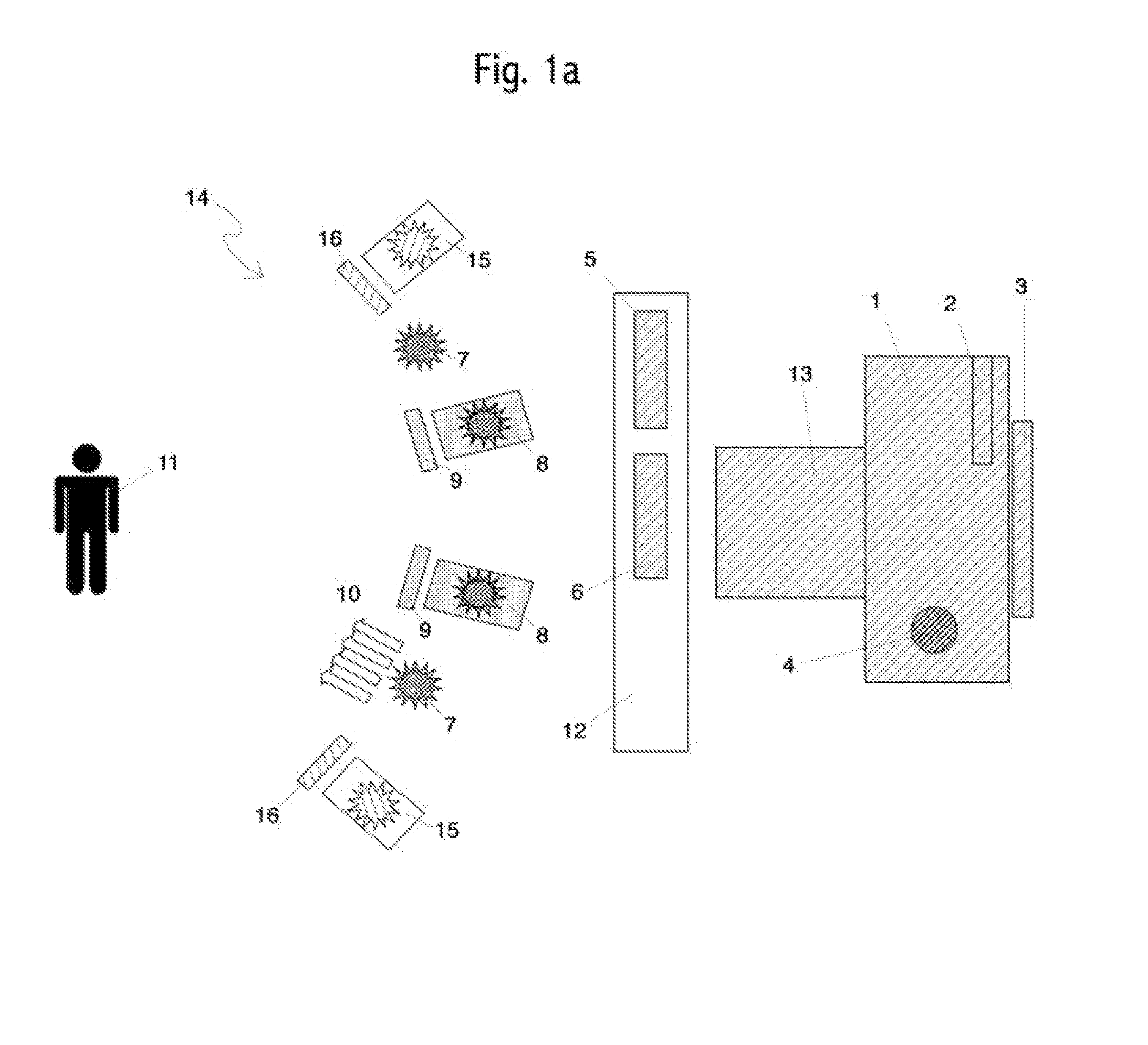
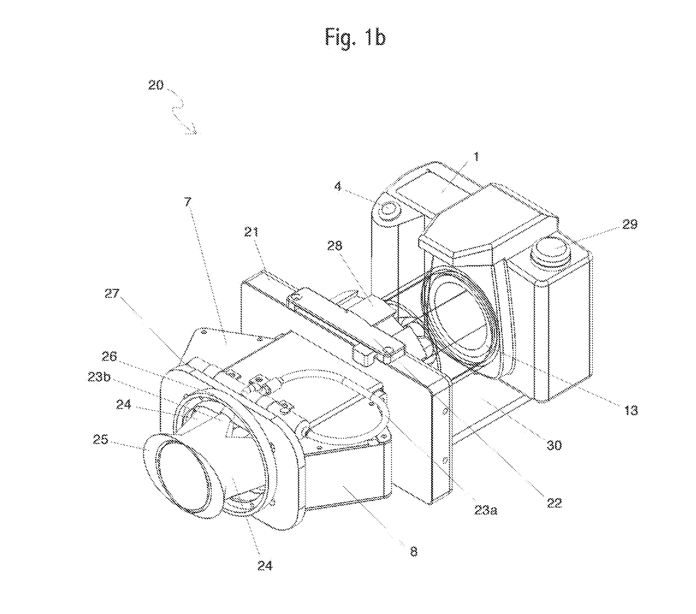
[0137]In one aspect, systems, compositions and methods are provided for imaging of cavity and / or tissue lesions. Various aspects described herein can be applied to any of the particular applications set forth below, alone or in combination, or for any other types of imaging systems. The embodiments described herein may be applied as a standalone system or method, or as part of an integrated medical diagnostic and / or treatment system. It shall be understood that different aspects can be appreciated individually, collectively, or in combination with each ot...
PUM
| Property | Measurement | Unit |
|---|---|---|
| wavelengths | aaaaa | aaaaa |
| wavelengths | aaaaa | aaaaa |
| wavelength band | aaaaa | aaaaa |
Abstract
Description
Claims
Application Information