Eureka AIR delivers breakthrough ideas for toughest innovation challenges, trusted by R&D personnel around the world.

Tumorspecific SPECT/MR(T1), SPECT/MR(T2) and SPECT/CT contrast agents

Inactive Publication Date: 2015-01-01
BBS NANOTECH
View PDF0 Cites 5 Cited by
  • Summary
  • Abstract
  • Description
  • Claims
  • Application Information

AI Technical Summary

Benefits of technology

This patent text describes a method of using self-assembled particles to diagnose tumors. These particles can target specific cells in the tumor and enhance the imaging process. The particles can also contain substances that make it easier to detect the tumor using radioactive labeling. The complexing agents used for labeling include several options such as DTPA and EDTA. Overall, this method improves the accuracy of tumor diagnosis.

Problems solved by technology

SPECT images, however, have limited spatial resolution and lack anatomical details for reference, making the precise localization of lesions difficult.

Method used


Image

  • Tumorspecific SPECT/MR(T1), SPECT/MR(T2) and SPECT/CT contrast agents
  • Tumorspecific SPECT/MR(T1), SPECT/MR(T2) and SPECT/CT contrast agents
  • Tumorspecific SPECT/MR(T1), SPECT/MR(T2) and SPECT/CT contrast agents

Examples


example 1

Preparation of Folated Poly-Gamma-Glutamic Acid (γ-PGA)

[0084]Folic acid was conjugated via the amino groups to γ-PGA using carbodiimide technique. γ-PGA (m=60 mg) was dissolved in water (V=100 ml) to produce aqueous solution. The pH of the polymer solution was adjusted to 6.0. After the dropwise addition of cold water-soluble 1-[3-(dimethylamino)propyl]-3-ethylcarbodiimide methiodide (CDI) (m=13 mg in 2 ml distilled water) to the γ-PGA aqueous solution, the reaction mixture was stirred at 4° C. for 1 h, then at room temperature for 1 h. After that, folic acid (m=22 mg in dimethyl sulfoxide, V=10 ml) was added droppwise to the reaction mixture and stirred 4° C. for 1 h, then at room temperature for 24 h. The folated poly-γ-glutamic acid (γ-PGA-FA) was purified by dialysis.

example 2

Preparation of Folated Poly-Gamma-Glutamic Acid

[0085]Synthesis of folated PGA was performed in a two steps process. First PEG amine was coupled to FA based on a well-known reaction describe elsewhere. JACS, 130 (2008) 114671 After that FA-PEG amine was conjugated via the amino groups to PGA using carbodiimide technique: γ-PGA (m=300 mg) was dissolved in water (V=300 ml) to produce aqueous solution at a concentration of 1 mg / ml. The pH of the polymer solution was adjusted to 6.0. After addition of 1-hydroxybenzotriazole hydrate (m=94 mg), the reaction mixture was sonicated for 5 min The reaction mixture was cooled to 4° C. and cold water-soluble 1[3-(dimethylamino)propyl]-3-ethylcarbodiimide hydrochloride (EDC) (m=445 mg in V=15 ml water) was added dropwise to the γ-PGA aqueous solution. The reaction mixture was stirred at 4° C. for 10 min, then folic acid-PEG-amine solution (m=100 mg in V=15 ml water) and triethylamine (V=324 μl) were added dropwise to the reaction mixture. The reac...

example 3

Preparation of Folated Poly-Gamma-Glutamic Acid Coated Gold Nanoparticles

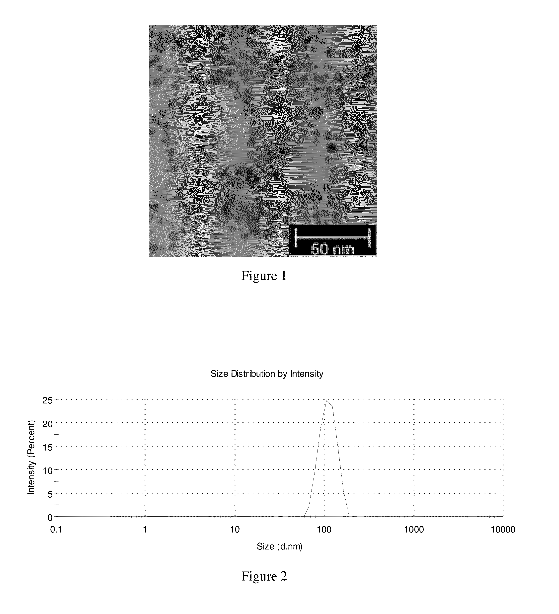
[0086]Folated PGA was dissolved in distilled water (V=10 ml) to produce a solution with a concentration of c=0.5 mg / ml. After the dropwise addition of solution of gold (III) chloride hydrate (V=500 μl, c=1.7 mg / ml), solution of sodium citrate tribasic dihydrate (V=75 μl, c=10 mg / ml) was added dropwise to the reaction mixture. Then solution of sodium borohydride (V=40 μl, c=1 mg / ml) was added to the reaction. The reaction mixture was stirred at room temperature for 4 h, after that it was purified by dialysis. (FIG. 1)


PUM


Abstract

The invention relates to cancer receptor-specific bioprobes for single photon emission computed tomography (SPECT) and computed tomography (CT) or magnetic resonance imaging (MRI) for dual modality molecular imaging. The base of the bioprobes is the self-assembled polyelectrolytes, which transport gold nanoparticles as CT contrast agents, or SPION or Gd(III) ions as MR active ligands, and are labeled using complexing agent with technetium-99m as SPECT radiopharmacon. Furthermore these dual modality SPECT / CT and SPECT / MR contrast agents are labeled with targeting moieties to realize the tumorspecificity.

Description

[0001]This application claims priority to U.S. provisional application Ser. No. 61 / 840,483, filed Jun. 28, 2013, the entire disclosure of which is hereby incorporated by reference herein.FIELD OF THE INVENTION[0002]The invention relates to cancer receptor-specific bioprobes for single photon emission computed tomography (SPECT) and computed tomography (CT) or magnetic resonance imaging (MRI) for dual modality molecular imaging. The base of the bioprobes is the self-assembled polyelectrolytes, which transport gold nanoparticles as CT contrast agents, or SPION or Gd(III) ions as MR active ligands, and are labeled using complexing agent with technetium-99m as SPECT radiopharmacon. Furthermore these dual modality SPECT / CT and SPECT / MR contrast agents are labeled with targeting moieties to realize the tumorspecificity.BACKGROUND OF THE INVENTION[0003]Combining two or more different imaging modalities using multimodal probes can be considerable value in molecular imaging, especially for c...

Claims


Application Information

Patent Timeline
IPC IPC(8): A61K51/12A61K51/06
CPCA61K51/065A61K51/1251A61K49/0002A61K49/126A61K49/1824A61K49/0428A61K49/085A61K51/1244
Inventor BORBELY, JANOSHAJDU, ISTVANBODN R, MAGDOLNACSIKOS, ZSUZSANNA
Owner BBS NANOTECH
Why Eureka
Eureka Blog
Learn More