Treatment of ischemic retinopathies
- Summary
- Abstract
- Description
- Claims
- Application Information
AI Technical Summary
Benefits of technology
Problems solved by technology
Method used
Image
Examples
example 1
HIF-1α Accumulation and Müller Glial Cell Injury / Activation Co-localize in the Ischemic Inner Retina in the OIR Model
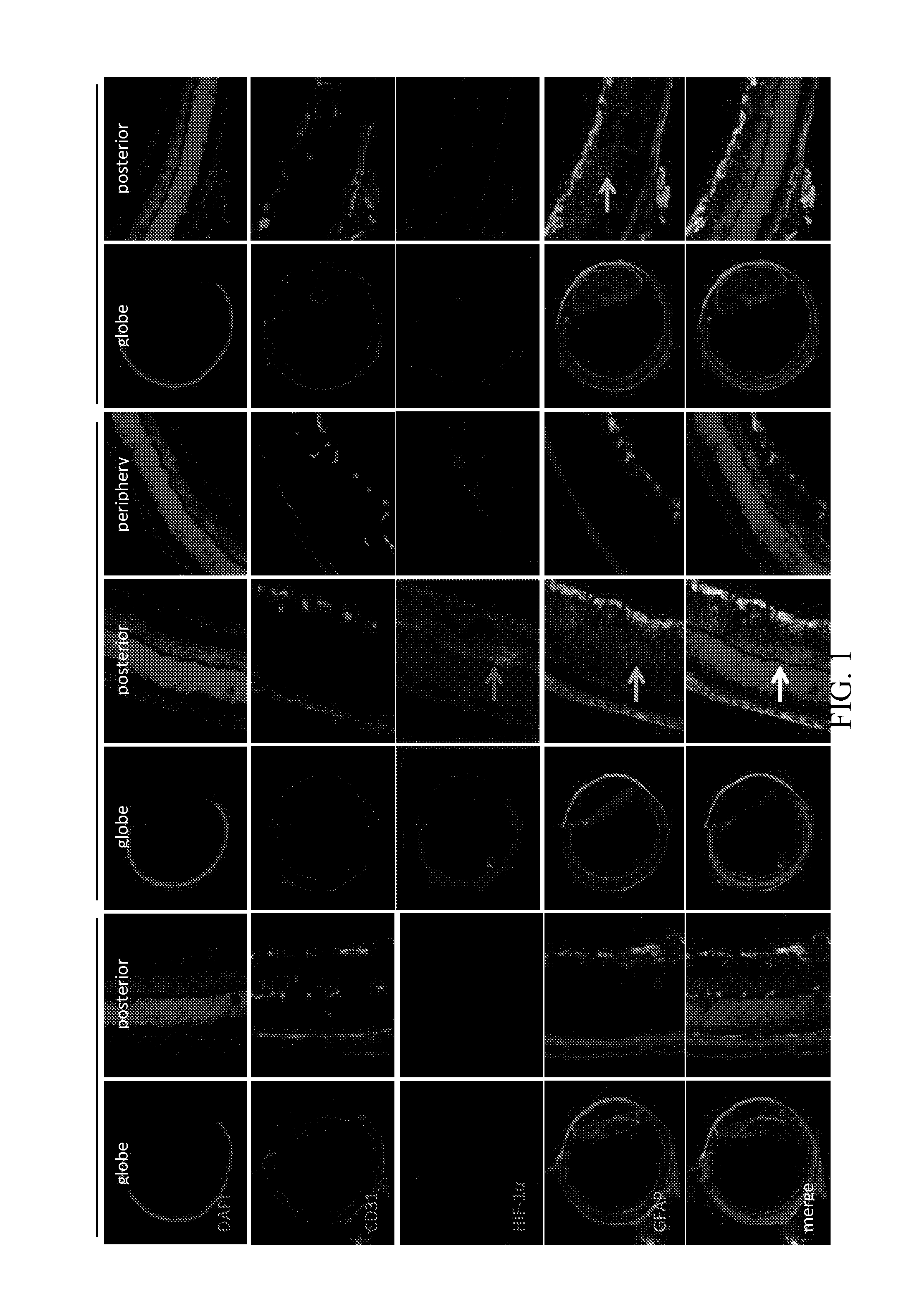
[0113]Microvascular complications in diabetic patients are caused by prolonged exposure to high glucose levels. Mouse models in which the hyperglycemic state is replicated have proven essential for studying the early stages of diabetic eye disease, to be sure. However, these models do not adequately reproduce the retinal nonperfusion that results in the release of growth factors that, in turn, promote the vascular permeability characteristic of patients with DME (1). Although no animal model has yet been found to demonstrate all of the microvascular complications associated with patients with diabetic eye disease, the oxygen induced retinopathy (OIR) mouse model faithfully reproduces the inner retina ischemia (nonperfusion) observed in these patients and has proven to be an important tool for studying the pathogenesis of ischemic retinopathies (13).
[0114]The inner re...
example 2
Hypoxia Upregulates HIF and VEGF in Injured Müller Glial Cells
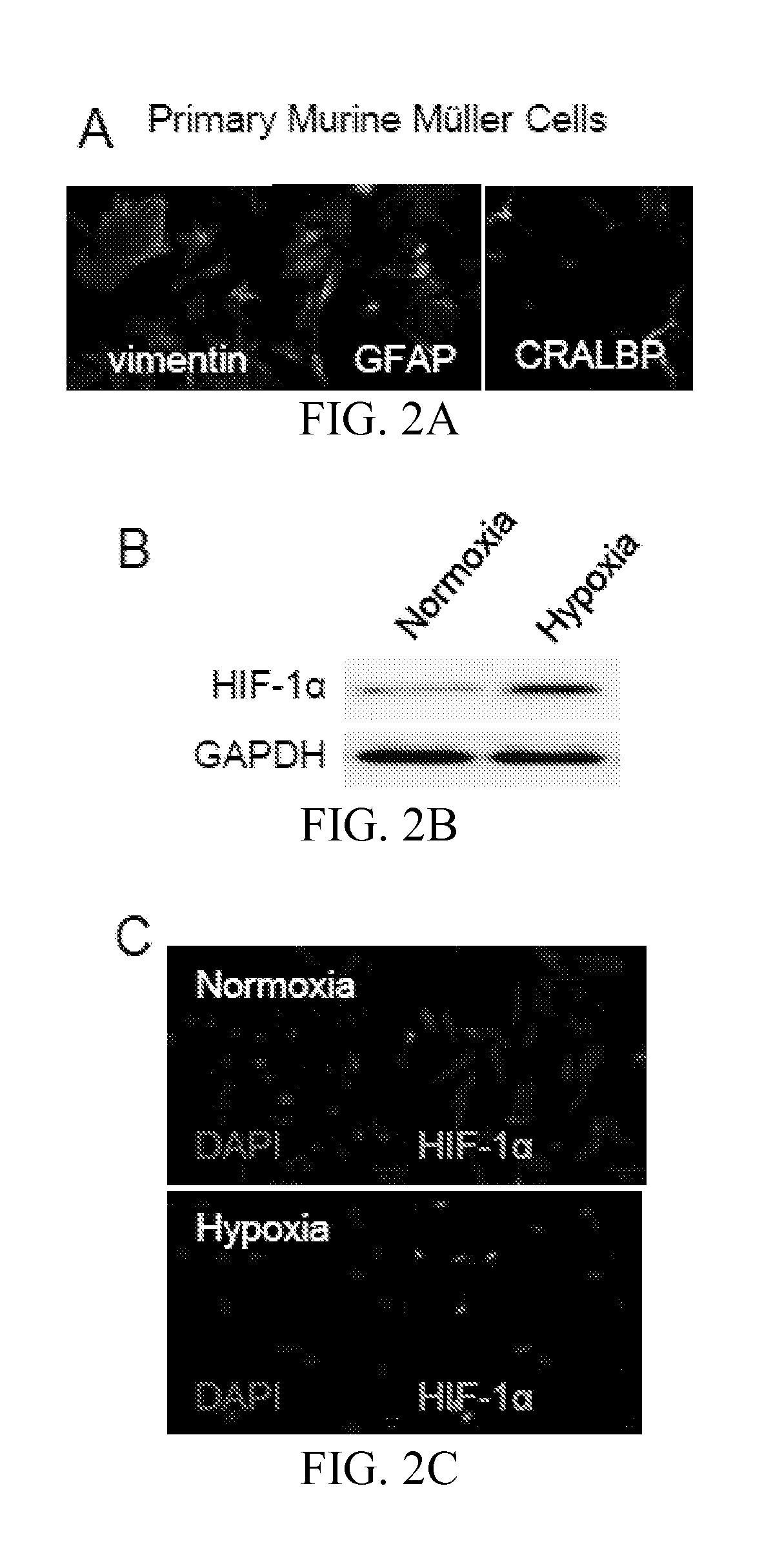
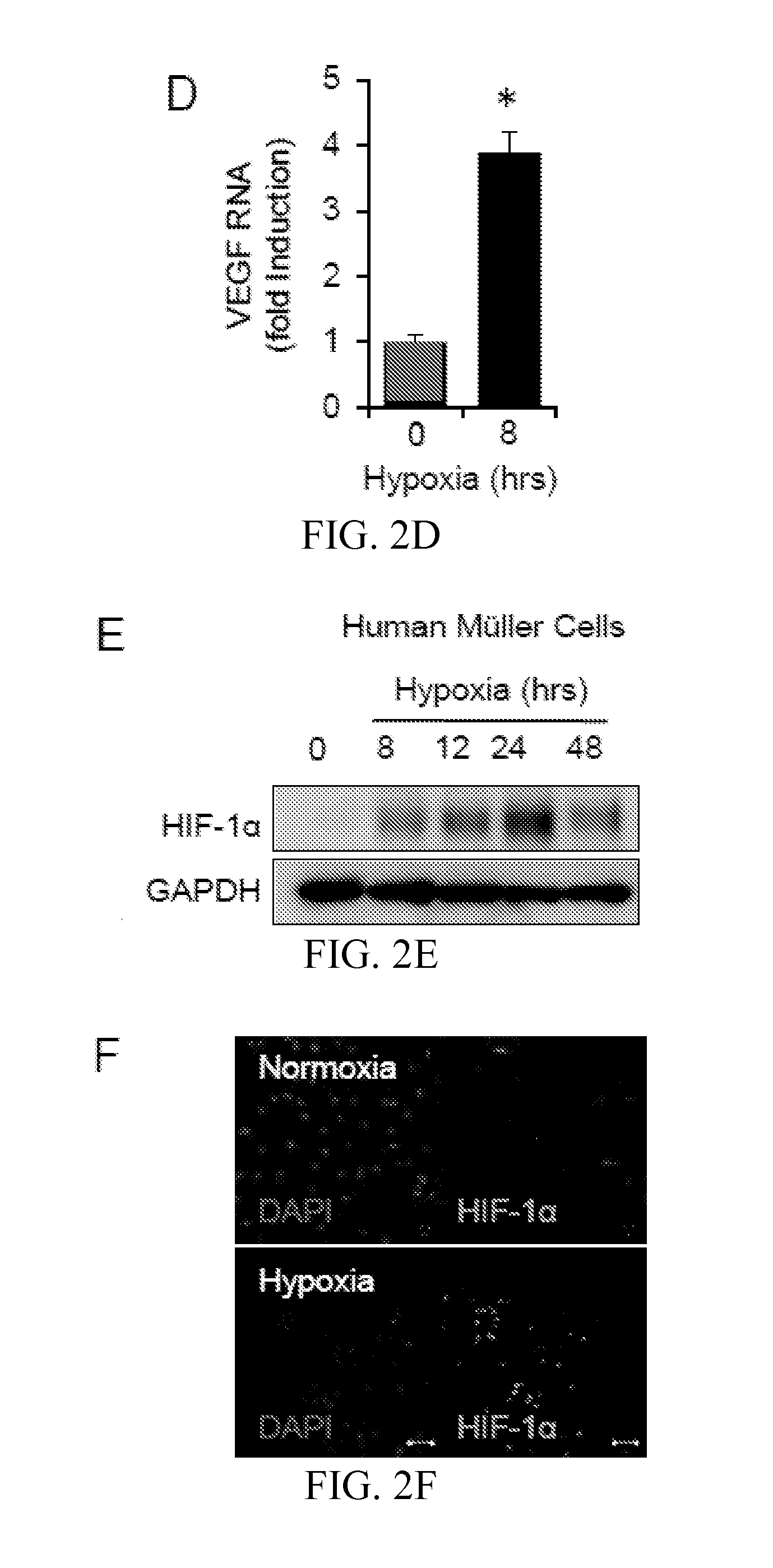
[0116]To directly assess the response of retinal Müller cells to hypoxia, we isolated primary Müller cell cultures (>95% pure) from the neurosensory retinas of P0-P5 C57BL / 6 mice (FIG. 2A). These cells maintained a Müller cell phenotype for over 8 passages, as demonstrated by the expression of key Müller cell markers, including vimentin, CRALBP and GFAP (FIG. 11). Primary murine Müller cells responded to hypoxia (3% O2) with an increase in HIF-1α protein stability and nuclear localization (FIGS. 2B and C), and an increase in the mRNA levels of the HIF-1 target gene, Vegf (FIG. 2D). To confirm a role for Müller cells in the hypoxic response in humans, we took advantage of the availability of a previously characterized immortalized human Müller (MIO-M1) cell line (18). Similar to primary murine Müller cells, exposure of MIO-M1 cells to hypoxia resulted in an increase in HIF-1α protein stability and nuclear localization (FIG...
example 3
Inhibition of HIF Blocks Edema in Ischemic Retinal Disease In Vivo
[0118]We next set out to determine whether inhibition of HIF-1α could reduce edema in ischemic retinopathies in vivo. Although the OIR model has been used extensively as a model for retinal neovascularization (14), we observed that this model results in increased vascular permeability, with leakage of plasma (FIG. 3A) and the plasma protein, albumin (FIG. 3B and C) into the interstitial tissue. The administration of digoxin to inhibit HIF-1α translation resulted in a decrease in vascular permeability (FIG. 3A-C and 12), demonstrating that HIF-1 is required for the promotion of vascular permeability, and supporting a therapeutic role for the inhibition of HIF-1α for the treatment of DME.
PUM
 Abstract
Description
Claims
Application Information
 - R&D
- Intellectual Property
- Life Sciences
- Materials
- Tech Scout
- Unparalleled Data Quality
- Higher Quality Content
- 60% Fewer Hallucinations
Browse by: Latest US Patents, China's latest patents, Technical Efficacy Thesaurus, Application Domain, Technology Topic, Popular Technical Reports.
© 2025 PatSnap. All rights reserved.Legal|Privacy policy|Modern Slavery Act Transparency Statement|Sitemap|About US| Contact US: help@patsnap.com


