Near infrared photonic prostatoscopy analyzer

a prostatoscopy and infrared technology, applied in the field of cancer detection systems and methods, can solve the problems of low accuracy, poor spatial resolution and contrast, and ineffective early-stage primary tumor detection, and achieve fast (near real-time) imaging and analysis, high sensitivity and accuracy, and non-invasive and safe detection

Active Publication Date: 2015-11-12
ALFANO ROBERT R +6
View PDF9 Cites 15 Cited by
  • Summary
  • Abstract
  • Description
  • Claims
  • Application Information

AI Technical Summary

Benefits of technology

[0010]The present invention provides a novel NIRPPA system for diagnostic function as a screening tool other than DRE, PSA and TRUS based on different absorption and scattering properties of light backscattered from cancerous and normal prostate tissues using native biomarkers of H2O, HbO2, and Hb in the four NIR tissue optical windows. The system provided by the present invention can overcome at least some of the problems associated with conventional screening techniques mentioned above. In particular, the present invention provides an optical system to detect 3D images and localize the 3D positions of abnormal prostate tissue hidden inside the normal prostate tissue through the human rectum in near-real-time. The present invention can also provide a system that has extended wide applications for the near real-time detection and 3D imaging and localization of tumor inside other organs such as rectum, colon, bladder, oral cavity and esophagus.
[0016]The present invention is based on specific design and arrangement of scanning light sources, coherence fibers for excitation and imaging, and an optical rectal probe with specific size to acquire a set of 2D images. As presented in our previous patent [2], there is a difference of water absorption between cancerous and normal prostate tissues, which can be used to image the difference of cancerous and normal prostate tissues with a large beam without scanning. However, without beam scanning, one can only detect cancerous tissue in two dimensions (2D) with a limited resolution. In the present invention, a scanning polarization imaging system is used to measure light intensity distributions caused by scattering and / or emission of cancerous and normal prostate tissues at the critical wavelength(s). By this way, the scanning optical imaging system can be used to acquire the multiple optical signals with multiple illumination (using scanning) and multiple detection (using CCD / CMOS cameras), which can be used for blind source analysis to identify and obtain the 3D images and positions of abnormal tissues with a higher resolution. In particular, the depth of cancerous tissue hidden inside the normal tissue can be obtained. In addition, this system, which applies the probe with a proper size, can be used to image the prostate through rectum to achieve the in vivo screening study.
[0020]The present invention can be extensively applied for the near real-time detection and 3D imaging of tumors inside other organs in human and animal bodies, such as rectum, colon, oral cavity and esophagus. The advantages of the systems described in the present invention are 3D imaging and location, high sensitivity and accuracy, fast (near real-time) imaging and analysis, and non invasive and safe detection.

Problems solved by technology

Although PSA test appears to have acceptable sensitivity for late stage cancer and disease with histopathologic features associated with tumor progression of a large volume, poorly differentiated cells and extracapsular penetration, its accuracy is limited as low as 28%-35%.
TRUS is no longer considered as a first-line screening test for prostate cancer because of its poor spatial resolution and contrast, but it does play a role in mapping the locations of the biopsy sampling.
Conventional oncology imaging methods for prostate cancer diagnosis still depend on bulk physical properties of cancer tissue and are not effective for early-stage primary tumors.
The major disadvantage of the optical imaging approaches is that high scattering of biological tissue causes most photons to diffuse.

Method used


Image

  • Near infrared photonic prostatoscopy analyzer
  • Near infrared photonic prostatoscopy analyzer
  • Near infrared photonic prostatoscopy analyzer

Examples


Embodiment Construction

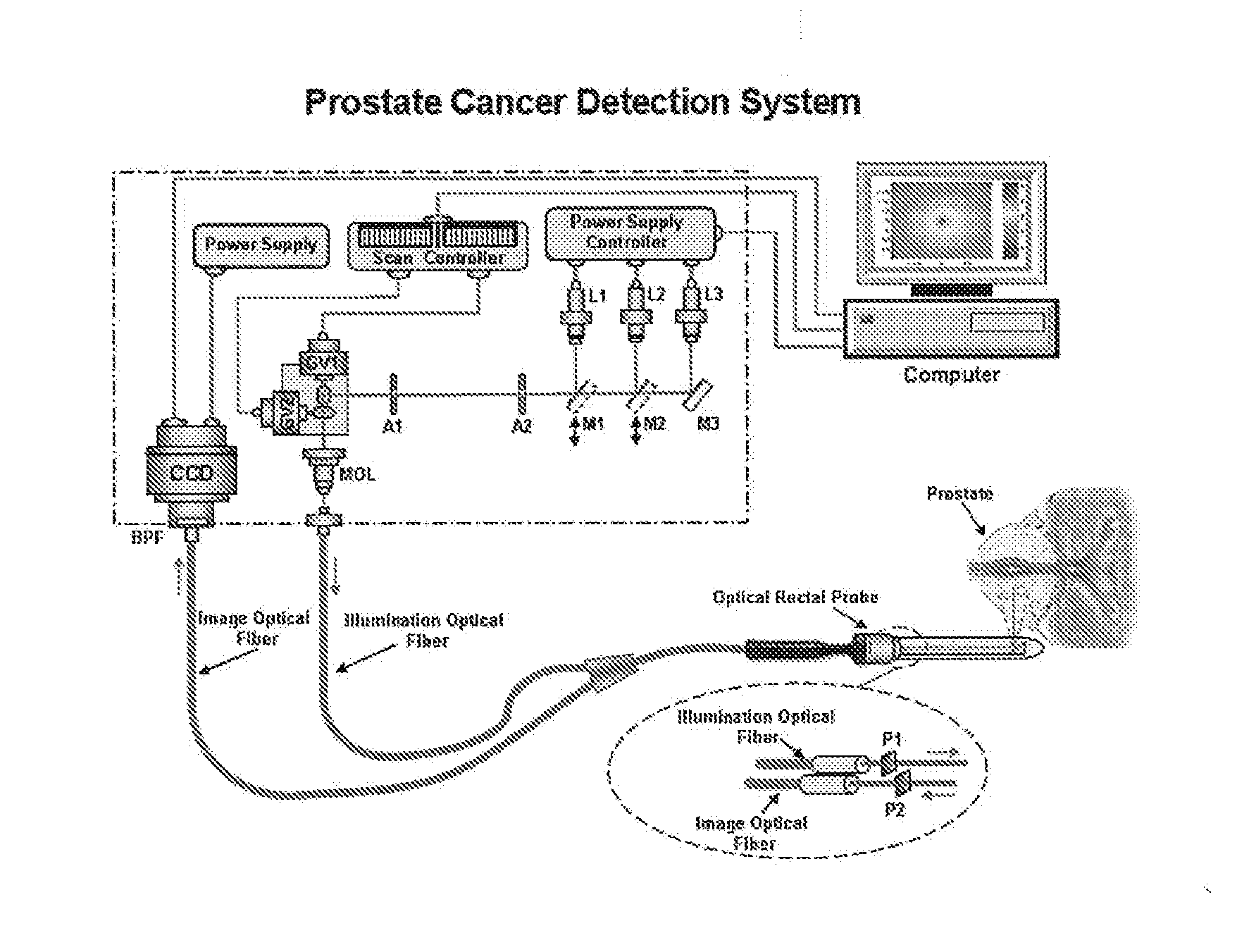
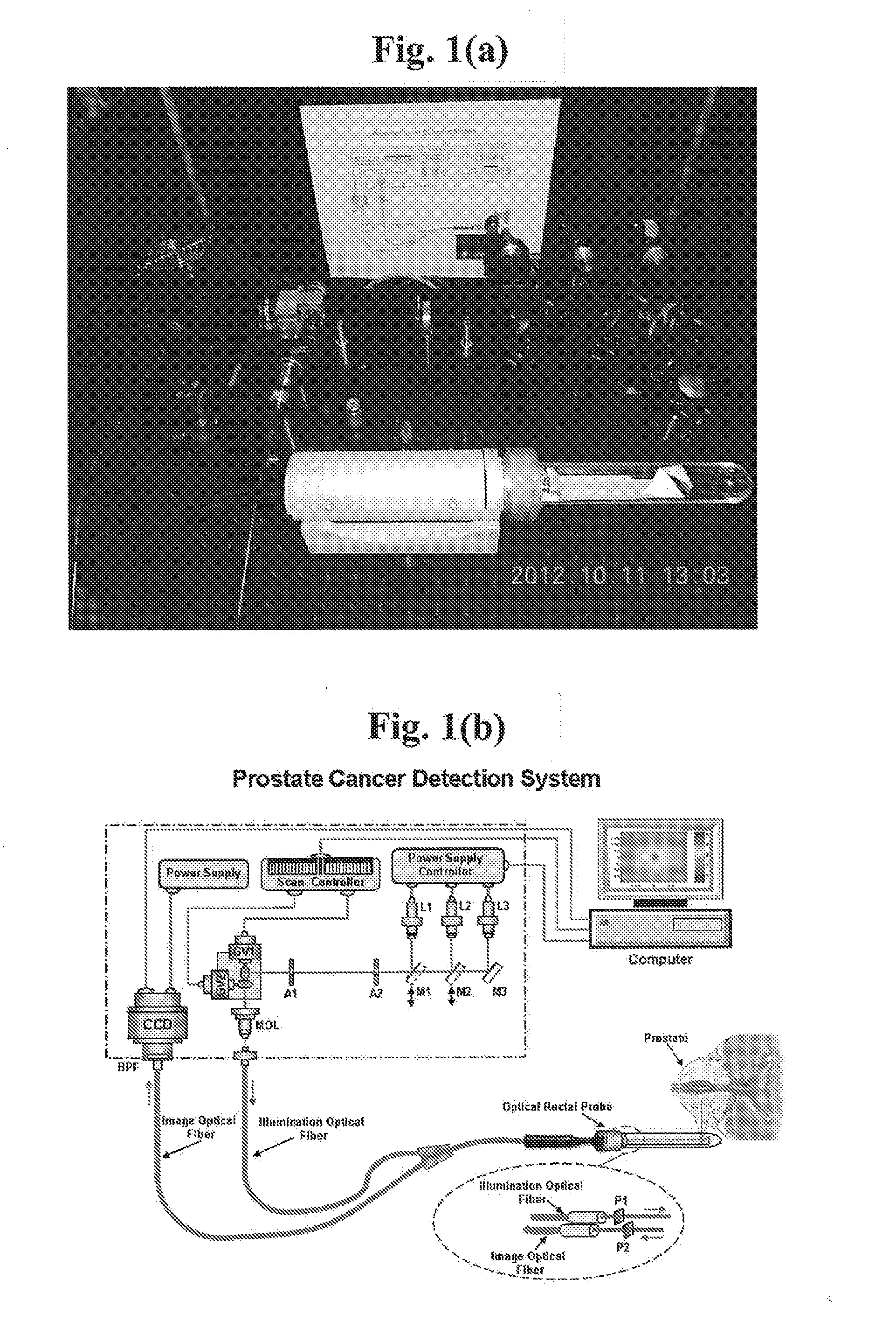
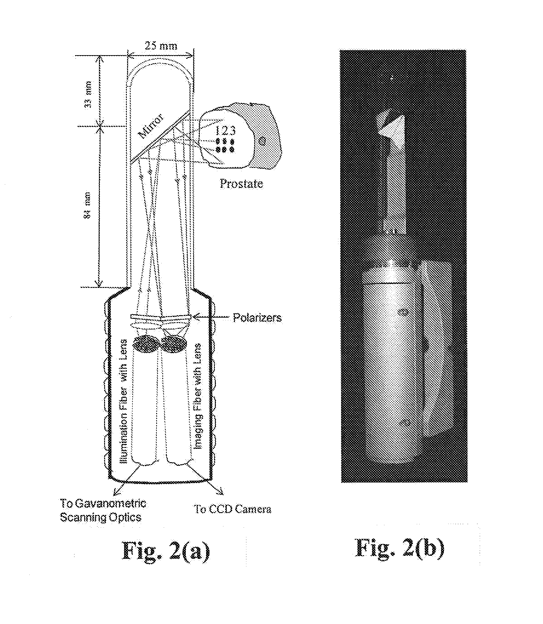
[0033]The present invention is directed to a novel NIRPPA system, which can be used for detecting cancerous tissue embedded in prostate and other deep organs using the four NIR tissue optical windows to improve livability of patients. In the application, the optical fiber-based rectal probe with a specific size can be inserted into a rectum. The illumination NIR light output from laser diodes / LEDs is directed to the scanning galvanometric mirrors. The beam output from the galvanometric mirrors is focused into a coherent optical fiber-bundle used for illumination. The output beam from the illumination fiber-bundle is first passed through a polarizer to generate linear or circular polarized illumination light, and then directed to a small reflection prism. Both of the polarizer and prism were located inside the optical rectal probe. The beam reflected from the prism is used to illuminate a prostate sample. This illumination beam can be scanned from point to point in the x- and y-direc...


PUM


Abstract

A rectal near infrared (NIR) scanning polarization imaging system uses NIR Photonic Prostatoscopy Analyzer (NIRPPA) for prostate cancer detection using light. The NIRPPA consists of a portable rectal NIR scanning polarization imaging unit and an optical fiber-based rectal probe capable of recording sets of 2D images of a prostate through rectum at different wavelengths and depths and obtaining a three dimensional (3D) image of the prostate and 3D locations of abnormal tissue inside the prostate. Diode lasers / light emission diodes (LEDs) with selected emitting wavelengths are used in the NIR spectral range from 650 nm to 2,400 nm corresponding to the four tissue optical windows (#I, 650 nm-950 nm; #II, 1,100 nm-1,350 nm; #III, 1,600 nm-1,870 nm; and #IV, 2,100 nm-2,300 nm). The fingerprint absorptions of water (H2O), Oxyhemoglobin (HbO2) and deoxyhemoglobin (Hb) in the prostate are used as native biomarkers for prostate cancer detection.

Description

BACKGROUND OF THE INVENTION[0001]1. Field of the Invention[0002]The invention generally relates to systems and methods of detecting cancer and, more specifically to an near infrared photonic prostatoscopy analyzer.[0003]2. Description of Prior Art[0004]The common screening tests for prostate cancer diagnosis are digital rectal examination (DRE), prostate specific antigen (PSA) blood test, and the transrectal ultrasound (TRUS) imaging. A value of PSA over 4.0 ng / ml is the commonly used threshold for further diagnostic evaluation. Although PSA test appears to have acceptable sensitivity for late stage cancer and disease with histopathologic features associated with tumor progression of a large volume, poorly differentiated cells and extracapsular penetration, its accuracy is limited as low as 28%-35%. During the DRE, a doctor inserts a lubricated gloved finger into the patient's rectum to feel for the enlargements and hard areas of prostate that might indicate prostate cancer. DRE has...

Claims


Application Information

Patent Timeline
Patent Type & Authority Applications(United States)
IPC IPC(8): A61B5/00A61B1/00A61B1/31
CPCA61B5/0086A61B1/00142A61B1/31A61B5/0066A61B5/4381A61B1/00165A61B1/00172A61B5/0062
Inventor ALFANO, ROBERT R.WANG, WUBAOPU, YANGBUDANSKY, YURYSORDILLO, LAURATANG, GUICHENEASTHAM, JAMES
Owner ALFANO ROBERT R
Why Eureka
Try Eureka