

Muse cells isolation and expansion
- Summary
- Abstract
- Description
- Claims
- Application Information
AI Technical Summary
Benefits of technology
Problems solved by technology
Method used
Image

Examples
example 1
[0062]This example describes methods used in EXAMPLES 2 and 3 below.
[0063]Isolation of Monomuclear Cells
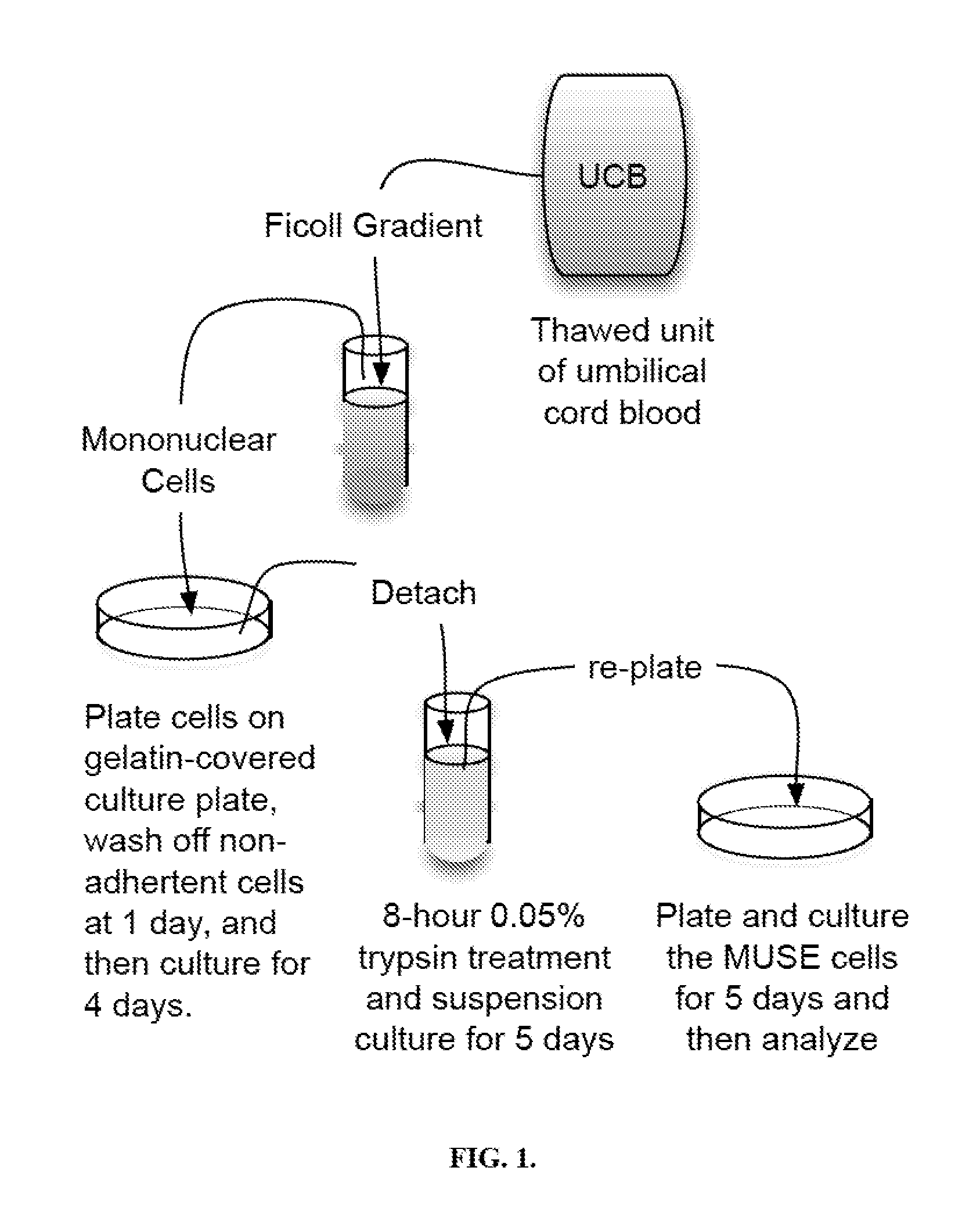
[0064]Mononuclear cells were isolated from umbilical cord blood by centrifugation in a Ficoll gradient, resulting in a buffy coat layer that contained the mononuclear cells. When cells were isolated from plasma depleted (PD) frozen units, osmotic shock was used to reduce the number of red blood cells and DNAase to prevent sticking of the cells to each other in the Ficoll gradient. PD usually yielded about a million cells per ml of thawed cord blood while thawed RCR units were 25 ml and contained 200-250 million mononuclear cells. About 40 million mononuclear cells were used.
[0065]Culturing
[0066]The cells were plated for 24 hours on gelatin-coated or other culture dishes to which mesenchymal cells attach and the non-adherent cells were washed off with phosphate-buffered saline. The cells were then in adherent culture in Minimal Essential Medium Eagle (MEM) Alpha Modifications, 10% ...
example 2
[0071]Mononuclear cells were isolated from a thawed unit of red cell reduced cord blood unit by Ficoll gradient centrifugation. The cells were then plated on gelatin-coated cell culture dishes, washed with phosphate buffered saline at 24 hours to remove non-adherent cells, and then cultured for 4 more days. After 5 days of adherent culture on gelatin-coated dishes, the umbilical cord blood mononuclear cells were then detached using a cell detachment solution and analyzed the cells by flow cytometry. It was found that after 5 days of adherent culture almost all the mononuclear cells expressed CD105, a mesenchymal cell marker.
[0072]The resulting flow cytometry scatterplots showed a variety of cells ranging from very low to very high side scatter and forward scatter. Application of propidium iodide (PI) and a phycoerythrin marker PE-Cy5,5-A to the cells showed a linear 45°“tail” of cells on scatterplots of PI / PE vs. PE. Since PI only gets into dead or dying cells, cells resulting this ...
example 3
[0083]The adherent mesenchymal cells obtained in the manner described in EXAMPLE 2 above were then exposed to 0.05% trypsin for 8 hours, cultured for 5 days in suspension cultures and then 5 days in adherent cultures. At the end of 10 days of culturing, the cells were detached with a non-trypsin detachment solution. Then, the above-described assays were performed to examine various markers.
[0084]CD105 expression was examined and related scatterplots were obtained. The obtained scatterplots included a graph showing a scatterplot of the cells distributed by side scatter (SSC-A) and forward scatter (FSC-A), and a graph of the PI / PE-Cy5,5A vs. PE-A distribution, showing a 45°“tail” of about 13% dead cells, which were eliminated from further analysis. The obtained scatterplots included a graph showing the isotype control and one showing the cells expressing CD105. It was found that almost all the cells (99.41%) express CD105, after eliminating the dead cells (i.e. high PI / PE ratio) from ...
PUM
 Abstract
Description
Claims
Application Information