Monomeric and bright infrared fluorescent proteins

a fluorescent protein and infrared technology, applied in the field of infrared fluorescent proteins, can solve the problems of limited use, poor penetration of excitation light, and limitations to the use of fluorescent proteins,

Active Publication Date: 2015-12-10
RGT UNIV OF CALIFORNIA
View PDF2 Cites 4 Cited by
  • Summary
  • Abstract
  • Description
  • Claims
  • Application Information

AI Technical Summary

Benefits of technology

[0180]In certain embodiments, the present invention provides a method of detecting a cancer cell in vivo, the method comprising administering to a patient with cancer a fusion protein of a variant polypeptide which has the amino acid sequence of SEQ ID NO:16, wherein the fusion protein binds to a cancer cell, and detecting the fluorescence of the variant polypeptide, wherein fluorescence of the polypeptide is greater than 10-fold higher than IFP1.4.
[0181]In certain embodiments, the present invention provides a method of detecting a cancer cell in vivo, the method comprising administering to a patient with cancer a fusion protein of a variant polypeptide which has the amino acid sequence of SEQ ID NO:18, wherein the fusion protein binds to a cancer cell, and detecting the fluorescence of the variant polypeptide, wherein fluorescence of the polypeptide is greater than 10-fold higher than IFP1.4.
[0182]In certain embodiments, the present invention provides a method of detecting a cancer cell in vivo, the method comprising administering to a patient with cancer a fusion protein of a variant polypeptide which has the amino acid sequence of SEQ ID NO:20, wherein the fusion protein binds to a cancer cell, and detecting the fluorescence of the variant polypeptide, wherein fluorescence of the polypeptide is greater than 10-fold higher than IFP1...

Problems solved by technology

However, despite the relative ease at expressing the GFPs, they offer limited use for in vivo imaging of whole animals (e.g., mice and humans) because of the low absorption/emission maxima (395 nm/509 nm).
Accordingly, although the availability of a wide variety of naturally occurring fluorescent proteins and spectral variants of the proteins has allowed for substantial advances, limitations to the use of fluorescent proteins remain.
In particular, the use of fluorescent proteins in intact animals such as mice has been hindered by poor penetration of excitation light.
For example, visibly ...

Method used


Image

  • Monomeric and bright infrared fluorescent proteins
  • Monomeric and bright infrared fluorescent proteins
  • Monomeric and bright infrared fluorescent proteins

Examples


example 1

Novel Truncation Mutant of a Bradyrhizobium sp. ORS278 Phytochrome

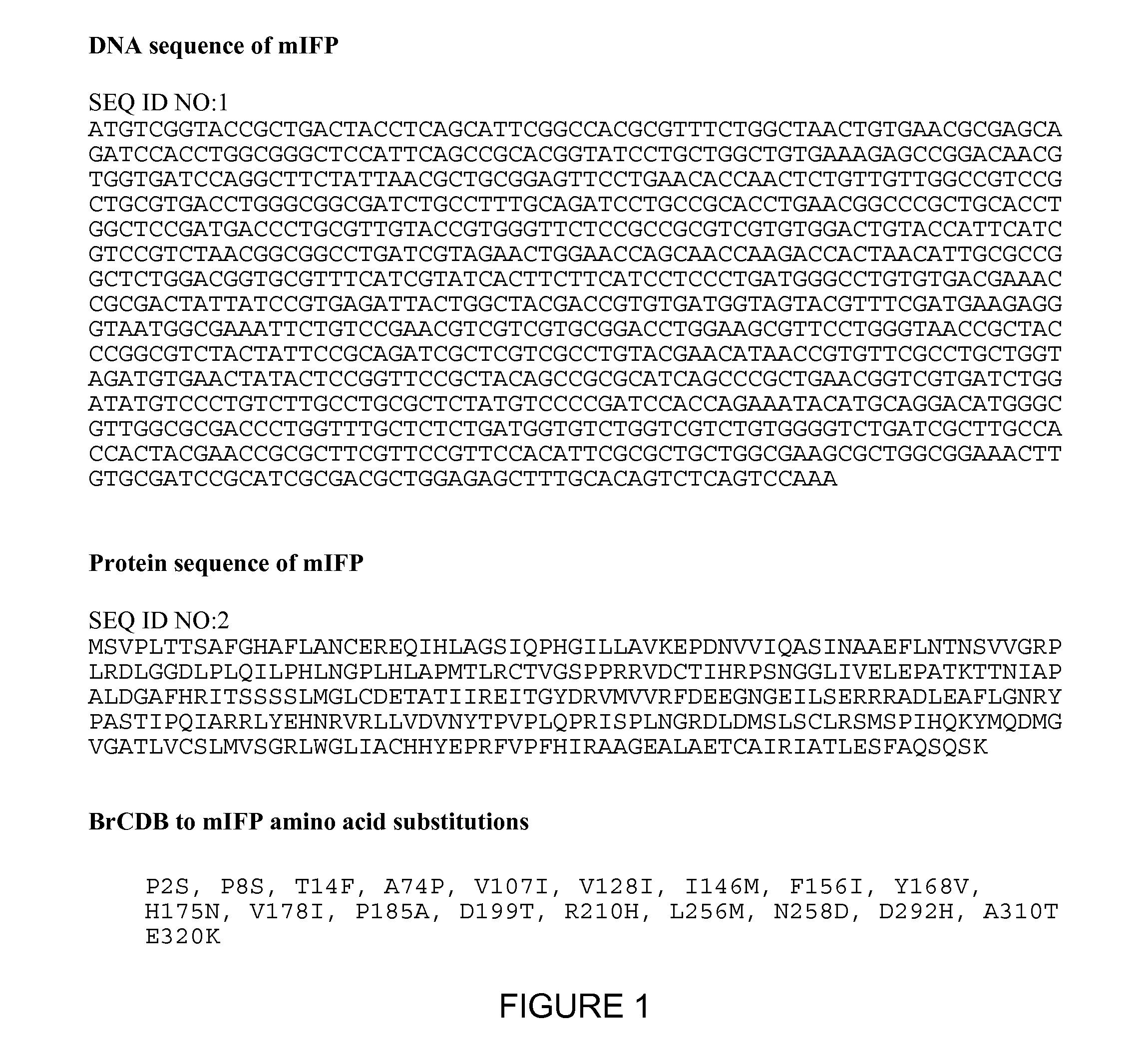
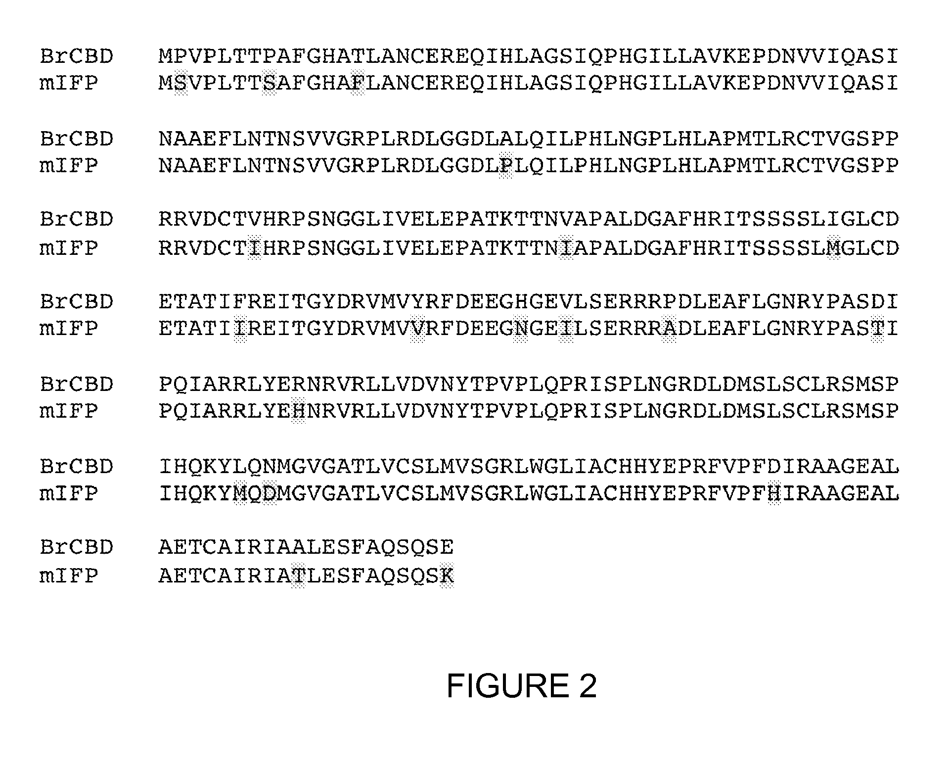
[0318]mIFP is a novel truncation mutant of the phytochrome BrBphP from the bacterium Bradyrhizobium sp. ORS278 (Giraud et al., Nature 417:202-205 (2002)). mIFP is monomeric, and contains 320 amino acids starting from the first N-terminal amino acid from BrBphP. mIFP includes 19 mutations introduced through directed evolution. The 19 amino acid substitutions of mIFP are P2S, P8S, T14F, A74P, V107I, V128I, I146M, F156I, Y168V, H175N, V178I, P185A, D199T, R210H, L256M, N258D, D292H, A310T, E320K.

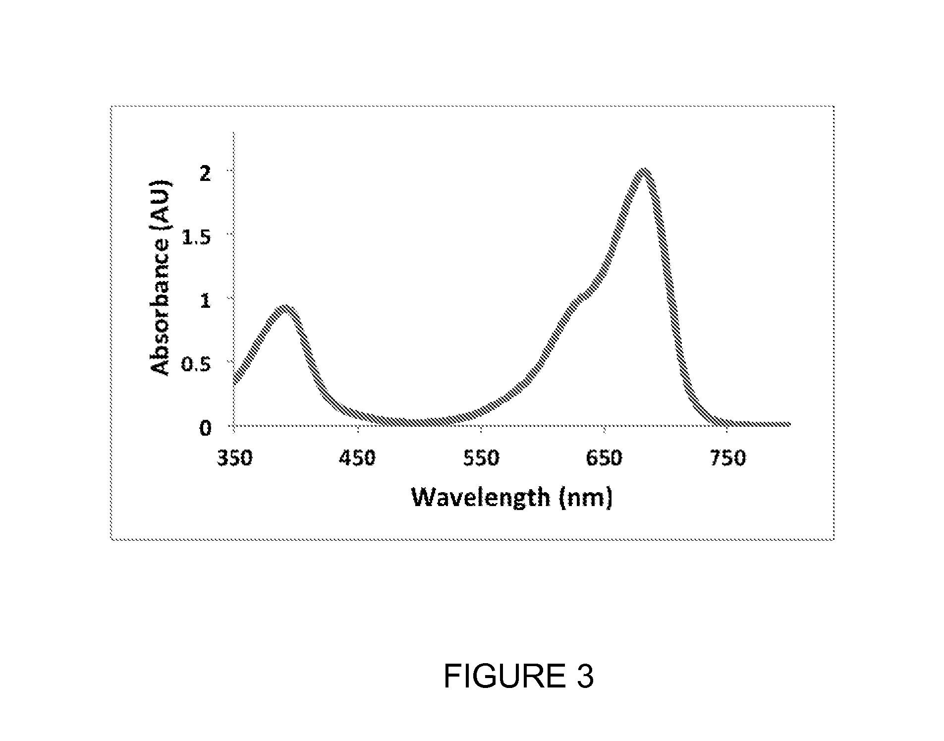
[0319]When expressed in either bacteria or mammalian cells, these mutant phytochromes spontaneously incorporate biliverdin, a ubiquitous intermediate in heme catabolism, and become fluorescent. The maximal absorbance wavelength of mIFP is 683 nm (FIG. 3). The maximal excitation and emission wavelengths are 683 nm and 704 nm, respectively (FIG. 4). mIFP shows approximately a ten-fold increase in brightness compared to IFP1.4. Gel ...

example 2

Additional Truncation Mutants

[0320]Additional truncation mutants were developed from BrBphP. The mutations are relative to wild-type BrCBD, which is truncated from full length BrBphP.

[0321]BrIFPv1 includes a D199M substitution. The amino acid sequence of BrIFPv1 is provided as SEQ ID NO:3, and the nucleotide sequence of BrIFPv1 is provided as SEQ ID NO:4.

[0322]BrIFPv2 includes the following substitutions: A74P, V107I, V128I, I146M, P185A, M199T, R210H, L256M, N258D, D292H, E320K. The amino acid sequence of BrIFPv2 is provided as SEQ ID NO:5, and the nucleotide sequence of BrIFPv2 is provided as SEQ ID NO:6.

[0323]BrIFPv3 includes the following substitutions: A74P, V107I, V128I, I146M, P185A, M199T, R210H, L256M, N258D, D292H, E320K, T14F, F156I, H175N, and A310T. The amino acid sequence of BrIFPv3 is provided as SEQ ID NO:7, and the nucleotide sequence of BrIFPv3 is provided as SEQ ID NO:8.

example 3

Mammalian Expression of mIFP Engineered from a Bacterial Phytochrome

[0324]mIFP is a genetically encoded tag for cellular and protein imaging. FIG. 6 shows mIFP as a genetically encoded tag for cellular and protein imaging. mIFP was expressed in a HeLa cell (FIG. 6, left panel), which co-expresses with EGFP (FIG. 6, right panel).

[0325]Because mIFP is monomeric, mIFP can be used as a protein fusion tag with no or little perturbation of the protein of interest. mIFP was fused to H2B, and localized to the nucleus (FIG. 7, left panel). In contrast, the same cell shows EGFP localization to both the cytosol and nucleus (FIG. 7, right panel). mIFP was also fused to LifeAct, which localized to F-acting filaments in a HeLa cell (FIG. 8).


PUM

PropertyMeasurementUnit
Temperatureaaaaaaaaaa
Fractionaaaaaaaaaa
Fractionaaaaaaaaaa

Abstract

The invention described herein features variants related to infrared fluorescent proteins, in particular to mutants of a phytochrome from the bacterium Bradyrhizobium sp. ORS278. The variants show approximately a ten-fold increase in brightness compared to other known infrared fluorescent proteins. The variants are monomeric, allowing them to be used as a protein tag without disrupting the function of the tagged protein of interest.

Description

FIELD OF THE INVENTION[0001]This invention relates to the discovery of novel infrared fluorescent proteins and methods of use thereof.BACKGROUND OF THE INVENTION[0002]The identification and isolation of fluorescent proteins in various organisms, including marine organisms, has provided a valuable tool to molecular biology. The isolated fluorescent proteins have been used in their natural state and have been modified to elicit excitation / emission shifts in order to maximize their utility in in vitro and in vivo imaging. Green Fluorescent Proteins (GFPs) are the most widely utilized of the fluorescent proteins.[0003]GFPs are involved in bioluminescence in a variety of marine invertebrates, including jellyfish such as Aequorea Victoria (Morise, H., et al., Biochemistry 13:2656-2662 (1974); Prendergast, F. G., and Mann, K. G., Biochemistry 17:3448-3453 (1978); Ward, W. W., Photochem. Photobiol. Rev. 4:1-57 (1979) and the sea pansy Renilla reniformis (Ward, W. W., and Cormier, M. J., Pho...

Claims


Application Information

Patent Timeline
IPC IPC(8): C07K14/195A61K49/00G01N33/52
CPCC07K14/195A61K49/0045G01N33/52C07K2319/00
Inventor YU, DANSHU, XIAOKUN
Owner RGT UNIV OF CALIFORNIA
Why Eureka
Try Eureka