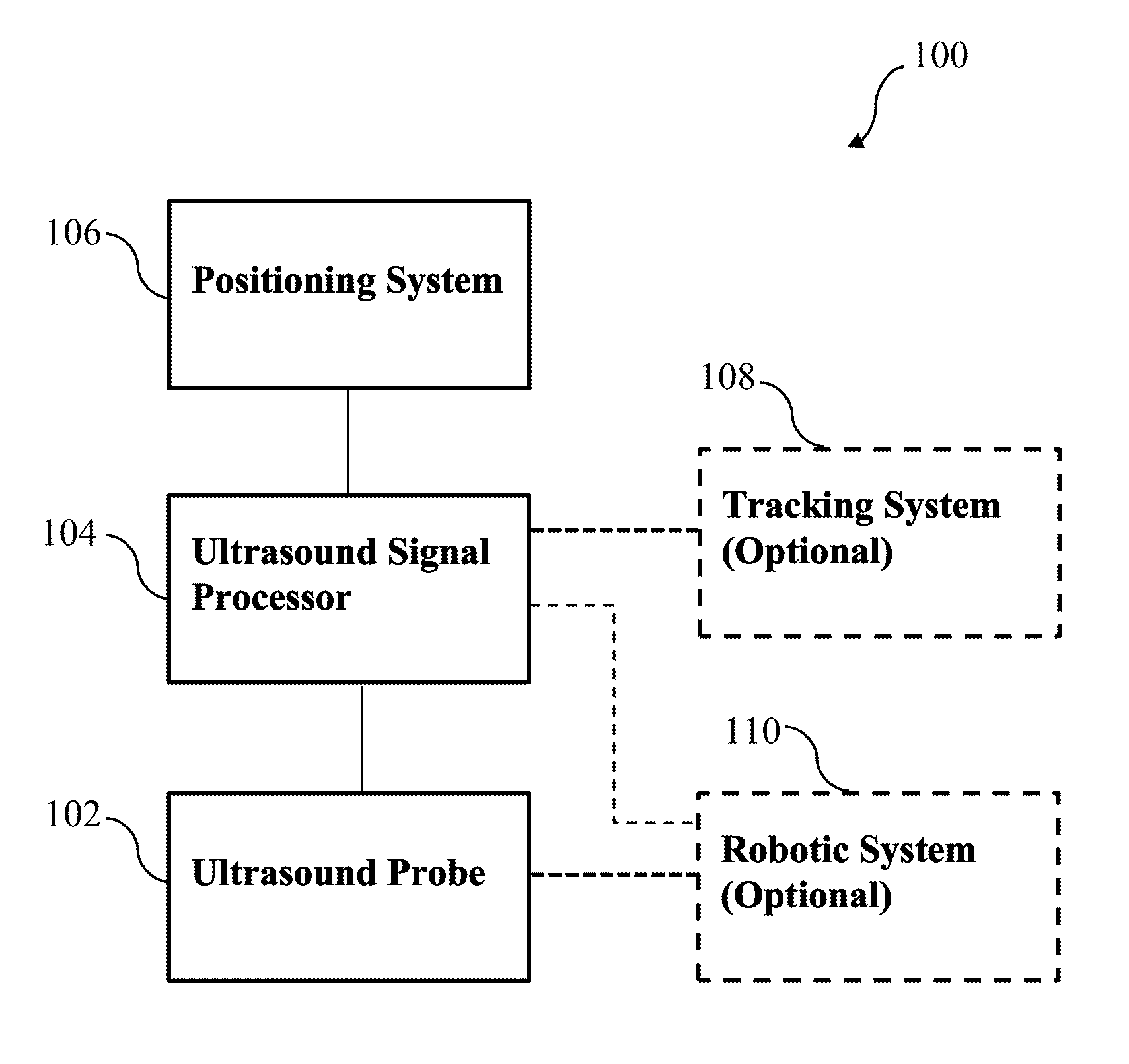
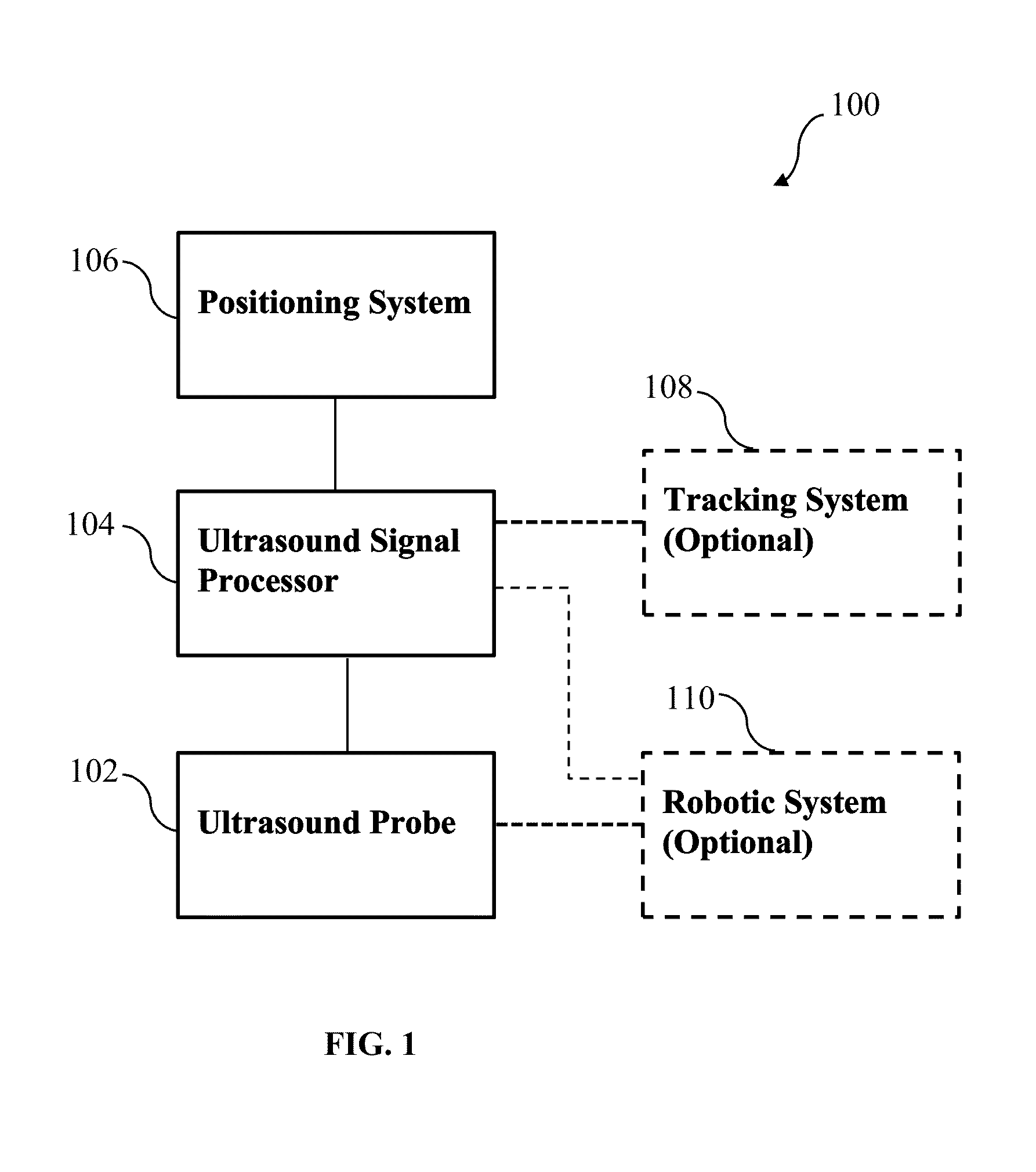
Synthetic aperture ultrasound system
a synthetic aperture and ultrasound technology, applied in the field of synthetic aperture ultrasound systems, can solve the problems of high cost, low flexibility for different usage requirements, and difficulty in achieving higher resolution in conventional synthetic aperture imaging
- Summary
- Abstract
- Description
- Claims
- Application Information
AI Technical Summary
Benefits of technology
Problems solved by technology
Method used
Image

Examples
example 1
Synthetic Aperture Ultrasound Imaging
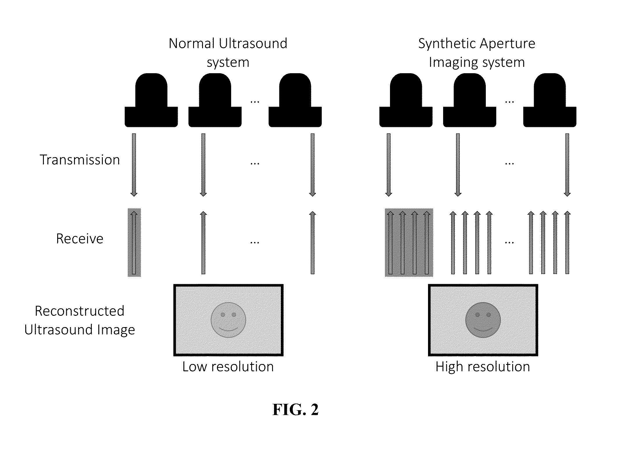
[0114]As described above, it is challenging to achieve higher resolution in conventional synthetic aperture imaging since the F number becomes too high in the deeper regions, and large arrays require huge costs and provide little flexibility for different usage requirements. To address these problems, some embodiment of the current invention expand the size of the available aperture by utilizing a robotic system. Having the ultrasound probe held by a robot arm allows rotational motion and translational displacement, which can generate imaginary elements, and the expanded aperture can be utilized in reconstruction. Since the F number can be reduced by widening the aperture, lateral resolution improvement can be expected. Therefore, some embodiments of the current invention provide higher resolution through an extended aperture implemented by a robotically controlled transducer. FIG. 2 illustrates the general idea of a synthetic aperture imaging sy...
example 2
Implementation of Swept Synthetic Aperture Imaging
[0144]Despite advancements in array construction, beamforming algorithms, and image post-processing, deep imaging targets still pose a challenge for modern ultrasound imaging. The resolution of an ultrasound image is limited by diffraction effects and largely dependent on three factors—transmit wavelength, imaging depth, and aperture extent. For a given target, imaging depth is fixed. The transmit wavelength is variable, but attenuation of the transmitted pulse through tissue increases with frequency, limiting the gains in resolution that can be made in practice. The aperture extent depends on the transducer used and is conventionally limited by the footprint of the array. We describe a system that extends the effective aperture and increases the imaging resolution using precisely tracked transducer motion.
[0145]Transducer motion has historically been used to extend the field of view (FOV) of an ultrasound image without altering the ...
example 3
Swept Synthetic Aperture Imaging: Ex Vivo Demonstration of Resolution Improvement
[0189]Ultrasound image quality is inherently limited by the dimensions of the imaging transducer. We describe an adaptively beamformed imaging system designed to image in vivo targets using free-hand motion of the transducer and analyze the potential benefits of such a system. Coherent summation of the recorded echo signals may be limited by clutter induced by aberration and reverberation from tissue layers overlying the target. We demonstrate the feasibility of the swept sensor imaging technique in the presence of these effects by imaging a point target through ex vivo abdominal tissue layers. Using an aperture synthesized from five lateral positions with half-aperture overlap without aberration correction, resolution was improved by 66% and the contrast-to-noise ratio of an anechoic lesion simulated using the experimentally-measured point spread function was increased by 18%. We conclude that the syst...
PUM
 Abstract
Description
Claims
Application Information
PatSnap Eureka turns technology decisions into work you can execute. Powered by our Innovation Knowledge Graph, it runs expert workflows across engineering, life sciences, materials and intellectual property. Get your review-ready output in minutes.


