
Cell sorting method

Active Publication Date: 2016-02-11
FOUND FOR BIOMEDICAL RES & INNOVATION
View PDF1 Cites 16 Cited by
  • Summary
  • Abstract
  • Description
  • Claims
  • Application Information

AI Technical Summary

Benefits of technology

The present invention allows for easier and less expensive purification of differentiated cells from a population of cells. This results in a safer and less tumorigenic source of cells for transplantation. The technical effect of this invention is the elimination of undifferentiated cells without the need for sorting.

Problems solved by technology

However, pluripotent stem cells such as iPS cell and ES cell often show canceration during their growth, and differentiated cells derived from pluripotent stem cell need to be carefully evaluated for the presence or absence of tumor formation caused by the remaining undifferentiated iPS cell and ES cell.
This poses the largest problem in transplantation of autologous iPSC-derived cells and tissues free of immunological barriers.
However, a direct influence of PEDF on undifferentiated cells has not been known well.

Method used


Image

  • Cell sorting method
  • Cell sorting method
  • Cell sorting method

Examples


reference example 1

Differentiation Induction from iPS Cell into RPE Cell

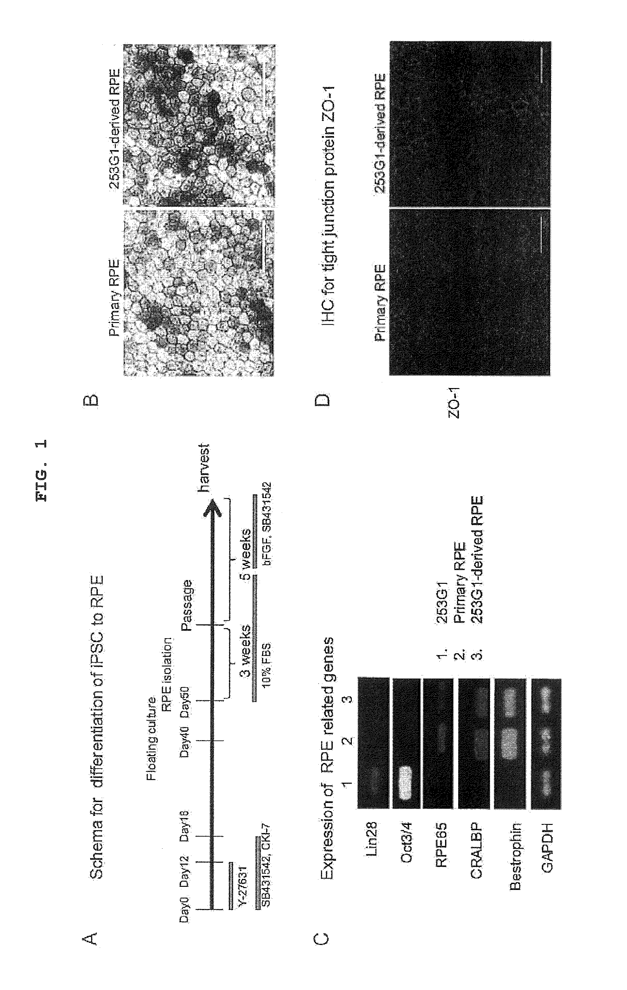
[0119]To establish robust differentiation protocol from pluripotent stem cell into retinal pigment epithelium (RPE), the differentiation protocol shown in FIG. 1A was used. To provide profile of reproducible iPSC-derived RPE, commercially available iPSC clone 253G1 (Nakagawa M, et al. (2008) Nat Biotechnol. 26:101-106; BioResource Center, RIKEN, Tsukuba) was used as a cell source for RPE differentiation. When cultured in a dish, RPE is a polygonal single layer cell showing sporadic pigment deposition. iPSC clone 253G1-derived RPE and primary RPE showed a similar morphology by microscopic observation (FIG. 1B). To examine whether iPSC-derived RPE shows gene expression characteristic of primary RPE, the expression of RPE65, CRALBP and bestrophin was analyzed by RT-PCR. While 253G1-derived RPE cells expressed mRNAs of RPE65, CRALBP and bestrophin, they did not express pluripotency-related genes such as Lin28, Oct3 / 4 and the like (FIG...

example 1

Inhibition of iPS Cell Growth by Co-Culture with iPSC-Derived RPE Cell

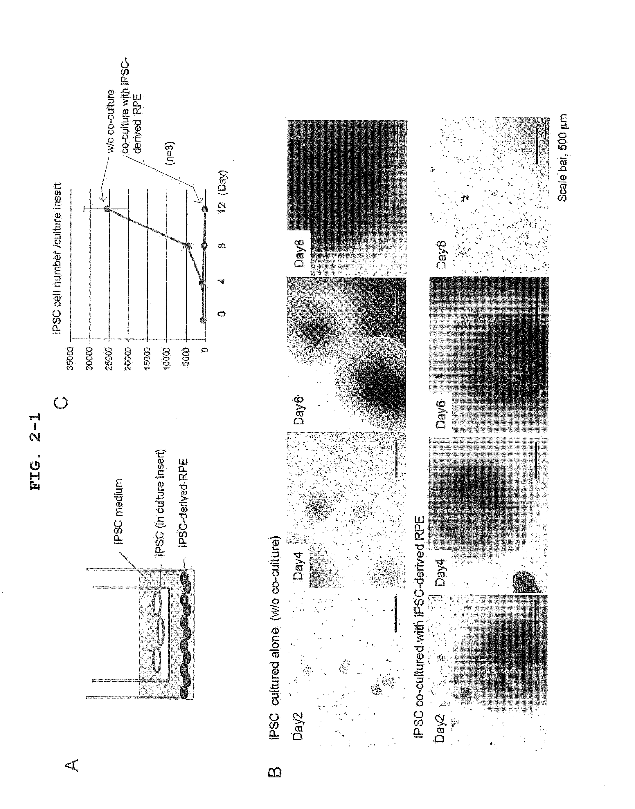
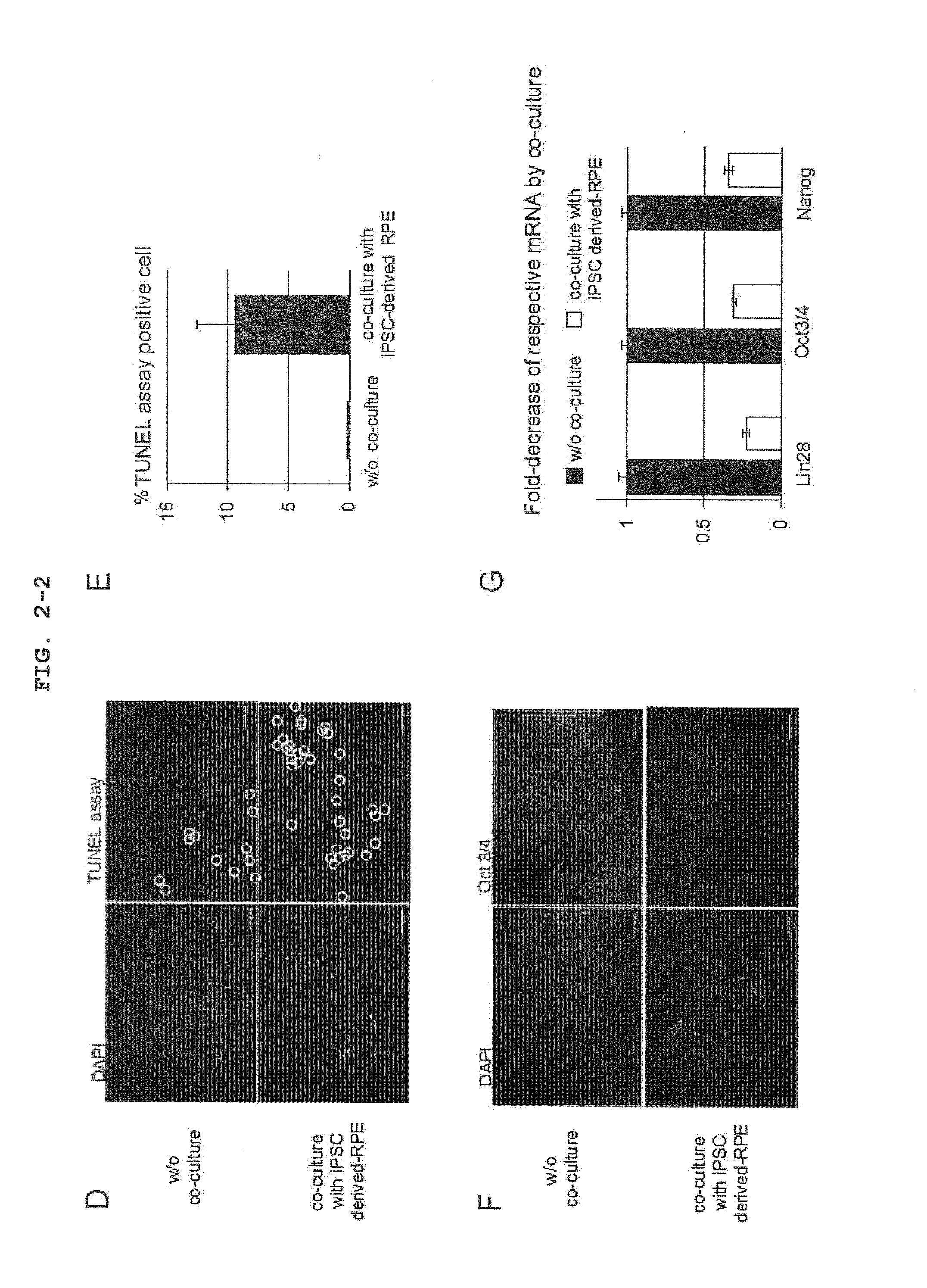
[0120]To examine an influence of a factor secreted from iPSC-derived RPE on iPS cell in vitro, the co-culture experiment shown in FIG. 2A was designed. That is, iPSC seeded on a culture insert (transwell, Corning) coated with Matrigel (BD) was co-cultured in an iPS cell medium (serum-free ReproFF added with bFGF) with iPSC-derived RPE seeded on a dish coated with CELL start (Invitrogen). iPSC in the culture insert was recovered every 4 days, and the cell number was counted. As a result, it was clarified that the growth of iPSC was markedly inhibited by co-culture with iPSC-derived RPE (FIGS. 2B, 2C). A similar trans-effect was observed even when iPSC was co-cultured with primary RPE (FIGS. 3A, B and C). The marked inhibition of the growth of iPSC co-cultured with iPSC-derived RPE was at least partially mediated by apoptotic cell death, as verified by the presence of obvious TUNEL assay positive cells (FIGS. 2D, 2E...

example 2

Induction of Apoptotic Cell Death of iPS Cell by PEDF

[0122]PEDF protein in an iPSC-derived RPE conditioned medium was detected by Western blotting using an anti-PEDF antibody (BioProducts, MD) (FIG. 4A). A fresh iPS cell medium free of co-culture was used as a control sample. The PEDF level in 24 hr of cell culture (24 hr after exchange with fresh medium) was measured by ELISA. Both the primary RPE conditioned medium and iPSC-derived conditioned medium contained a considerable amount of PEDF (exceeding 1 μg / ml) (FIG. 4B).

[0123]Therefore, an influence of PEDF on the growth of iPSC was tested. An anti-PEDF neutralizing antibody (BioProducts, MD) was added to the co-culture system, and the growth of iPSC in the culture insert was examined. The growth of 253G1 co-cultured with 253G1-derived RPE was markedly inhibited in the presence of control IgG, but such inhibition of cell proliferation was considerably blocked in the presence of an anti-PEDF antibody (FIG. 4C). Although the dose and...


PUM


Abstract

The invention provides a method of removing or reducing undifferentiated cells from a differentiated cell population contaminated or having a risk of contamination with undifferentiated cells by contacting a pigment epithelium-derived factor with the differentiated cell population to induce apoptosis of the undifferentiated cells. The invention also provides an agent for cell transplantation therapy, containing a differentiated cell population substantially free of an undifferentiated cell, which is obtained by the method, as well as an agent for inducing apoptosis of an undifferentiated cell, containing a pigment epithelium-derived factor, and combined use of the aforementioned agent for cell transplantation therapy and the aforementioned agent for inducing apoptosis.

Description

TECHNICAL FIELD[0001]The present invention relates to a method of sorting cells, more specifically, a method of sorting / purifying differentiated cells by removing or reducing undifferentiated cells from a differentiated cell population contaminated or having a risk of contamination with undifferentiated cells. The present invention also relates to an undifferentiated cell elimination agent for the aforementioned method, and an agent for a cell transplantation therapy, which is obtained by the aforementioned method, composed of tumorigenicity risk-free, uniform and safe differentiated cells.BACKGROUND ART [0002]Clinical application of regenerative medicines has been studied recently, and induction of differentiation of pluripotent stem cells such as iPS cell and ES cell into desired cells has been actively pursued. In fact, clinical tests using ES cell-derived RPE (retinal pigment epithelium) cell for Stargardt disease and dry age-related macular degeneration has already been started...

Claims


Application Information

Patent Timeline
IPC IPC(8): C12N5/00
CPCC12N5/0081A61K35/545A61P43/00C12N5/0696
Inventor KANEMURA, HOSHIMIGO, MASAHIROKAWAMATA, SHINNISHISHITA, NAOKI
Owner FOUND FOR BIOMEDICAL RES & INNOVATION
Learn More