Multi-scale complex systems transdisciplinary analysis of response to therapy
a complex system and multi-scale technology, applied in the field of multi-scale complex systems transdisciplinary analysis of the response to therapy, can solve the problems of poor prediction power, poor resolution, and many therapies that are initially effective, and achieve the effects of improving the predictive power, and improving the accuracy of diagnosis
- Summary
- Abstract
- Description
- Claims
- Application Information
AI Technical Summary
Benefits of technology
Problems solved by technology
Method used
Image
Examples
example 1
Immunostaing for Ki-67 of Ductal Carcinoma Cells
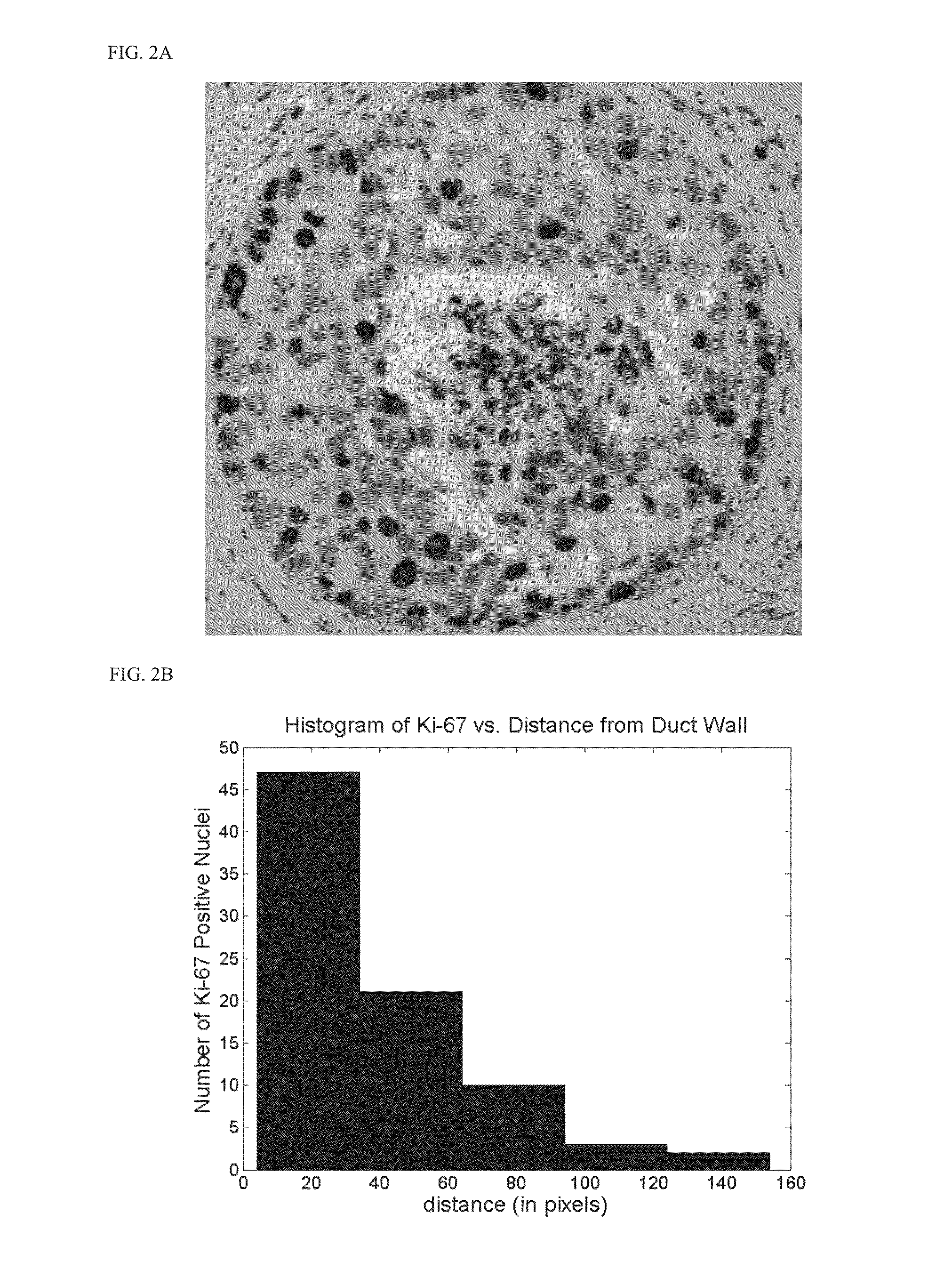
[0213]Samples of ductal carcinoma in situ in the breast from full mastectomies were immunostained for Ki-67 (a nuclear protein marker for cell proliferation) in ex vivo (FIG. 2). Proliferation occurred most frequently along the duct wall where nutrients and growth factors are most common. Much rarer proliferation was observed along the perinecrotic boundary towards the duct center where oxygen and nutrient levels were lowest (FIG. 2, top). To further quantify this, the distance between the duct wall and the Ki-67 positive nuclei were measured and plotted this as a histogram (FIG. 2, bottom). As seen in the graph, an excellent correlation between distance from the duct wall and the proliferation activity was observed, indicating a functional relationship between a microenvironmental quantity (e.g., oxygen), an intracellular protein (Ki-67), and cell phenotype (proliferation).
[0214]A similar correlation has been observed between internal...
example 2
Structural Model of an MSCS
[0215]RP1 module collects data on p53 activation in response to diverse forms of cellular stress to mediate a variety of anti-proliferative processes. Stress data include molecular events or extracellular stimuli associated with DNA damage, hypoxia, aberrant oncogene expression to promote cell-cycle checkpoints, DNA repair, cellular senescence, or apoptosis. Presence of anticancer agents activating p53 is recorded. Presence of agents promoting apoptosis or senescence is also recorded. Loss of p53 function is checked. RP2 module collects data on treatment with drug in p53 null system. The module checks if the loss causes decreased perturbation of pathways associated with cell death relative to ARF null. RP3 module analyzes treatment with drug in p53 null system to see if it has normal growth character. The module checks ARF level to see if it is increased. If increased, the module checks if there is correlation between increase apoptosis and increased drug ...
example 3
Structural Model of an MSCS
[0216]First, a cell model is built based on known information on p53 pathways. Protein binding assays on p53 pathway components and proteins known or suspected to be proximal to the pathway are conducted to collect up-to-date data on the cellular model. Differences in phosphorylation states measured by phospho-flow from those expected in the p53 pathway are added to the model. Updated network / pathway components are introduced to the Inferelator and its predictions are validated by comparing predicted dynamics to be measured. The model's phenotypic predictions, such as growth rate and pathway activates, are incorporated as input to the tumor model. At tumor-level model, any differences in measured tumor growth, vasculature, necrosis, and heterogeneity are used to update the tumor model. Updated tumor model parameters (such as change in growth rate, physical and spatial characteristics of the cell, invasiveness and mobility, etc.) are extracted and are used ...
PUM
 Abstract
Description
Claims
Application Information
 - R&D
- Intellectual Property
- Life Sciences
- Materials
- Tech Scout
- Unparalleled Data Quality
- Higher Quality Content
- 60% Fewer Hallucinations
Browse by: Latest US Patents, China's latest patents, Technical Efficacy Thesaurus, Application Domain, Technology Topic, Popular Technical Reports.
© 2025 PatSnap. All rights reserved.Legal|Privacy policy|Modern Slavery Act Transparency Statement|Sitemap|About US| Contact US: help@patsnap.com


