
Immunochromatographic Diagnosis Kit

a technology of immunochromatographic and diagnostic kit, which is applied in the direction of instruments, chemical methods analysis, analysis using chemical indicators, etc., can solve the problems of insufficient reproducibility, and achieve the effects of improving analysis sensitivity, reproducibility of examination results, and rapid diagnosis

Active Publication Date: 2016-05-05
ASAHI KASEI FIBERS CORPORATION
View PDF3 Cites 2 Cited by
  • Summary
  • Abstract
  • Description
  • Claims
  • Application Information

AI Technical Summary

Benefits of technology

The invention is an immunochromatographic diagnostic kit that uses prescribed chromogenic particles and a sample pad made of regenerated cellulosic fibers to increase the speed and sensitivity of diagnosis. The sample pad allows the chromogenic particles to flow more easily, reducing interference with the pores of the porous structures and increasing the reproducibility of examination results. The kit also allows for a constant concentration of chemical agents to facilitate flow of the chromogenic particles, ensuring that all particles contribute to the examination and improving analysis sensitivity and reproducibility.

Problems solved by technology

However, for quantitation in immunochromatography it is ideal to not only have proportionality between antigen concentration and examination result signal strength at certain antigen concentrations, but also to have reproducibility of test results, i.e. to obtain a signal of the same intensity when measuring antigen of the same concentration, but it is the general consensus that such reproducibility is still inadequate.

Method used


Image

  • Immunochromatographic Diagnosis Kit
  • Immunochromatographic Diagnosis Kit
  • Immunochromatographic Diagnosis Kit

Examples


example 1

Preparation of Antibody-Sensitized Colored Cellulose Particles

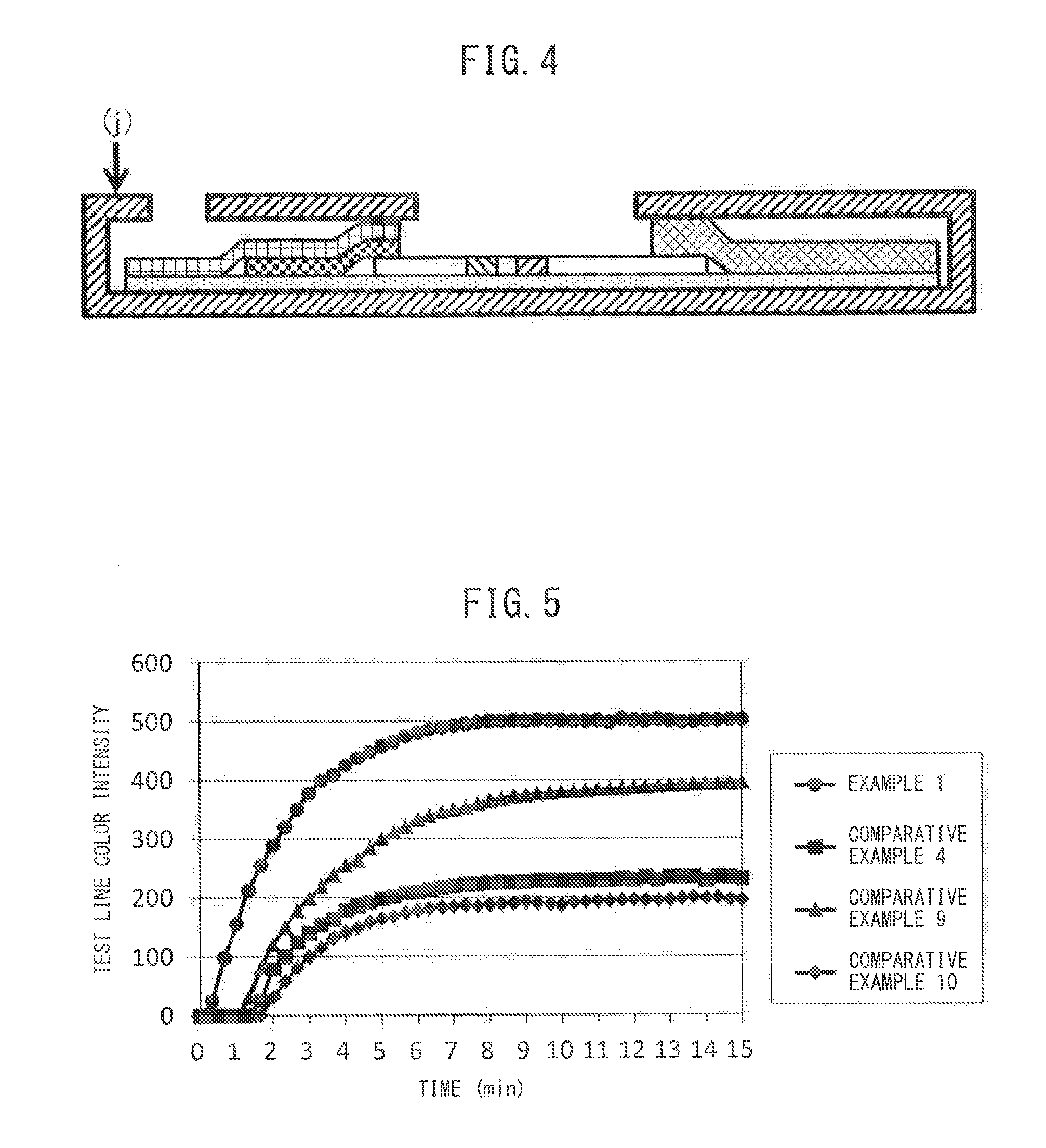
[0086]A 120 μl portion, of 1.0 wt % colored cellulose particles 1 (average particle diameter: 352 nm, color intensity: 2.9 ABS, colorant proportion: 49%), prepared by a known method, was placed in a 15 ml centrifuge tube, and then 240 μl of Tris-buffering solution (50 mM, pH 7.0) and 120 μl of 0.1% anti-hCG-α mouse antibody (10-C25C, product of Fitzgerald) were added and the mixture was stirred for 10 seconds with a vortex. It was then placed in a dryer adjusted to 37° C. and allowed to stand for 120 minutes. Next, 14.4 ml of blocking solution (100 mM boric acid, pH 8.5) containing 1.0 wt % casein (030-01505 by Wako Pure Chemical Industries, Ltd.) was added, and the mixture was further allowed to stand for 60 minutes in a dryer at 37° C. A centrifugal separator (6200 by Kubota Corp.) and a centrifugal separation rotor (AF-5008C by Kubota Corp.) were then used for centrifugation at 10,000 g for 15 minutes, and upon precipi...

example 2

[0092]An immunochromatographic diagnostic kit was prepared by the same method as Example 1, except that the chromogenic particles were colored cellulose particles 2 (average particle diameter: 588 nm, color intensity: 2.7 ABS, colorant proportion: 45%), and the performance was evaluated. The results are shown in Table 1 below.

example 3

[0093]An immunochromatographic diagnostic kit was prepared by the same method as Example 1, except that the chromogenic particles were colored cellulose particles 3 (average particle diameter: 790 nm, color intensity: 2.6 ABS, colorant proportion: 42%), and the performance was evaluated. The results are shown in Table 1 below.


PUM

PropertyMeasurementUnit
thicknessaaaaaaaaaa
particle diameteraaaaaaaaaa
volume-average median diameteraaaaaaaaaa

Abstract

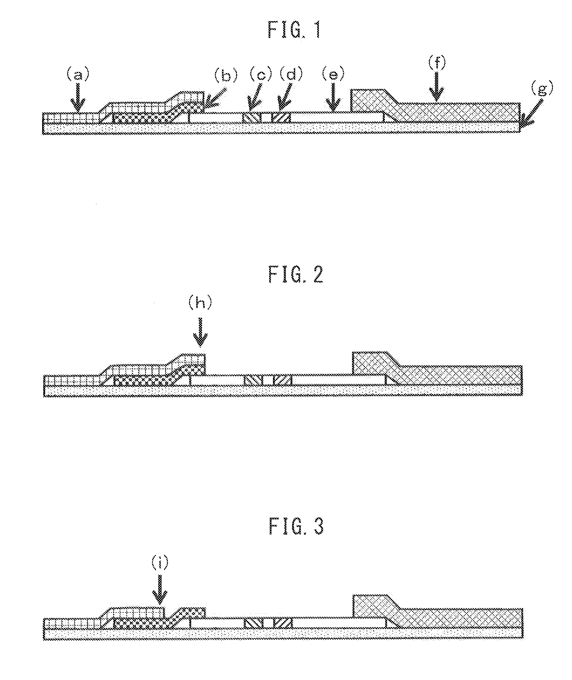
Provided is an immunochromatographic diagnostic kit which renders prompt diagnosis possible, has high analysis sensitivity, and gives test results with excellent reproducibility. The immunochromatographic diagnostic kit includes: a conjugate pad that contains a conjugate containing chromogenic particles which have an average particle diameter of 100-1,000 nm and attain a color intensity of 1.0-10.0, 10-90 wt % of the particles being derived from cellulose and 90-10 wt % of the particles being derived from a colorant; and a sample pad for an in vitro diagnostic reagent, the sample pad being constituted of nonwoven fabric that has a basis weight of 10-150 g / m2 and a thickness of 0.07-1.00 mm and comprises regenerated cellulosic fibers.

Description

TECHNICAL FIELD[0001]The present invention relates to an immunochromatographic diagnostic kit containing a sample pad for an in vitro diagnostic reagent, and more specifically it relates to a lateral flow-type immunochromatographic diagnostic kit.BACKGROUND ART[0002]In recent years, convenient examination reagents, diagnostic reagents and diagnostic kits have been developed for conducting various examinations for the presence of pathogenic infection by viruses, bacteria or the like, the presence or absence of pregnancy, the presence or absence of cancer markers, and the presence or absence of specific raw materials or hazardous substances such as residual pesticides in foods, in short periods of time. These utilize specific reactions between the different substances to be examined and substances that specifically react with the substances to be examined. In particular, a large number of assay methods have been developed as immunoassays using antigen-antibody reaction between antigen...

Claims


Application Information

Patent Timeline
Patent Type & Authority Applications(United States)
IPC IPC(8): G01N33/543
CPCG01N33/54386G01N33/558G01N33/54388
Inventor HORII, ATSUSHISHIOMI, YOSHIYUKIOGAWA, TAKAYUKIMATSUSE, TAKESHISUGINO, MAKI
Owner ASAHI KASEI FIBERS CORPORATION
Learn More