Acceleration and enhancement methods and system for ultrasound scatterer structure visualization

a technology of ultrasound scatterer and enhancement method, which is applied in the field of acceleration enhancement method and ultrasound scatterer structure visualization system, can solve the problems of unsuitable quantitative analysis of scatterer properties, tissue structure, and unsuitable b-mode images of conventional ultrasonic b-mode, and achieves the preservation of the accuracy of the ultrasound scatterer structure image, avoid computation time consumption, and improve the quality and resolution

Active Publication Date: 2016-05-12
AMCAD BIOMED CORP
View PDF11 Cites 4 Cited by
  • Summary
  • Abstract
  • Description
  • Claims
  • Application Information

AI Technical Summary

Benefits of technology

[0005]The present invention relates to a method for ultrasound scatterer structure visualization. The present invention reflects the tissue characteristics and enhances the quality and the resolution of ultrasound scatterer structure images, in particular for homogeneous tissues. The present invention maintains the accuracy of the ultrasound scatterer st

Problems solved by technology

Conventional ultrasonic B-mode images qualitatively describe tissue structures but are unsuitable for quantitative analyses of scatterer properties.
Nevertheless, due to that the backscattered signals are actually dependent on the shape, size, density, and other properties of the scatterers

Method used


Image

  • Acceleration and enhancement methods and system for ultrasound scatterer structure visualization
  • Acceleration and enhancement methods and system for ultrasound scatterer structure visualization
  • Acceleration and enhancement methods and system for ultrasound scatterer structure visualization

Examples


Embodiment Construction

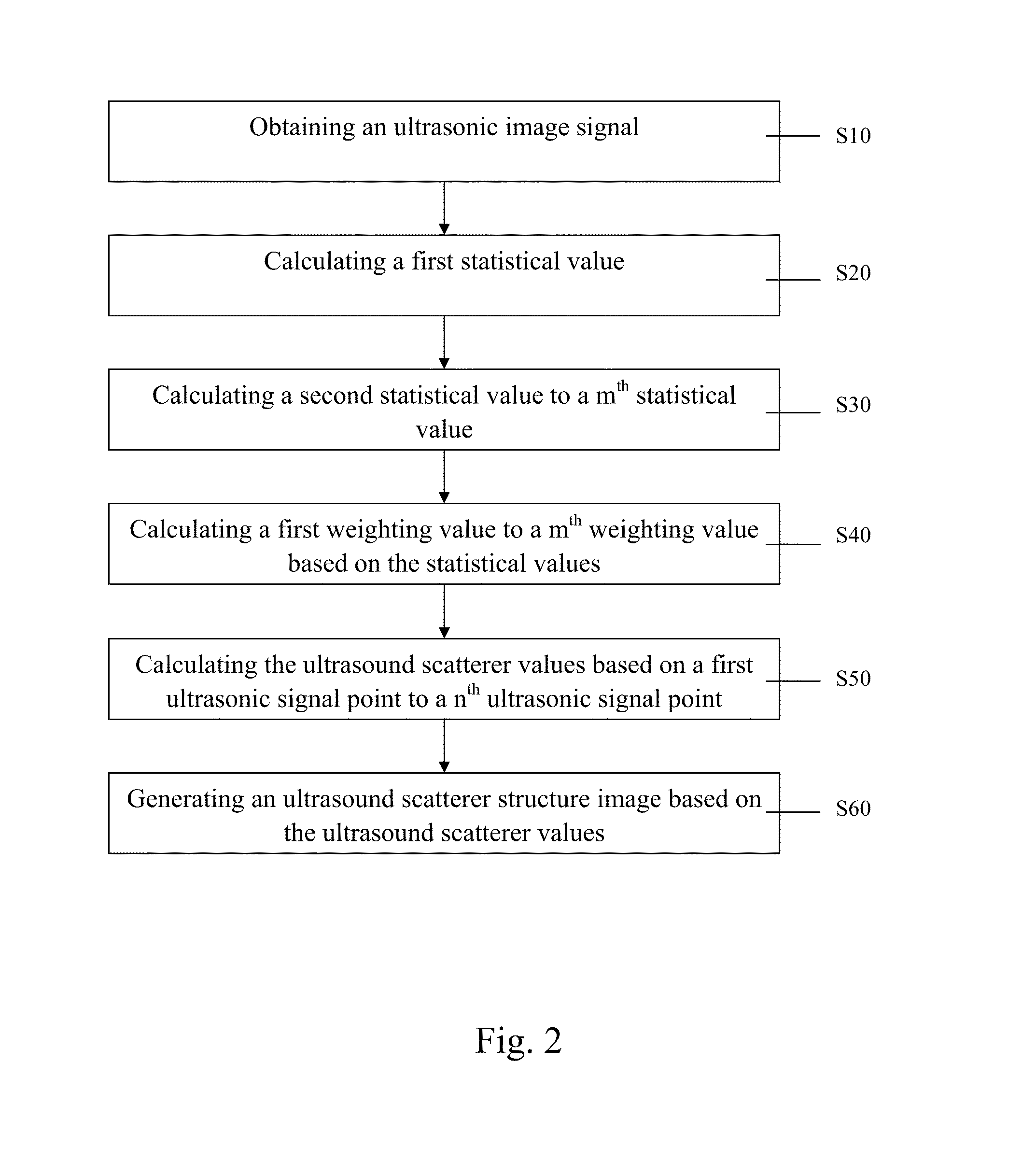
[0023]The present invention discloses an acceleration and enhancement method for ultrasound scatterer structure visualization. It is understood that the method provides merely an example of the many different types of functional arrangements that may be employed to implement the operation of the various components of an ultrasound device, a computer system connected to the ultrasound device, a multiprocessor computing device, and so forth. The execution steps of the present invention may include application specific software which may store in any portion or component of the memory including, for example, random access memory (RAM), read-only memory (ROM), hard drive, solid-state drive, magneto optical (MO), IC chip, USB flash drive, memory card, optical disc such as compact disc (CD) or digital versatile disc (DVD), floppy disk, magnetic tape, or other memory components.

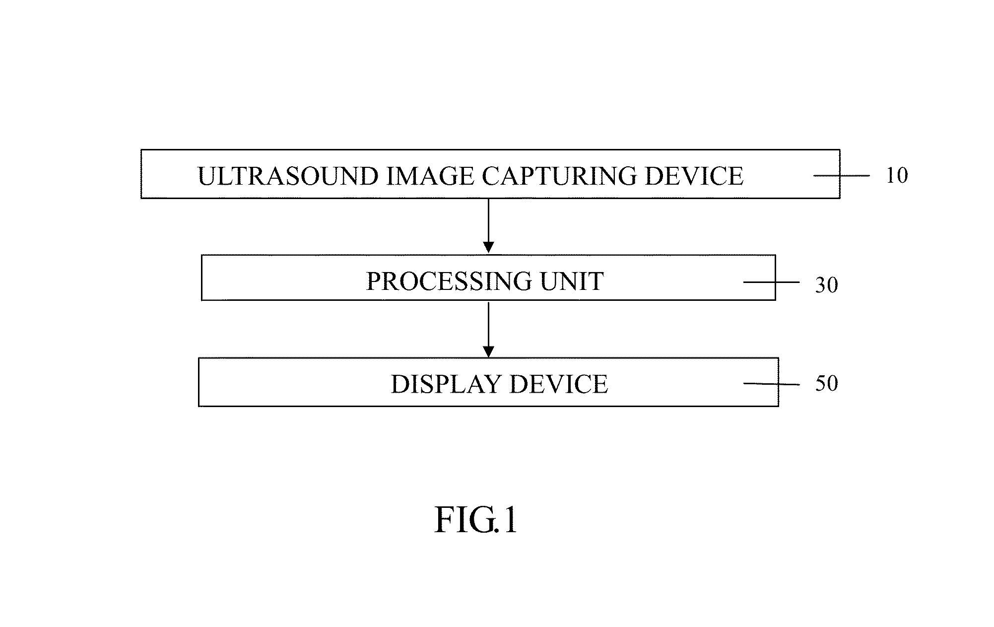
[0024]For embodiments, the computer system comprises a display device, a processing unit, a memory, an input devi...


PUM


Abstract

The present invention provides an acceleration and enhancement methods for ultrasound scatterer structure visualization. The method includes: obtaining an ultrasonic image, calculating all values of the ultrasonic signal points in each mth window centered at a nth signal point to obtain a plurality of original statistical values anxm, obtaining a plurality of mth statistical values by averaging value of original statistical values in the same window, calculating a plurality of mth weighting values based on the statistical values by different weighting formulas, multiplying each weighting value with the original statistical values corresponding to the various size of windows, summing up to obtain an ultrasound structure scatterer value of the nth ultrasonic signal point, and generating an ultrasound scatterer structure image based on a matrix of the ultrasound scatterer values. The present invention further combined interpolation method can reduce the computation time and retain the 80% accuracy.

Description

FIELD OF THE INVENTION[0001]The invention relates to a method and system for ultrasound scatterer structure visualization. In particular, the invention relates to an acceleration and enhancement method and system for ultrasound scatterer structure visualization.BACKGROUND OF THE INVENTION[0002]Conventional ultrasonic B-mode images qualitatively describe tissue structures but are unsuitable for quantitative analyses of scatterer properties. The scattering phenomenon is occurred when the incident wavelength is greater than the size of the scatterer in a tissue. The generated backscattered signals would form speckle, which exhibits a granular pattern of white and dark spots in the ultrasonic B-mode image. To avoid the influence of the speckle effect on the image quality, many methods were proposed to reduce the speckle appearance in the B-mode image. Nevertheless, due to that the backscattered signals are actually dependent on the shape, size, density, and other properties of the scatt...

Claims


Application Information

Patent Timeline
IPC IPC(8): G06T7/00A61B8/00A61B8/14
CPCG06T7/0034A61B8/14A61B8/461G06T2207/30024G06T7/0026G06T2207/10132G06T7/0014G01S15/8977G01S7/52047
Inventor TSUI, PO-HSIANGHO, MING-CHIHCHEN, CHIUNG-NEINCHEN, ARGONCHEN, JIA-JIUNWANG, YU-HSINHUANG, KUO-CHEN
Owner AMCAD BIOMED CORP
Why Eureka
Try Eureka