
Proteins and protein conjugates with increased hydrophobicity
- Summary
- Abstract
- Description
- Claims
- Application Information
AI Technical Summary
Benefits of technology
Problems solved by technology
Method used
Image

Examples
example 1
[0065]The GLP-1 C-terminal cysteine mutein (GLP-1 [7-36] plus an added cysteine at position 37) was cloned as a fusion to a larger polypeptide that contained a self-cleaving autoproteolytic sequence between GLP-1 and its fusion partner. The fusion protein was expressed in E. coli using an IPTG inducible system under the control of T7 polymerase.
example 2
[0066]The GLP-1 fusion protein was isolated from cell lysates under denaturing conditions, renatured, cleaved (autoproteolysis), and further purified using cationic chromatography. Characterization assays to confirm the peptides identity included RP-HPLC analysis, SDS-PAGE, and mass spectrometry. Commercially available synthetic cysteine mutein of GLP-1 was used as the control for these assays.
example 3
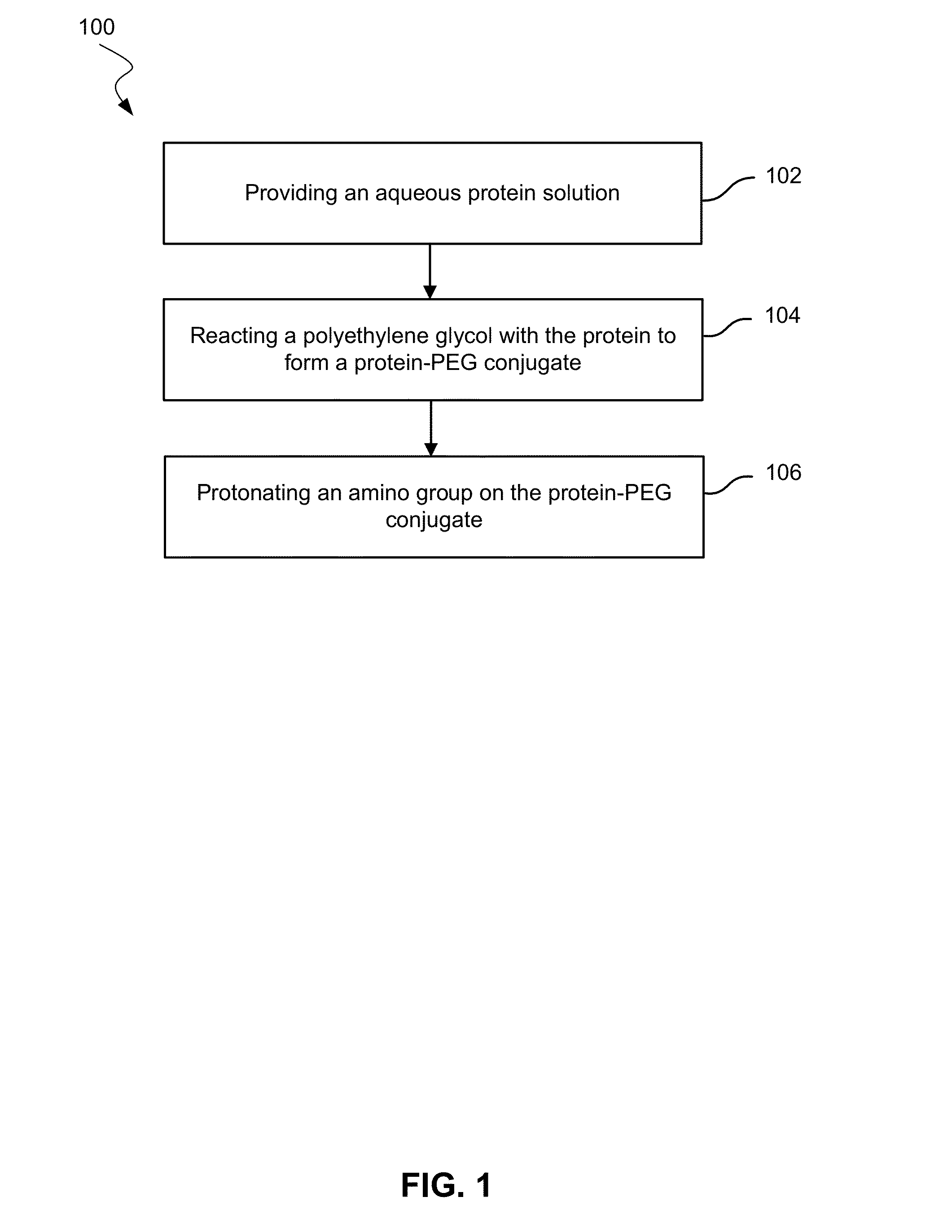
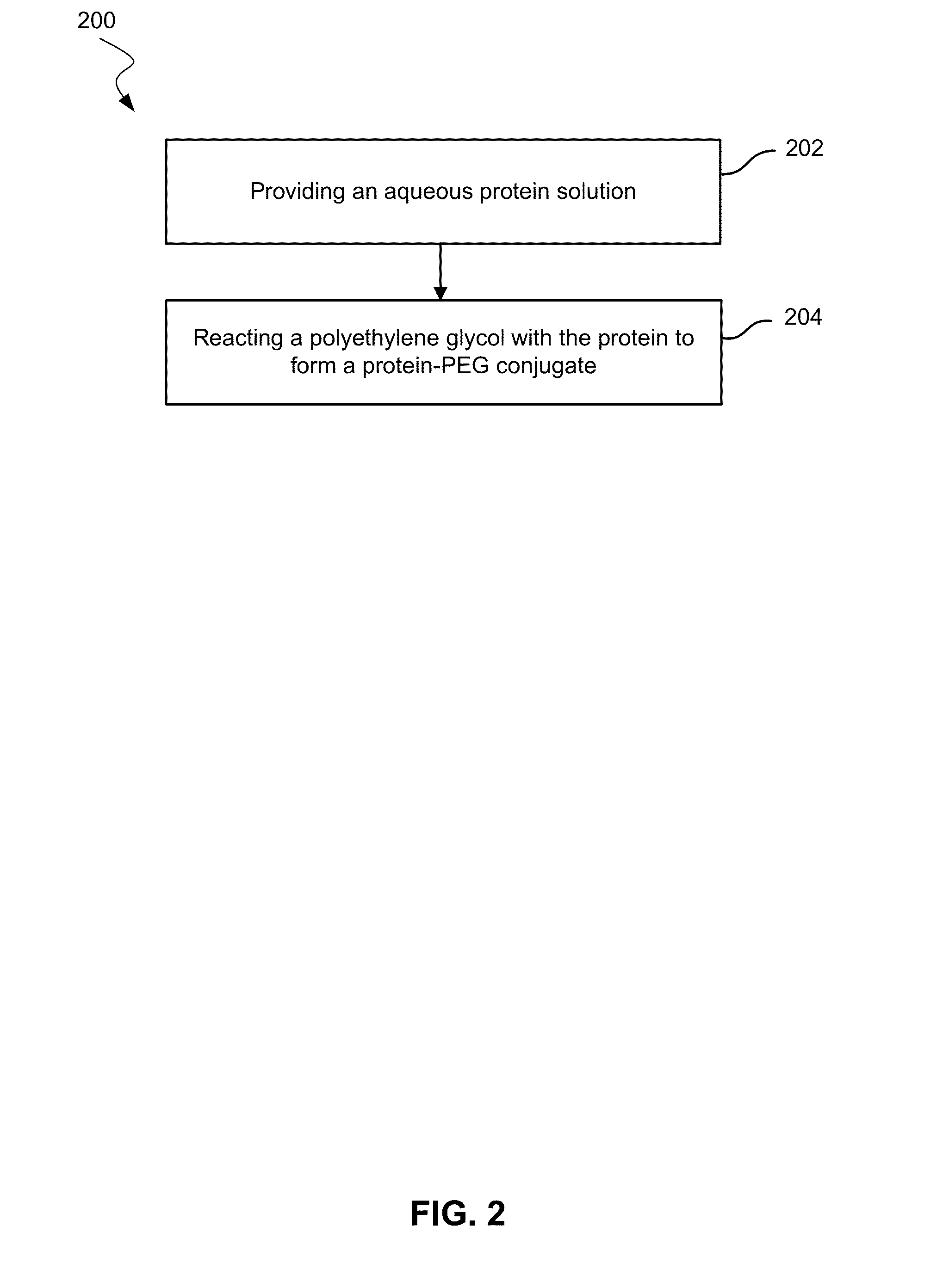
[0067]The GLP-1 cysteine mutein (produced either by a recombinant or synthetic chemical process) was PEGylated with a 5 kDa or 10 kDa cysteine-reactive maleimide-PEG by the following process. The peptide was first dissolved in 20 mM sodium phosphate buffer, pH 7.5 at a concentration of 1-5 mg / mL, and an equal molar amount of the maleimide PEG reagent was added. The reaction was allowed to continue overnight. The PEGylated GLP-1 peptide was purified using cation exchange chromatography (SP-HP Sepharose) with an equilibration buffer of 10 mM sodium acetate at pH 3.5 and a step elution buffer of 0.02% ammonium bicarbonate. The product-containing fractions were pooled, dialyzed against 0.02% ammonium bicarbonate, and lyophilized. The concentration of purified PEGylated peptide was determined by UV spectroscopy or by Bradford protein assay. Additional analytical assays performed post-PEGylation include SEC-HPLC analysis, SDS-PAGE, mass spectral analysis, N-terminal analysis, and endotoxi...
PUM
| Property | Measurement | Unit |
|---|---|---|
| Mass | aaaaa | aaaaa |
| Mass | aaaaa | aaaaa |
| Molecular weight | aaaaa | aaaaa |
Abstract
Description
Claims
Application Information