Prosthesis for Partial and Total Joint Replacement

a partial and total joint replacement technology, applied in the field of prosthetic joints, can solve the problems of post-traumatic arthritis, patients experiencing pain and loss of range of motion, and affecting the function of the entire arm, and affecting the function of the elbow joint in younger age groups

Active Publication Date: 2016-06-09
KAUFMANN ROBERT A
View PDF9 Cites 1 Cited by
  • Summary
  • Abstract
  • Description
  • Claims
  • Application Information

AI Technical Summary

Problems solved by technology

In post-traumatic settings, where surgical efforts have failed to restore adequate alignment, post-traumatic arthritis may set in and become symptomatic with patients experiencing pain and a loss of range of motion.
Whether idiopathic, inflammatory or post-traumatic, elbow arthritis is often painful and may interfere with function of the entire arm.
Whereas degenerative joint diseases usually affect patients after the fifth decade of life, traumatic conditions may cause elbow joint impairment in younger age groups.
Elbow fractures such as of the distal humerus or olecranon often exhibit intra-articular involvement, which can lead to permanent cartilage damage despite the best surgical reconstruction efforts.
This is particularly true when anatomic reconstruction of the joint surfaces is not successful or if osteochondral portions are missing as may occur in open injuries.
High energy injuries such as Motorcycle accidents may damage the distal humerus of younger patients and leave the ulna and radius relatively uninjured.
However, the results of these early efforts were characterized by implant loosening, instability and poor clinical results.
This improved the stability of the construct, but still resulted in rates of loosening of up to 50%.
The linked design offers superior stability and dislocations of these joints are rare, however, the constrained design transfers forces through the bone-cement or bone-implant interface which then results in significant loosening rates.
Understandably, this reliance on soft tissues to maintain stability results in higher rates of instability and joint dislocation compared to linked designs.
These characteristics prevent the use of an unlinked prosthesis in situations where soft tissue integrity is poor or extensive bone loss has occurred.
Total elbow arthroplasty may not be an ideal solution for younger patients.
On occasion, the distal humerus is severely injured with relative sparing of the proximal ulna or radius.
In some instances, such as high energy motor vehicle accidents, the distal humerus is not salvageable.
However, radiographic signs of attrition indicate that this implant may not be ideally suited for this role.
Even though this offers a quick, reliable, and relatively durable bone-to-implant fixation, there are significant limitations.
The most noteworthy is that the implant and cement mantle is difficult to remove if this is required such as in the face of an infection or peri-prosthetic fracture.
It is when eradicating an infection, however, when the entire cement mantle needs to be accounted for and eliminated, that the surgical effort to accomplish this becomes quite involved.
Damage to the soft tissues as well as neurovascular structures can occur and, even in the best of circumstances, significant bone stock is removed along with the cement.
The use of cement has the potential added drawback of causing thermal osteonecrosis.
This high temperature may damage the surrounding bone and potentially compromise its ability to heal after implant installation.
It may also hinder new bone growth advantageous to helping secure the implant after installation.
Elbow arthroplasty carries serious potential complications.
The soft tissue envelope that surrounds the elbow is thin, thereby making this location vulnerable to this complication.
Given that the cement must be removed in addition to the implant in order to eradicate the infection, the likelihood of having reasonable bone stock remaining after the implant and bone cement has been removed is low.
Realizing the low survival rate of the second implant, the decision to re-implant a new arthroplasty is often met with significant resistance.
Peri-prosthetic loosening may occur after implantation and often requires revision surgical efforts.
In general, the ulnar components are at the highest risk for aseptic loosening and this risk is associated with the quality of fixation.
Uncemented elbow arthroplasty in rheumatoid arthritis patients was described utilizing the Kudo prosthesis which demonstrated a high rate of aseptic loosening (7 of 49) within the ulnar component ultimately leading to an inability to recommend this implant without the use of cement fixation.
Not all fractures require surgical treatment and immobilization may be sufficient in some cases.
A significant hardship when treating periprosthetic fractures involves the cement mantle that surrounds the implant.
As it lies within the fractured bone and exhibits no healing capability, the cement often has to be removed and then the void filled with bone graft.
If significant loss of bone stock is present within the distal humerus or proximal ulna, successful surgical revision may not be possible leading to salvage options such as an arthrodesis or leaving the elbow flail or even consideration of an amputation.
Other complications may occur that are unrelated to the method of component fixation.
Bone cement implantation syndrome (BCIS) is a unique problem associated with cement fixation of an implant.
Bone cement implantation syndrome (BCIS) is poorly understood and yet is an important cause of intraoperative mortality and morbidity in patients undergoing cemented hip arthroplasty and may also be seen in the postoperative period in a milder form causing hypoxia and confusion.
This unconstrained device requires that the patient's natural soft tissues are functional.
Furthermore, threaded attachments are subject to loosening if not further secured by some additional means.
Although it is often difficult for a surgeon to know whether a hemiarthroplasty or total arthroplasty will be required prior to commencing surgery, very few of the implants described above may be used for either type of surgery.
However, none of these methods has included any type of ligament reconstruction that would essentially reproduce that which was present in the elbow prior to injury or deterioration.
All mechanical devices are subject to wear.

Method used


Image

  • Prosthesis for Partial and Total Joint Replacement
  • Prosthesis for Partial and Total Joint Replacement
  • Prosthesis for Partial and Total Joint Replacement

Examples


Embodiment Construction

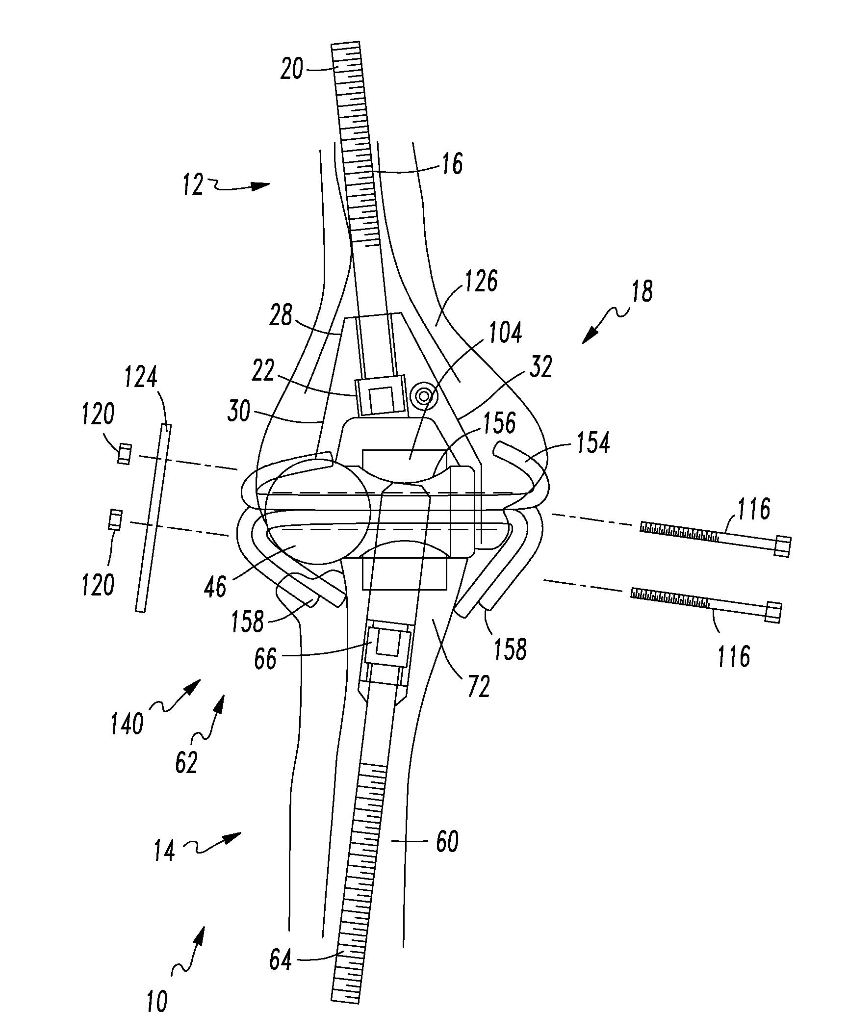
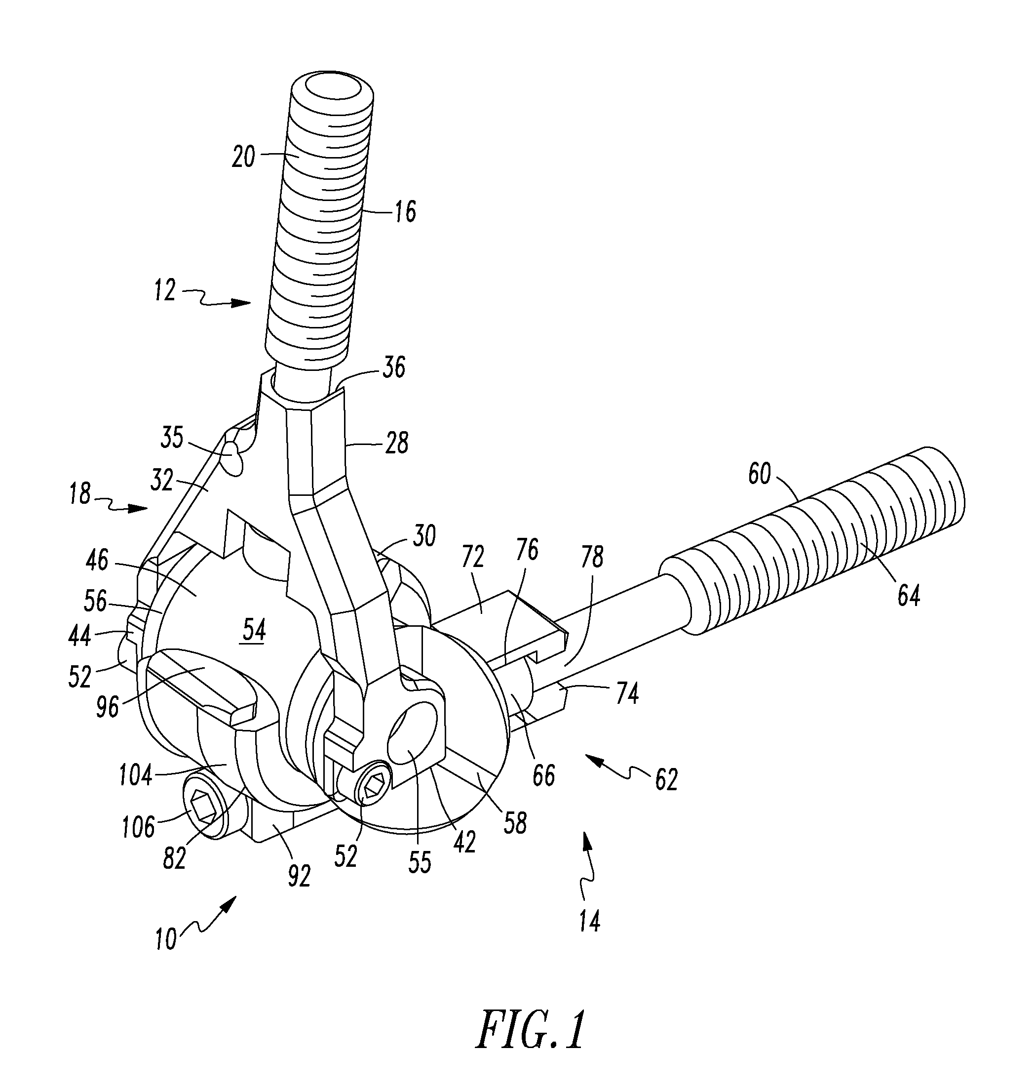
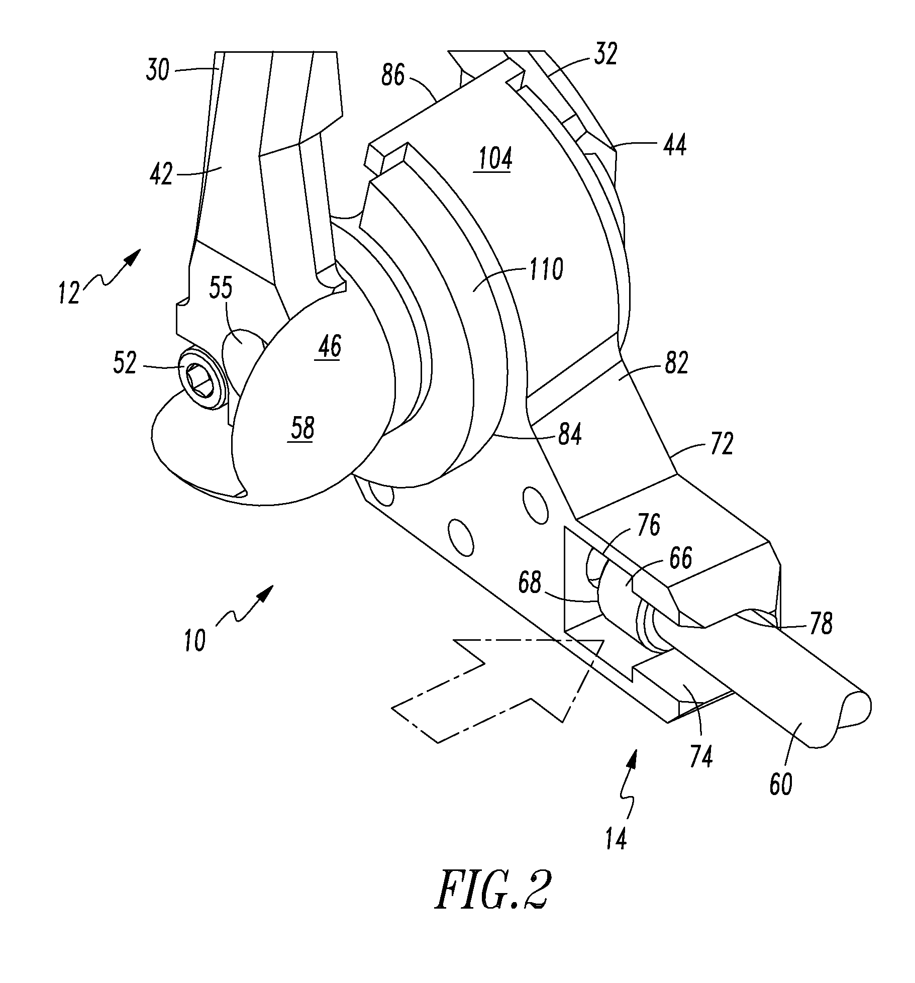
[0123]Referring to the drawings, an example of a prosthetic joint 10 is illustrated. As shown in FIG. 1, the illustrated example of the prosthetic joint 10 is a hinge joint, with the specific example illustrated being an elbow joint. The prosthetic joint 10 includes a first component 12, which in the illustrated example is a humeral component utilized for reconstruction of the distal end of a humerus. The prosthetic joint 10 further includes a second component 14, which in the illustrated example is an ulnar components for use in reconstructing the proximal end of an ulna.

[0124]The humeral component 12 includes an intramedullary stem 16 that is rotatably and removably secured to a connection portion 18. The intramedullary stem 16 is structured for uncemented, mechanical securing within the intramedullary canal of the humorous. The illustrated example of the intramedullary stem 16 includes a threaded portion 20 disposed at one end, that is structured to engage a portion of the intram...


PUM


Abstract

A prosthetic joint is secured to the bones forming the original joint by utilizing strictly mechanical fasteners, for example, a threaded rod engaging a tapped intramedullary canal. Cross locking members may be provided. The need for bone cement is avoided. The prosthetic joint may be used to replace one end of one bone forming the joint, utilizing the naturally occurring end of the other bone. Alternatively, both bone ends may be replaced with prosthetic joint portions. The decision to replace one or both bone ends may be made mid-surgery. The prosthetic joint portions are secured together utilizing ligament reconstruction members made from portions of the patient's tendons or allograft tendons. A bearing forming the interface between the two joint portions is designed to wear in order to protect the remaining components from wear, and to be easily replaced in relatively simple future surgeries.

Description

CROSS REFERENCE TO RELATED APPLICATION[0001]This application is a divisional of U.S. patent application Ser. No. 14 / 046,924, which was filed on Oct. 5, 2013, and entitled “Prosthesis for Partial and Total Joint Replacement.TECHNICAL FIELD[0002]The present invention relates to prosthetic joints. More specifically, a prosthetic joint having an improved connection to the medullary canals is provided. Additionally, a prosthetic joint that may be used for either partial or total joint replacement is provided.BACKGROUND INFORMATION[0003]There are three bones in the elbow: the humerus, the ulna, and the radius. These three bones articulate in the ulnohumeral, radiohumeral and radioulnar joints. Each of the joint surfaces is covered with articular cartilage and motion occurs at these articulations with distinct motion profiles. The cartilage allows the bones to slide easily against one another as the joint moves through its range of motion. The ulnohumeral joint is most essential for transm...

Claims


Application Information

Patent Timeline
Patent Type & Authority Applications(United States)
IPC IPC(8): A61F2/38A61F2/30
CPCA61F2/3804A61F2/30A61F2220/0008A61F2002/3813A61F2002/3809A61F2/0811A61F2002/0829A61F2002/0852A61F2002/0888A61F2002/30301A61F2002/30433A61F2002/3085A61F2/38A61F2/08A61B17/7233A61F2002/30624
Inventor KAUFMANN, ROBERT A.
Owner KAUFMANN ROBERT A
Why Eureka
Try Eureka