Method For Creating Endometriotic Cells And Endometriosis Model Animal

Inactive Publication Date: 2016-07-07
KYOTO UNIV +1
View PDF2 Cites 2 Cited by
  • Summary
  • Abstract
  • Description
  • Claims
  • Application Information

AI Technical Summary

Benefits of technology

The patent describes a method to produce an endometriotic-like cell that can be used to study the condition of endometriotic tissue. The cell has been shown to express markers of differentiation and inflammation found in endometriotic tissue. It also has the ability to produce estrogen from androgens. An animal model has been created using the cell to confirm the presence of an endometriotic tumor.

Problems solved by technology

Endometriosis is a disease which develops only in primates including humans, and it is difficult to construct a disease model of an endometriosis through use of generally used experimental animals, such as mice.
In addition, an in vitro experiment using a tissue or a cell extirpated from a patient with endometriosis has problems in that a material is not regularly available and there is a large individual difference.
Further, there is no established cell line having properties unique to endometriosis, and hence it has hitherto been difficult to continuously perform basic research and drug screening for elucidation of pathology.
However, the cell line cannot be said to faithfully reproduce a condition of endometriosis because of, for example, its deficiency in estrogen receptor-expressing ability.
However, this report is a report on research which is not aimed at inducing differentiation to a pathological endometriotic cell, but is aimed at inducing differentiation to a normal endometrial cell, and does not suggest induction of differentiation to an endometriotic cell.

Method used


Image

  • Method For Creating Endometriotic Cells And Endometriosis Model Animal
  • Method For Creating Endometriotic Cells And Endometriosis Model Animal
  • Method For Creating Endometriotic Cells And Endometriosis Model Animal

Examples


example 1

Production of Endometriotic-Like Cells

[0053]In this Example, endometriotic-like cells were produced from mesenchymal stem cells. Thus, procedures of the production are described in detail.

[0054]Step (1) Culture of Mesenchymal Stem Cells

[0055]In this step, 2×10⁵ human bone marrow-derived primary cultured mesenchymal stem cells (Lonza, Cat. No. PT-2501) were added to 10 ml of a low-carbon-source proliferation culture medium (shown below), inoculated into a culture vessel having a diameter of 100 mm and having a surface coated with collagen type I (derived from rat tail, BD Biosciences, Cat. No. 354236), and cultured for 6 days to a confluency of from 70% to 80% at 37° C. in the presence of 5% CO2. The culture medium was exchanged every 2 days to 3 days.

[0056]In this Example and Experimental Examples shown below, and Examples shown below, the “low-carbon-source proliferation culture medium” refers to a culture medium prepared by supplementing low-glucose Dulbecco's Modified Eagle's Med...

experimental example 1-1

Analysis of Cells Induced to Differentiate in Step (2)

[0063]In this Experimental Example, the cells induced to differentiate in the step (2) were confirmed and analyzed for their cell morphology, expression of endometrial differentiation markers, and expression of endometriosis-related inflammatory factors (genes and proteins).

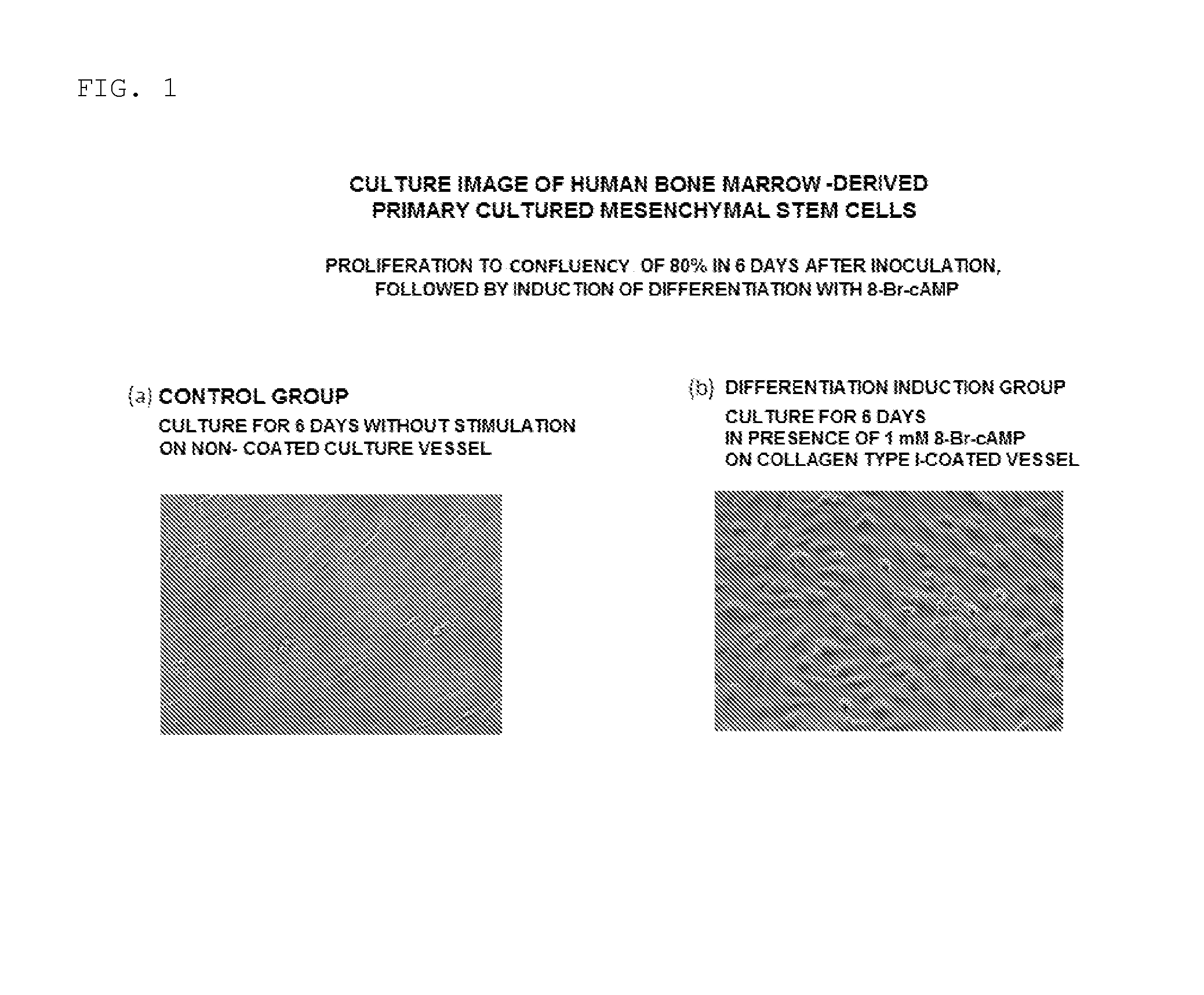
[0064](i) Cell Morphology

[0065]The morphology of the cells on day 6 after the start of the induction of differentiation in the step (2) was observed with a light microscope (magnification: 10×). As a result, as shown in FIG. 1(b), it was observed that, in the cells induced to differentiate with the medium supplemented with 8-Br-cAMP, the cells protruded to exhibit a round shape as compared to the control shown in FIG. 1(a), and had morphology similar to that of normal endometrial stromal cells decidualized in vitro.

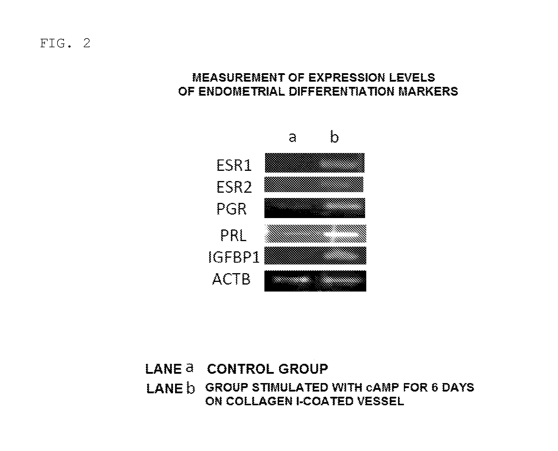
[0066](ii) Expression of Endometrial Differentiation Markers

[0067]The cells on day 6 after the start of the induction of differentiation in the ste...

experimental example 1-2

Analysis of Endometriotic-Like Cells Produced in Step (3)

[0072]In this Experimental Example, the cells produced in the step (3), i.e., the cells induced to differentiate in the step (2) and then cultured for 2 days (total number of days for culture: 14 days) with a low-carbon-source proliferation culture medium free of 8-Br-cAMP through medium exchange again were analyzed.

[0073](i) Cell Morphology

[0074]Cell morphology was observed with a light microscope (magnification: 10×) in the same manner as in the above-mentioned section (i) of Experimental Example 1-1. The morphology of a control group (i.e., cells obtained after human mesenchymal stem cells have been cultured for 14 days with a low-carbon-source proliferation culture medium free of 8-Br-cAMP on a culture vessel not coated with extracellular matrix) is shown in FIG. 5(a), and the morphology of a differentiation induction group is shown in FIG. 5 (b). As a result, as shown in FIG. 5 (b), it was confirmed that, when the cells w...


PUM


Abstract

Provided is a method of producing a cell having properties of endometriosis (hereinafter referred to as “endometriotic-like cell”) from a human mesenchymal stem cell. Also provided are a method of producing an animal model exhibiting human endometriosis, the method including transplanting the cell to an animal, and an animal model of endometriosis. The method of producing an endometriotic-like cell comprises a step of culturing a human mesenchymal stem cell induced to differentiate by allowing a differentiation inducing agent to act on the cell, through use of a low-carbon-source culture medium free of a differentiation inducing agent, in a culture vessel coated with extracellular matrix.

Description

TECHNICAL FIELD[0001]The present invention relates to a cell having properties of endometriosis. The present invention also relates to a method of producing an animal model exhibiting human endometriosis, and to an animal model of endometriosis.[0002]The present application claims priority from Japanese Patent Application No. 2013-192596, which is incorporated herein by reference.BACKGROUND ART[0003]Endometriosis is defined as a “disease in which a tissue having a form and a function identical or similar to those of endometrium ectopically grows and causes symptoms (Japan Society of Obstetrics and Gynecology, 1993),” and is a disease which is still unknown in many points including its development mechanism. Endometriosis is a disease which develops only in primates including humans, and it is difficult to construct a disease model of an endometriosis through use of generally used experimental animals, such as mice. In addition, an in vitro experiment using a tissue or a cell extirpa...

Claims


Application Information

Patent Timeline
IPC IPC(8): A01K67/027C12N5/071
CPCA01K67/0271C12N5/0682C12N2506/1353C12N2501/01A01K2207/15C12N2533/90A01K2207/12A01K2267/0331C12N2500/34A01K2267/035C12N5/0663C12N2533/54
Inventor KAJITANI, TAKASHI
Owner KYOTO UNIV
Why Eureka
Try Eureka