System and method for analyzing tissue intra-operatively using mass spectrometry

a mass spectrometry and tissue technology, applied in the field of intra-operative diagnosis of tissue samples, can solve the problems of difficult intra-operative determination of tumor margins, high complexity of cancer, and high difficulty in diagnosis and treatmen

Inactive Publication Date: 2016-11-24
THE BRIGHAM & WOMEN S HOSPITAL INC
View PDF9 Cites 49 Cited by
  • Summary
  • Abstract
  • Description
  • Claims
  • Application Information

AI Technical Summary

Problems solved by technology

Cancer presents many highly complex issues in clinical medicine.
As a severely malignant and invasive tumor, breast cancer is a leading cause of death in cancerous women.
However, the accurate intra-operative determination of a tumor margin is challenging when planning and performing a breast-conserving surgery.
Although accurate tumor size assessment may be available, the lack of real-time imaging in conjunction with surgical procedures relative to these techniques affects the surgery success rate and oftentimes leads to the need for further operations, giving the risk of local recurrence of breast cancer after lumpectomy and leading to a higher incidence of mastectomy.
However, instead of providing real time imaging, this technique still requires the surgery to be interrupted.
Ultrasonography has been applied intra-operatively in breast cancer excision, but it is unreliable in detecting nonpalpable tumor or ductal carcinoma in situ lesions.
However, the incorporation of radioactive or fluorescent labels presents a disadvantage not only to the patient but also to the operative personnel repeated exposure.
These ancillary tests, however, often require days to perform and results become available long after surgery is completed.
This approach is time consuming, requiring nearly 30 minutes between the moment a tissue is biopsied and the time the pathologist's interpretation is communicated back to the surgeon.
Despite the advances noted above, the diagnostic use of ultrasonic and mechanical probes has gone largely undeveloped and potential advantages over traditional methods of tissue sampling have often gone unrecognized.
As discussed above, routine intra-operative distinction between tumor and normal breast tissue is currently not possible in breast conserving surgery.
This limitation affects the success of many surgical procedures.

Method used


Image

  • System and method for analyzing tissue intra-operatively using mass spectrometry
  • System and method for analyzing tissue intra-operatively using mass spectrometry
  • System and method for analyzing tissue intra-operatively using mass spectrometry

Examples


Embodiment Construction

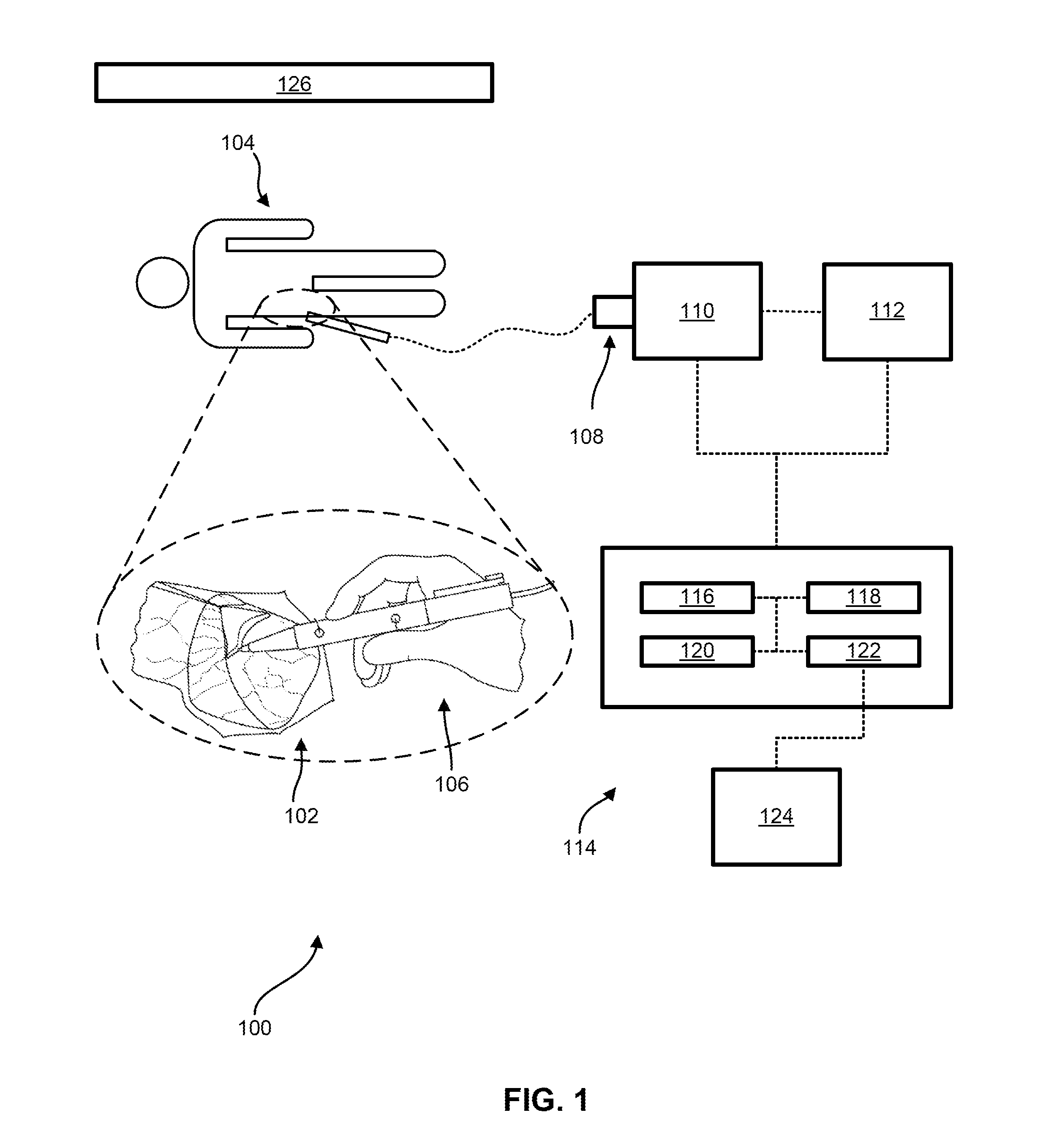
[0081]As discussed above, routine intra-operative distinction between tumor and normal breast tissue is currently not possible in breast conserving surgery. This limitation affects the success of many surgical procedures. For example, considering just one common cancer surgery, in breast cancer surgery, up to about forty percent (40%) of operations require more than one operative procedure.

[0082]Mass spectrometry imaging (MSI) has been applied to investigate the molecular distribution of proteins, lipids and metabolites without the use of labels. In particular, desorption electrospray ionization (DESI) allows direct tissue analysis with little or no sample preparation. Therefore, with the advantage of easy implementation, DESI mass spectrometry imaging (DESI-MSI) has great potential in the application of intra-operative tumor assessment. As described herein, imaging includes spatially encoded information correlated with the surgical site and / or the tissue histology itself. However, ...


PUM

PropertyMeasurementUnit
lengthaaaaaaaaaa
frequencyaaaaaaaaaa
frequencyaaaaaaaaaa

Abstract

A system and method for ample analysis including acquiring a tissue sample, preparing the tissue sample for mass spectrometry imaging, conducting a mass spectrometry procedure on the tissue sample to produce an image, analyzing the image to determine the presence or absence of a biomarker; and generating a report indicating a presence or absence of cancer.

Description

CROSS-REFERENCE TO RELATED APPLICATIONS[0001]This application claims priority to U.S. Provisional Patent Application No. 61 / 894,595, filed Oct. 23, 2013, the entire contents of which are incorporated herein by reference.BACKGROUND[0002]The invention relates generally to intra-operative diagnostics of sample tissues. More specifically, the invention relates to the use of mass spectrometry for the detection of specific biomarkers.[0003]Cancer presents many highly complex issues in clinical medicine. For example, consider just one of the many different and varied types of cancer, such as breast cancer. As a severely malignant and invasive tumor, breast cancer is a leading cause of death in cancerous women. Surgical removal of a cancerous tumor is usually the initial treatment of breast cancer, either by lumpectomy or mastectomy. Most women have a preference for the less invasive lumpectomy, for example, because of the cosmetic appearance. However, the accurate intra-operative determina...

Claims


Application Information

Patent Timeline
Patent Type & Authority Applications(United States)
IPC IPC(8): G01N33/483G01N33/574A61B90/00A61B34/20A61B10/02A61B17/32
CPCG01N33/4833A61B10/02A61B17/320068A61B90/36A61B34/20G01N2800/52G01N33/57496A61B2218/007A61B2034/2051A61B2090/374A61B2090/3762A61B90/37A61B2017/32007G16C20/20H01J49/26
Inventor AGAR, NATHALIE
Owner THE BRIGHAM & WOMEN S HOSPITAL INC
Why Eureka
Try Eureka