Synthetic peptides, enzymatic formation of pericellular hydrogels/nanofibrils, and methods of use

Active Publication Date: 2017-02-09
BRANDEIS UNIV
View PDF2 Cites 2 Cited by
  • Summary
  • Abstract
  • Description
  • Claims
  • Application Information

AI Technical Summary

Benefits of technology

[0016]The accompanying Examples demonstrate the unexpected observation of the selective formation of nanonets/hydrogels of small D-peptide derivatives in the pericellular space of cancer cells. As demonstrated in the accompanying Examples, enzyme-catalyzed molecular nanofibers: (i) form selectively on target cells that express an ectoenzyme; (ii) inhibit target cell migration, target cell survival, target cell growth, and the passage of intracellular signaling molecules to or from the target cell; (iii) e

Problems solved by technology

Cancer remains a major ch

Method used


Image

  • Synthetic peptides, enzymatic formation of pericellular hydrogels/nanofibrils, and methods of use
  • Synthetic peptides, enzymatic formation of pericellular hydrogels/nanofibrils, and methods of use
  • Synthetic peptides, enzymatic formation of pericellular hydrogels/nanofibrils, and methods of use

Examples


example 1

Phosphatase-Catalyzed Dephosphorylation of D-2a Results in Localized Self-Assembly of Pericellular Hydrogels / Nanonets on the Surface of Cancer Cells

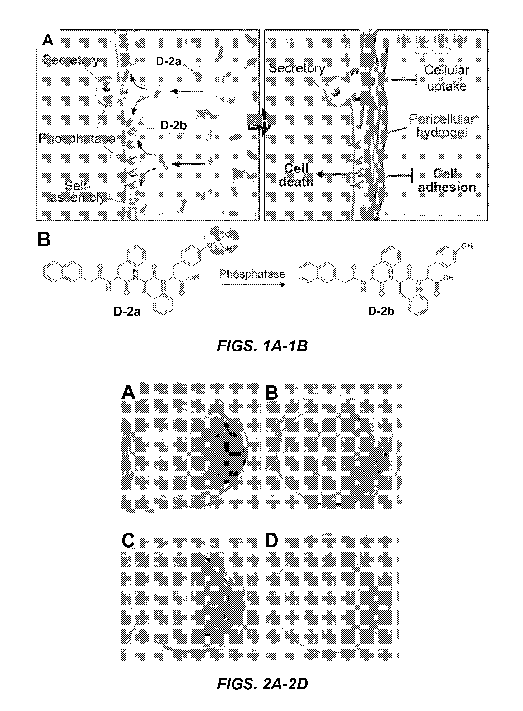
[0143]Napthalene capped tripeptide precursor molecule D-2a and hydrogelator molecule D-2b, synthesized according to Yang et al. (Small 3:558-562 (2007), which is hereby incorporated by reference in its entirety), are comprised of the tripeptide D-Phe-D-Phe-D-Tyr (FIG. 1B). The D-Tyr of D-2a is phosphorylated (FIG. 1B). Alkaline-phosphatase (“ALP”) catalyzed dephosphorylation of precursor molecule D-2a (0.20 wt % / 2.77 mM) forms hydrogelator molecule D-2b (0.18 wt % / 2.77 mM), which self assembles to form the nanofibrils of a hydrogel.

[0144]Unexpectedly, treatment of a confluent layer of HeLa cells with either 560 or 280 μM D-2a (2 hours at 37° C.) results in the formation of a hydrogel-like soft material (hydrogel) on the cell surface (FIGS. 2A, 2B). Little hydrogelation was observed when cells were treated with 140 μM D-2a (FIG. 2C).

[0145...

example 2

The Phosphatase Gradient Promotes Dynamic Pericellular Hydrogelation

[0146]HeLa cell conditioned medium (“CM”) dephosphorylates D-2a to form D-2b nanofibrils. CM treated with either 560 or 280 μM D-2a for 48 hours completely transforms from a solution to a hydrogel (FIGS. 4A, 4B). Similar to experiments conducted with HeLa cells incubated with either 140 μM D-2a or 560 μM D-2b for two hours (FIGS. 2C-2D), CM containing either 140 μM D-2a or 560 μM D-2b fails to form a hydrogel following 48 hours of incubation (FIGS. 4C-4D). These results confirm that the secretory phosphatases present in HeLa cell conditioned medium are sufficient to convert D-2a to D-2b, thereby contributing to the formation of a hydrogel.

[0147]Since surface and secretory phosphatases are present on or near the cell membrane, the concentration of phosphatases is expected to be high in the pericellular space, resulting in the selective formation of pericellular hydrogels following incubation with D-2a. The homogenous...

example 3

Pericellular Hydrogels Consist of Networks of Nanofibrils which Form Nanonets

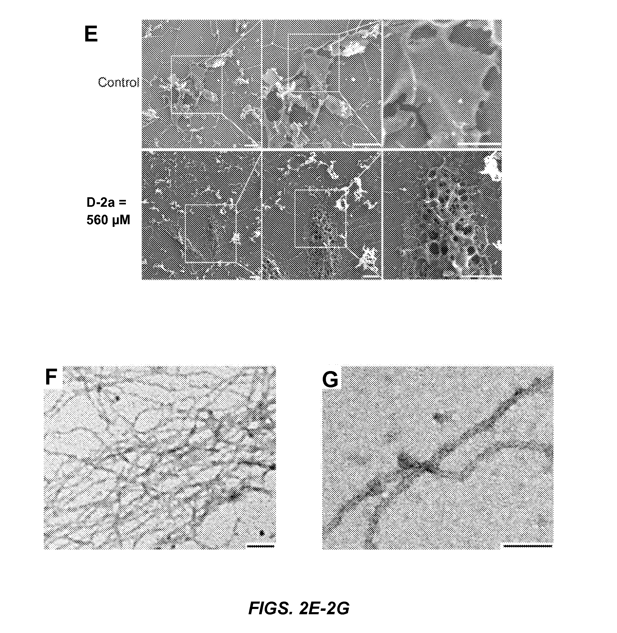
[0148]SEM reveals that the surface of the HeLa cells incubated with D-2a differs from the surface of untreated HeLa cells. Since the organic solvents traditionally used for cell fixation and washing cell monolayers (i.e., alcohol or acetone) destroy the supramolecular structure of the nanofibrils within the hydrogels, cell monolayer samples were instead freeze-dried for SEM analysis. While untreated HeLa cells exhibit a smooth surface (FIG. 2E, top), cells treated with 560 μM D-2a are covered in porous structures (i.e., nanonets) (FIG. 2E, bottom). Additionally, cells treated with 280 μM D-2a display fiber-like structures attached closely to the cell surface (FIG. 6). These results coincide with the pericellular hydrogelation observed on HeLa cells treated with 280 and 560 μM D-2a (FIGS. 2A, 2B).

[0149]Negative stained TEM of the pericellular hydrogel on HeLa cells treated with 280 μM D-2a reveals that the h...


PUM

PropertyMeasurementUnit
Molar densityaaaaaaaaaa

Abstract

Disclosed are peptides that contain up to about 35 amino acids, including a plurality of aromatic amino acid residues and either (i) an amino acid residue that is phosphorylated or sulfated, or (ii) an amino acid comprising an ester-moiety linked via peptide bond, or both (i) and (ii), wherein the peptide is capable of self-assembly to form nanofibrils in the presence of an enzyme that hydrolyzes the phosphate group, the sulfate group, or the ester-moiety. These peptides are enzymatically responsive hydrogelators, and they can be used to form pericellular hydrogels/nanofibrils upon exposure to target cells that secrete or express a surface bound ectoenzyme having hydrolase activity suitable to induce peptide gelation. These materials, and compositions containing the same, can be used for in vitro and in vivo cellular imaging, treating cancerous conditions, collecting a secretome from a cell upon which the pericellular hydrogels/nanofibrils form, and screening the collected secretome.

Description

[0001]This application claims the priority benefit of U.S. Provisional Patent Application Ser. No. 61 / 977,484 filed Apr. 9, 2014, and 62 / 105,871 filed Jan. 21, 2015, each of which is hereby incorporated by reference in its entirety.[0002]The present invention was made with support from the National Institutes of Health under grant R01CA142746. The U.S. government has certain rights in this invention.FIELD OF THE INVENTION[0003]The present invention relates generally to enzymatically responsive peptide hydrogelator precursors, the enzymatically activated hydrogelators and hydrogels formed therefrom, and use of the precursors to form pericellular hydrogels / nanofibrils upon exposure to target cells that secrete or express a surface bound ectoenzyme having hydrolase activity suitable to induce peptide gelation. These materials, and compositions containing the same, can be used for in vitro and in vivo cellular imaging, treating cancerous conditions, collecting a secretome from a cell up...

Claims


Application Information

Patent Timeline
IPC IPC(8): C07K5/087G01N33/58G01N33/573A61K49/00
CPCC07K5/0812G01N33/573G01N33/582A61K49/0056C07K5/06078C07K5/1013C07K5/1016C07K14/001
Inventor XU, BINGSHI, JUNFENGKUANG, YI
Owner BRANDEIS UNIV
Why Eureka
Try Eureka