Segmentation of cardiac magnetic resonance (CMR) images using a memory persistence approach

a cardiac magnetic resonance and segmentation technology, applied in image enhancement, image analysis, instruments, etc., can solve the problems of time-consuming and tedious manual delineation of anatomical structures, limited by inter- and intra-observer variability, and the difficulty of accurate lv and rv segmentation, so as to increase the flexibility of the proposed cs-fcm algorithm

Inactive Publication Date: 2017-04-20
AGENCY FOR SCI TECH & RES +1
View PDF15 Cites 17 Cited by
  • Summary
  • Abstract
  • Description
  • Claims
  • Application Information

AI Technical Summary

Benefits of technology

[0018]A weighting parameter may be used to adjust the weight of the spatial distance against the intensity feature, which increases the flexibility of the proposed CS-FCM algorithm.
[0019]Experimental results of using embodiments of the second aspect of the invention to process real CMR images have demonstrat...

Problems solved by technology

However, manual delineation of the anatomical structures is time-consuming and tedious (even a trained clinician takes 20 minutes to do this), and limited by inter- and intra-observer variability.
Although ventricle segmentation methods have improved over the last few decades, accurate LV and RV segmentation is still acknowledged as a difficult problem, especially for RV segmentation (due to the high anatomical complexity and poorly-delineated ventricular boundaries of the RV).
However, such models need to be constructed or learned from man...

Method used


Image

  • Segmentation of cardiac magnetic resonance (CMR) images using a memory persistence approach
  • Segmentation of cardiac magnetic resonance (CMR) images using a memory persistence approach
  • Segmentation of cardiac magnetic resonance (CMR) images using a memory persistence approach

Examples


Embodiment Construction

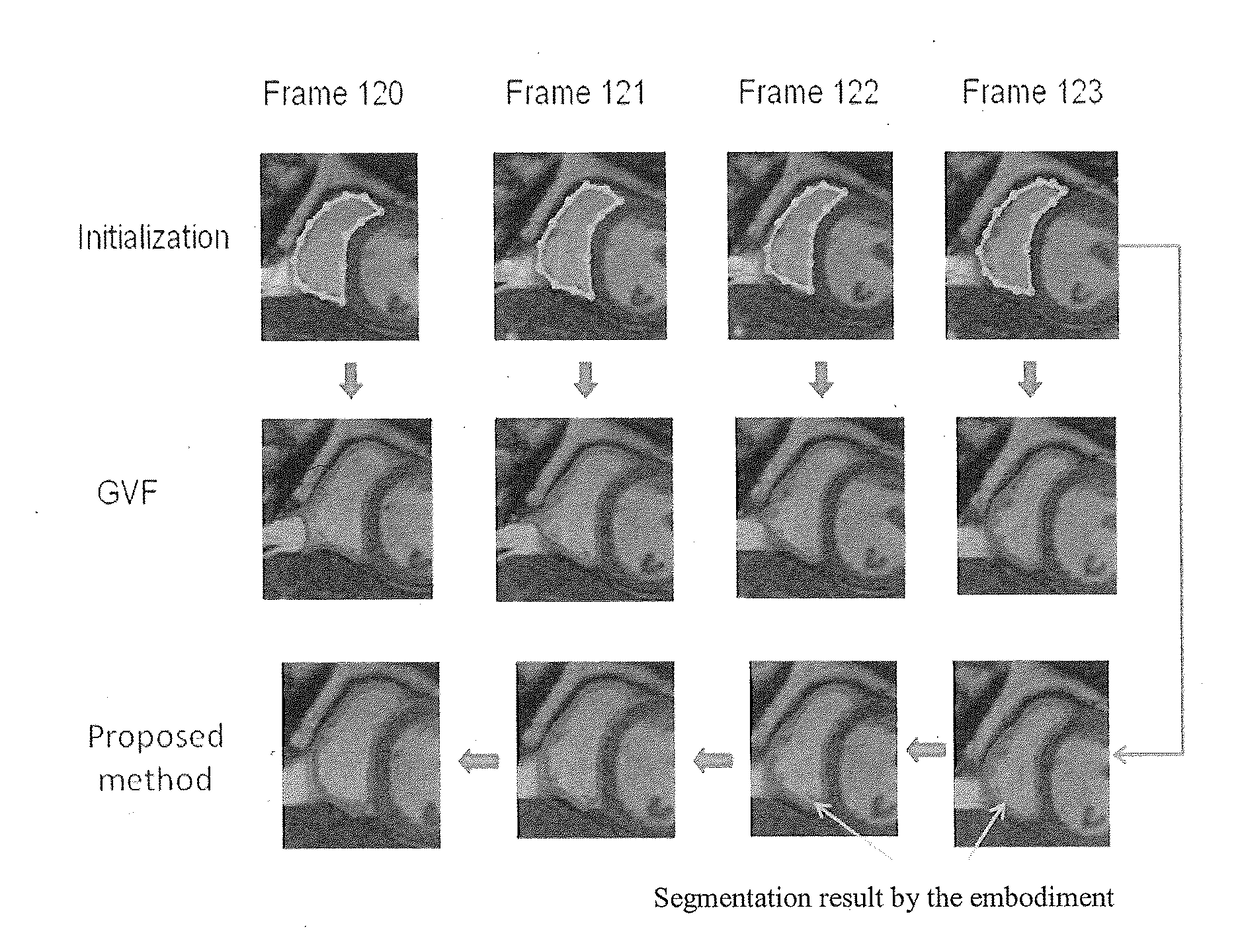
1. Embodiment of the “Memory Persistence” Aspect of the Invention

[0031]The following explanation denotes a sequence of two-dimensional (2D) CMR image frames from a single cardiac cycle as:

It(x,y) t=1,2, . . . ,N

where It(x,y) denotes the 2D image frame at time t, and N is the total number of frames in a single cardiac cycle.

[0032]From these images it is intended to generate a contour, and to produce corresponding segmented binary images in which the intensity of the pixels within the contour is set to 255, and the intensity of other pixels is set to 0. The segmented binary images are denoted by

Bt(x,y) t=1,2, . . . ,N

where Bt(x,y) denotes the 2D binary image from the segmentation of the image frame It(x,y) at time t.

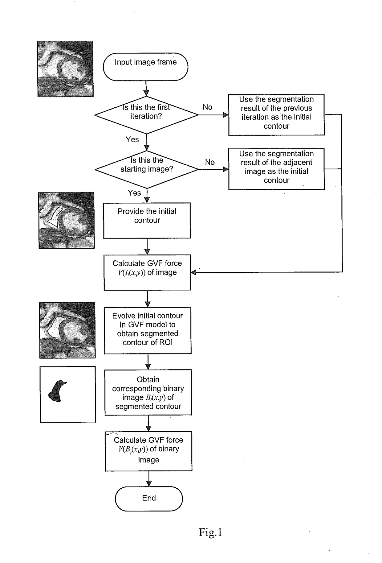
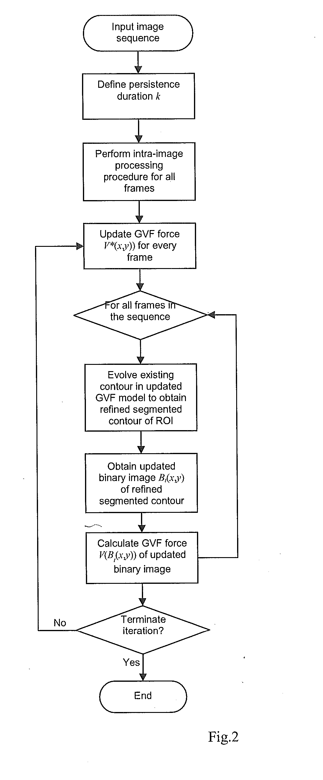
[0033]To segment a given frame at time t, instead of solely using the information from the current image It(x,y) (as did most of the existing methods), the proposed framework incorporates into the segmentation techniques information from segmented binary images of previo...


PUM


Abstract

A method is proposed for identifying an anatomical structure within a spatial-temporal image (i.e. a series of frames captured as respective times). A current frame of spatial-temporal medical image is processed using information from one or more previous and/or subsequent temporal frames, to aid in the segmentation of an object or a region of interest (ROI) in a current frame. The invention is applicable to both two- and three-dimensional spatial-temporal images (i.e., 2D+time or 3D+time), and in particular to cardiac magnetic resonance (CMR images). An initialisation process for this method segments the left ventricle (LV) in a CMR image by a fuzzy c-means (FCM) clustering algorithm which employs a circular shape function as part of the definition of the dissimilarity measure.

Description

FIELD OF THE INVENTION[0001]The present invention relates to methods and apparatus for identifying anatomical structures within medical images, especially spatial-temporal medical images (that is, a series of two- or three-dimensional images (“frames”) captured at successive respective times). The invention relates particularly to identification of anatomical structures within cardiac magnetic resonance (CMR) images of the heart of a subject. Nevertheless, the invention is sufficiently generic to be applicable for identification of dynamic anatomical or biological structures from other imaging modalities that produce time-series images, such as computed tomography and ultrasound images of the heart and the lungs, and cell transport images from digital microscopy.BACKGROUND OF THE INVENTION[0002]Spatial-temporal medical images such as those obtained from Cardiac Magnetic Resonance (CMR) images provide important information for diagnosis and treatment of cardiovascular diseases in a n...

Claims


Application Information

Patent Timeline
IPC IPC(8): G06T7/00G06T7/11
CPCG06T7/0012G06T2207/30048G06T2207/10088G06T7/11G06T2207/20116G06T7/12G06V10/267
Inventor YANG, XULEISU, YIYEO, SI YONGZHONG, LIANGTAN, RU SAN
Owner AGENCY FOR SCI TECH & RES
Why Eureka
Try Eureka