Compositions and methods for treating bone, joints and cartilage

a technology of cartilage and collagen fibers, applied in the field of collagen and joint composition and methods, can solve the problems of collagen fibers becoming susceptible to degradation and degeneration, loss of cartilage, and loss of collagen

Active Publication Date: 2017-06-22
REPLICEL LIFE SCI
View PDF2 Cites 2 Cited by
  • Summary
  • Abstract
  • Description
  • Claims
  • Application Information

AI Technical Summary

Benefits of technology

[0018]The details of one or more embodiments are set forth in the description below. Other features, objects and advantages will be apparent from the description,

Problems solved by technology

During onset of osteoarthritis the amount of fluid in the cartilage increases, and furthermore there is a loss of collagen.
Thereby the collagen fibers become susceptible to degradation and degenerate.
Also injuries to adjacent ligaments and menisci, as a result of accidents or surgeries may end up in osteoarthritis where ligamentous deterioration or instability is the triggering factor.
As a consequence of those causes, a process is initiated leading to loss of cartilage.
Later on the bone surfaces become less well protected by cartilage, bone is then exposed and damaged.
This results in decreased movement secondary to pain, regional muscles atrophy, and lax ligamen

Method used


Image

  • Compositions and methods for treating bone, joints and cartilage
  • Compositions and methods for treating bone, joints and cartilage

Examples


example 1

Tissue Sampling

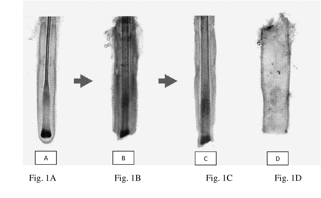
[0047]A skin biopsy from the occipital area of the scalp is obtained from a subject as follows. Briefly, once an appropriate area of the scalp has been selected, it is shaved with hair clippers, ensuring some stubble remains. The biopsy area is then thoroughly disinfected and anaesthetized. Once anesthesia has taken effect, a 4-10 mm deep punch biopsy is gently removed from the biopsy site and the incision closed with sutures which can be removed 12-14 days later. The skin biopsy is then packaged under aseptic conditions into a pre-labelled biopsy tube containing biopsy transport medium, composed of DMEM / Hams F12 with antibiotics.

example 2

Isolation and Cultivation of NBDS Cells

[0048]A sterility test is performed on the medium in which the biopsy has been transported to ensure the sample is free from contamination, or alternatively, if the sample is contaminated to ensure that medium with antibiotics is subsequently utilized. The biopsy is then washed several times to remove the biopsy transportation medium and any debris to prepare the tissue for subsequent processing. Hair follicles are processed in Hams F10 by cutting away the skin epithelium with a sterile scalpel and “plucking” or dissecting the whole hair follicle unit from the surrounding dermal tissue using sterile forceps. The hair follicle is gripped with a forceps as close as possible to the skin surface and the follicle exposed by pulling up on the hair in the hair follicle unit. Follicles in the anagen phase (growing phase of the hair cycle, indicated by the visible outer root sheath, and DSC of the hair bulb) are selected for further processing.

[0049]NBD...

example 3

Preparation and Administration of NBDS Cells into a Joint

[0052]The skin over the joint to be treated is first prepared for injection by application of a topical analgesia (e.g., EMLA-cream) for approximately one hour. Thereafter, the skin is washed and disinfected. NBDS cells, prepared as described above, are then injected into the affected joint as a bolus or in a repetitive manner.

[0053]Alternatively, NBDS cells may be injected by arthroscopic guidance directly into the synovial fluid, or damaged cartilage or menisci, or ligaments, or into the bone defects arising from osteoarthrosis in order to fill the entire surface of the desired treatment area.


PUM

PropertyMeasurementUnit
Compositionaaaaaaaaaa
Antimicrobial propertiesaaaaaaaaaa
Biodegradabilityaaaaaaaaaa

Abstract

Briefly stated, the present invention provides compositions and methods for treating or preventing joint injuries utilizing hair follicle derived Non-Bulbar Dermal Sheath (“NDBS”) cells. Within one aspect of the invention methods are provided for isolating NBDS cells, comprising the steps of: (a) preparing vital hair; (b) cleaving the hair prepared in step (a) to remove the hair follicle bulb; (c) isolating Non-Bulbar Dermal Sheath tissue; and (d) cultivating the isolated Non-Bulbar Dermal sheath tissue to produce NBDS cells. Within one embodiment of the invention the vital hair is obtained by full skin biopsy from the occipital scalp of a subject.

Description

CROSS-REFERENCE TO RELATED APPLICATION[0001]This application claims the benefit under 35 U.S.C. §119(e) of U.S. Provisional Patent Application No. 61 / 939,187 filed Feb. 12, 2014, which application is incorporated herein by reference in its entirety.FIELD OF THE INVENTION[0002]The present invention relates to compositions and methods for treating joints and adjacent bone or cartilage as well for various medical purposes, and more specifically, to compositions comprising of autologous or allogenic non-bulbar dermal sheath (NDBS) cells for use in the treatment and repair of joints, including for example, joint injuries and of osteoarthritis, as well as for various other medical purposesBACKGROUNDDescription of the Related Art[0003]A joint is a body part at which bones (two or more) interconnect. Joints are designed to allow movements and mechanical support. They are classified by their structure and function.[0004]The structural classification describes how bones connect to each other,...

Claims


Application Information

Patent Timeline
IPC IPC(8): A61K35/36C12N5/071A61K45/06A61K35/16A61K31/728
CPCA61K35/36A61K35/16C12N5/0627A61K45/06A61K31/728A61K38/18A61K38/39A61P19/00
Inventor HOFFMANN, ROLFMCELWEE, KEVIN JOHN
Owner REPLICEL LIFE SCI
Why Eureka
Try Eureka