Apparatus for ultrasound flow vector imaging and methods thereof

a vector imaging and ultrasound technology, applied in the field of ultrasonic imaging systems, can solve the problems of significant pitfall, prone to error, and method flaws of ultrasonic color flow imaging

Active Publication Date: 2017-08-03
VERSITECH LTD
View PDF7 Cites 2 Cited by
  • Summary
  • Abstract
  • Description
  • Claims
  • Application Information

AI Technical Summary

Benefits of technology

[0014]The foregoing and other objects and advantage of the present invention will become more apparent upon reference to the following specification and annexed drawings in which:
[0015]FIGS. 1A and 1B illustrate data acquisition in practicing the present invention based upon the use of plane wave transmissions and parallel beam-forming (both performed from multiple angles);
[0016]FIGS. 2A and 2B illustrates an arrangement for flow vector estimation according to the present invention for every pixel position and its corresponding set of slow-time signals from all Tx-Rx angle pairs;
[0017]FIGS. 3A and 3B are quantitative flow visualizations according to the present invention utilizing dynamic rendering of color-encoded particle projectiles;
[0018]FIGS. 4A, 4B and 4C illustrate a set up for an anthropomorphic carotid bifurcation phantom study and an image of the co-planar geometry of a carotid bifurcation of a healthy and a diseased patient, respectively;
[0019]FIGS. 5A and 5B illustrate a color static map of estimated flow vectors and a graph of velocity versus position from the proximal wall of vessel for vessels at three different depths;

Problems solved by technology

Despite its popular role in clinical screening, ultrasound color flow imaging is known to possess method flaws (Evans 2010).
In particular, as its operating principle is typically based on axial Doppler estimation, it is prone to error if the beam-flow angle (i.e. angle between the ultrasound propagation axis and the flow trajectory) varies over the vasculature (Evans D H, Jensen J A, Nielsen M B. Ultrasound color Doppler imaging.
This issue represents a significant pitfall in diagnostic scenarios where the vasculature is not in straight-tube form, such as the bifurcation geometry found in the carotid arteries (Ku D N. Blood flow in arteries.
In these cases, it can be challenging for sonographers to properly interpret color flow images (Arning C, Eckert B. The diagnostic relevance of colour Doppler artefacts in carotid artery examinations.
While each of these approaches has its own merit, they are all known to yield erroneous flow vector estimates under certain scenarios.
Notably, Doppler / phase-shift estimation is prone to aliasing artifacts when tracking fast flow, whereas speckle tracking and directional cross-correlation have difficulty in following out-of-plane motion (Hansen L K, Udesen J, Oddershede N, Henze L, Thomsen C, Jensen J A, Nielsen M B. In vivo comparison of three ultrasound vector velocity techniques to MR phase contrast angiography.

Method used


Image

  • Apparatus for ultrasound flow vector imaging and methods thereof
  • Apparatus for ultrasound flow vector imaging and methods thereof
  • Apparatus for ultrasound flow vector imaging and methods thereof

Examples


Embodiment Construction

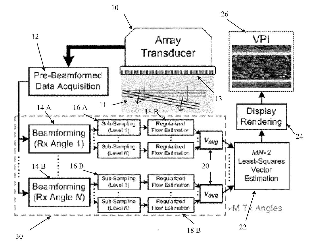
[0028]An arrangement of apparatus for carrying out the vector projectile imaging (VPI) of the present invention is illustrated in FIG. 10. This arrangement carries out the steps of (1) data acquisition, (2) regularized flow vector estimation (including a first stage of multi-level sub-sampling and a second stage of flow vector derivation) and (3) dynamic visualization.

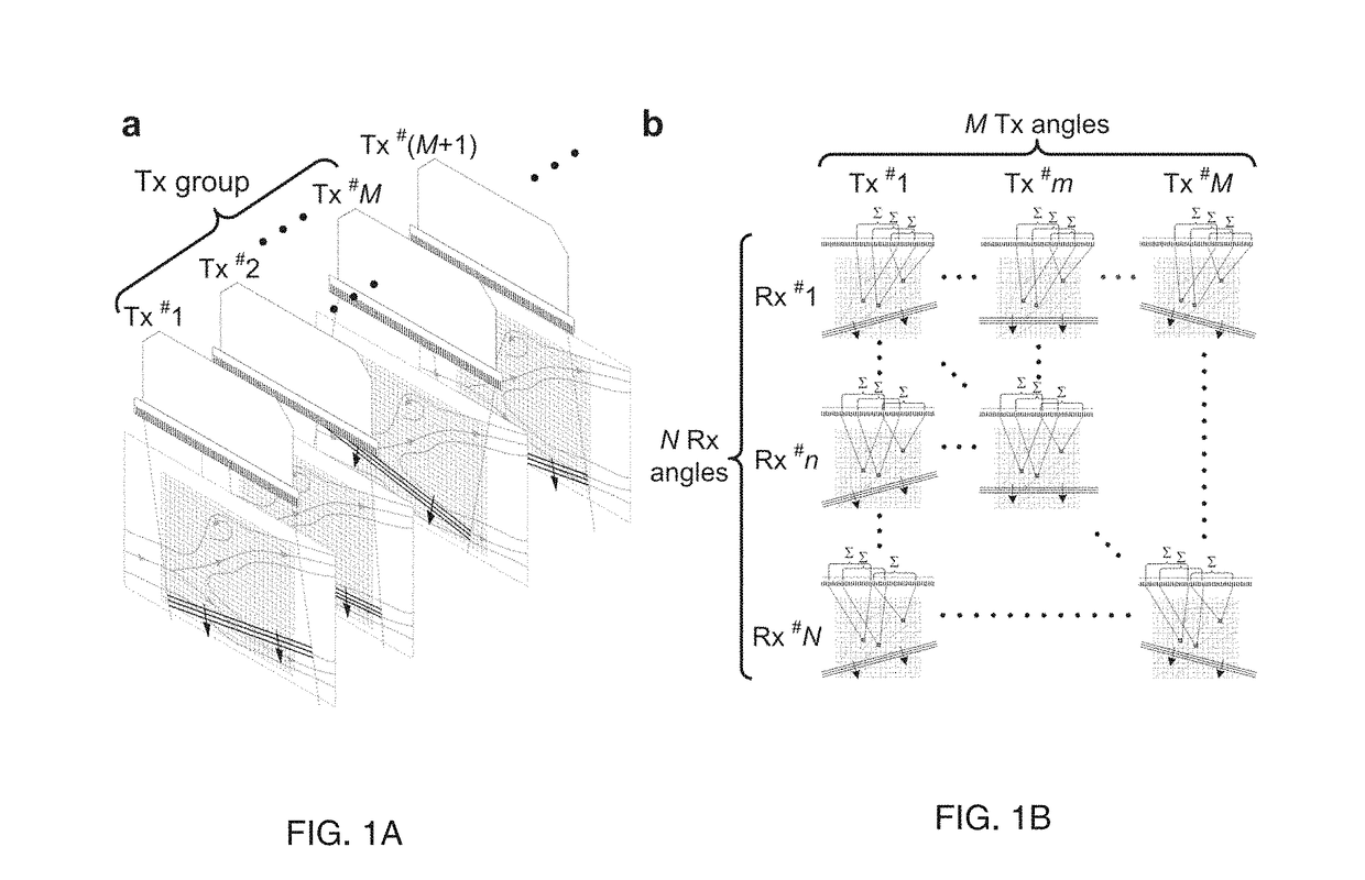
[0029]To achieve data acquisition an ultrasonic array transducer 10 is positioned on the outside of the patient's body adjacent the vasculature in which the fluid (e.g., blood) is to be imaged. The array 10 transmits a series of ultrasonic unfocused, steered plane waves 11 into the tissue at a high rate. The transmission angle with respect to the transducer surface 13 is changed after each wave. The transducer 10 also receives the waves reflected from the tissue at different receive steering angles and stores it in Pre-beam-Formed Data Acquisition device 12. This data is in the form of frames for each angle.

[0030]Each ...


PUM


Abstract

Apparatus and methods of use are provided for complex flow imaging and analysis that is non-invasive, accurate, and time-resolved. It is particularly useful in imaging of vascular flow with spatiotemporal fluctuations. This apparatus is an ultrasound-based framework called vector projectile imaging (VPI) that can dynamically render complex flow patterns over an imaging view at millisecond time resolution. The VPI apparatus and methods comprise: (i) high-frame-rate broad-view data acquisition (based on steered plane wave firings); (ii) flow vector estimation derived from multi-angle Doppler analysis (coupled with data regularization and least-squares fitting); and (iii) dynamic visualization of color-encoded vector projectiles (with flow speckles displayed as adjunct).

Description

[0001]The present application claims priority to U.S. Provisional Application No. 61 / 905,974 filed on Nov. 19, 2013, which is incorporated herein in its entirety.TECHNICAL FIELD[0002]The present invention is generally directed to an ultrasonic imaging system, and more particularly to an ultrasonic system for imaging vascular flow. This imaging system can be used for non-invasively visualizing the flow of blood in a human blood vessel.BACKGROUND OF THE INVENTION[0003]Non-invasive visualization of flow dynamics in human arteries is widely considered to be of high diagnostic importance as it may foster clinical detection of abnormal vascular conditions (Steinman D A, Taylor C A. Flow imaging and computing: large artery hemodynamics. Ann. Biomed. Eng., 2005; 33: 1704-1709). For instance, monitoring flow patterns in the carotid arteries has long been implicated in useful in stroke prognosis (Donnan G A, Fisher M, Macleod M, Davis S M. Stroke. Lancet, 2008; 371: 1612-1623; Shields R C. Me...

Claims


Application Information

Patent Timeline
Patent Type & Authority Applications(United States)
IPC IPC(8): A61B8/06A61B8/08A61B8/00A61B8/02
CPCA61B8/06A61B8/02A61B8/488A61B8/5223A61B8/4494A61B8/5207A61B8/461A61B8/48A61B8/5215G01S15/8984A61B8/469G01S7/52073G01S7/52085G01S15/8915A61B8/12A61B8/0891G16H50/30G16H50/20G01S15/58G06T7/20
Inventor YU, ALFRED CHEUK HANGYIU, YAT SHUN
Owner VERSITECH LTD
Why Eureka
Try Eureka