Use of charge-balanced imaging agents for determining renal function

a charge-balanced imaging and renal function technology, applied in the field of optically imaging tissues or cells, can solve the problems of inability to obtain suitable fluorophores as imaging agents, inability to achieve in vivo properties of most current fluorophores, undesirable fluorescence throughout the gastrointestinal tract, etc., and achieve superior clinical imaging characteristics and improved in vivo properties.

Active Publication Date: 2017-10-12
BETH ISRAEL DEACONESS MEDICAL CENT INC +1
View PDF1 Cites 2 Cited by
  • Summary
  • Abstract
  • Description
  • Claims
  • Application Information

AI Technical Summary

Benefits of technology

[0005]The invention is based, at least in part, on the discovery that if one balances, or almost balances, the overall charge on an imaging agent molecule, then the resulting charge-balanced molecule has improved in vivo properties that lead to superior clinical imaging characteristics.
[0011]The charge-balanced imaging agents of the invention are particularly advantageous because their behavior in vivo is believed to contribute to superior optical imaging properties. More specifically, the charge-balancing is believed to impart good biodistribution and clearance properties to the agents, and reduce undesirable non-specific binding. These in vivo properties help improve the signal-to-background ratio of imaged tissues, leading to higher resolution imaging.

Problems solved by technology

However, the availability of suitable fluorophores as imaging agents has been a primary hindrance.
Most current fluorophores contemplated for use as imaging agents fail in connection with their in vivo properties.
For example, known fluorophores tend to clear through the liver, which results in undesirable fluorescence throughout the gastrointestinal tract.
And in some cases, known fluorophores suffer from significant non-specific background uptake in normal tissues, resulting in a low signal-to-background ratio.

Method used


Image

  • Use of charge-balanced imaging agents for determining  renal function
  • Use of charge-balanced imaging agents for determining  renal function
  • Use of charge-balanced imaging agents for determining  renal function

Examples


example 1

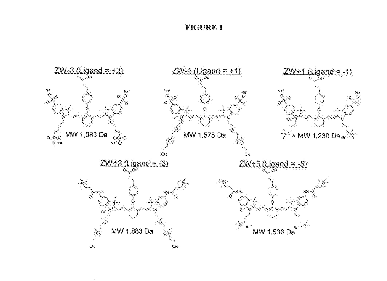
[0229]FIG. 1 depicts certain dyes that can be used in the imaging methods of the invention. Note that for molecules ZW−3, ZW−1, ZW+1, ZW+3, and ZW+5, a targeting ligand would have to be introduced having a +3, +1, −1, −3, and −5 charge, respectively, to achieve neutrality.

example 2

on of Dye ZW+1

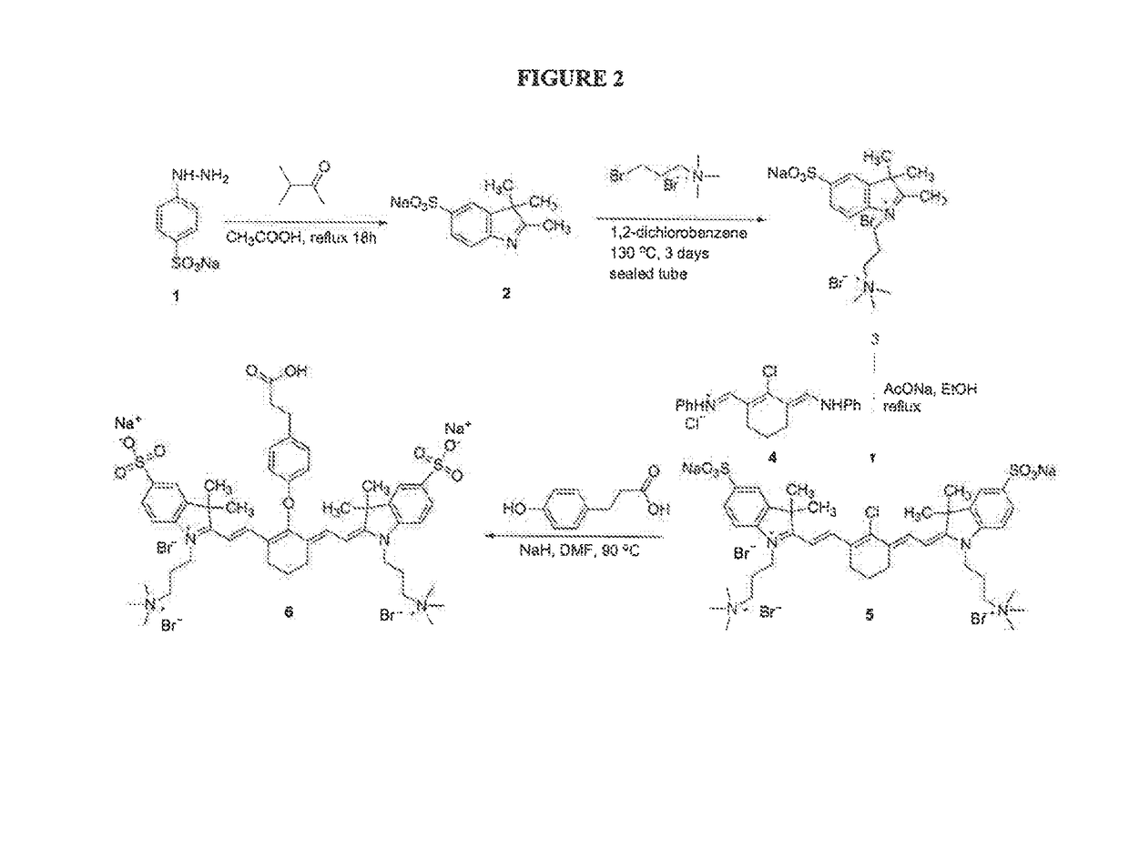
[0230]Dye ZW+1 (see FIG. 1) was made according to the synthetic procedure depicted in FIG. 2. Sodium 4-hydrazinylbenzenesulfonate (1) was reacted with 3-methylbutan-2-one to form the 2,3,3-trimethyl-3H-indole derivative (2). The indole nitrogen was then capped by reaction with 3-bromo-N,N,N-trimethylpropan-1-aminium bromide, and the product (3) reacted with the 2-chloro-3-((phenylamino)methylene)cyclohex-1-enypmethylene)benzenaminium compound (4) to yield the Cyanine dye (5). A linking group was attached by reaction of (5) with 3-(4-hydroxyphenyl)propanoic acid to thin’ the ZW+1 dye (6). See also Mojzych, M. et al. “Synthesis of Cyanine Dyes” Top. Heterocycl. Chem. (2008) 14:1-9; Sysmex Journal International (1999), Vol. 9, No. 2, pg 185 (appendix); Strekowski, L. et al. Synthetic Communications (1992), 22(17), 2593-2598; Strekowski, L. et al. J. Org. Chem. (1992) 57, 4578-4580; and Makin, S. M. et al. Journal of Organic Chemistry of the USSR (1977) 13(6), part 1, 1093...

example 3

on of Dye ZW+5

[0231]Dye ZW+5 (see FIG. 1) can be made according to the synthetic procedure depicted in FIG. 3. The first step involves alkylation of indolenine 7. The resultant quaternary salt 8 can be allowed to react with 3-aminopropanoyl chloride (in the form of an ammonium salt to protect the amino group). Then the terminal amino group can be quaternized with excess of methyl iodide to give the bis-quaternary salt 9. The subsequent steps 9→10→11 are analogous to the synthesis described in Example 2 above. Ion exchange can be used to prepare final products with a single counter anion such as chloride or bromide.

[0232]The presence of a quaternary ammonium cation close to the aromatic ring may inhibit formation of the final dye. Accordingly, a short spacer between the aromatic ring and the ammonium group of ZW+5 can be introduced.

[0233]Purification can be accomplished through silica gel chromatography or reverse-phase chromatography under both high and low-pressure conditions. Size...


PUM

PropertyMeasurementUnit
pHaaaaaaaaaa
molecular weightaaaaaaaaaa
wavelengthaaaaaaaaaa

Abstract

The present invention relates to a method for detecting renal disease and for assessing the efficacy of dialysis treatment using imaging agents having desirable in vivo properties.

Description

CROSS REFERENCE TO RELATED APPLICATIONS[0001]This application is a Continuation-in-Part of U.S. patent application Ser. No. 14 / 681,068 filed on Apr. 7, 2015, which application is a Continuation of application U.S. patent application Ser. No. 13 / 148,137 filed on Oct. 13, 2011, which application is a National Stage of International PCT Patent Application No. PCT / US2010 / 023305, filed Feb. 5, 2010, which application claims the benefit of U.S. Provisional Patent Application No. 61 / 150,522 filed on Feb. 6, 2009. The entire contents of these patent applications are incorporated herein by reference in their entirety.FEDERALLY SPONSORED RESEARCH OR DEVELOPMENT[0002]This invention was made with Government support under grant #CA115296 awarded by NIH. The Government has certain rights in the invention.FIELD OF THE INVENTION[0003]The present invention relates to methods of optically imaging tissues or cells using imaging agents having desirable in vivo properties that result in improved signal-...

Claims


Application Information

Patent Timeline
Patent Type & Authority Applications(United States)
IPC IPC(8): A61K49/00C07D209/42G01N33/58C07D209/02C09B23/01C09B23/08A61B6/00C07D255/02
CPCA61K49/0032A61B6/00C07D209/42G01N33/582C07D209/02C09B23/0066C09B23/086C07D255/02A61B5/0059C09B67/0025G01N2800/347
Inventor FRANGIONI, JOHN V.
Owner BETH ISRAEL DEACONESS MEDICAL CENT INC
Why Eureka
Try Eureka