Method for serum-free culture of chondrocytes and serum-free culture medium
a chondrocyte and serum-free technology, applied in the field of serum-free culture of chondrocytes and serum-free culture medium, can solve the problems of inability to successfully culture chondrocytes, inability to e.g. a transplant, and inability to obtain chondrocytes, etc., and achieve good transplant results
- Summary
- Abstract
- Description
- Claims
- Application Information
AI Technical Summary
Benefits of technology
Problems solved by technology
Method used
Image
Examples
example 1
[0063]Digestion of Tissue—Preparation of Culture
[0064]Primary human chondrocytes were isolated from a fresh sample of articular cartilage, and the sample was fractionated. The obtained fractionated sample was then immersed in a 0.1% to 0.3% collagenase solution, and subjected to enzymatic digestion at 37° C. for 4 to 5 hours. The obtained cells were collected by centrifugation for 5 minutes at 1,200 rpm.
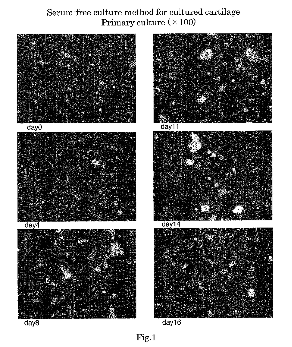
[0065]A mixture obtained by adding 100 ng / mL FGF, 0.5 μM SAG, 400 ng / mL HC, 5 ng / mL IGF, and 5 μg / mL insulin to DMEM medium was used as a culture medium. The cultured cells previously obtained were seeded at a density of 1000 cells / cm2 and cultured in an environment of 37° C. and 5% CO2.
[0066]FIG. 1 is a photograph substituted for drawings showing the cultured chondrocytes from the 0th day to 16th day of culture in Example 1. The figure shows that cartilage tissues gradually increased.
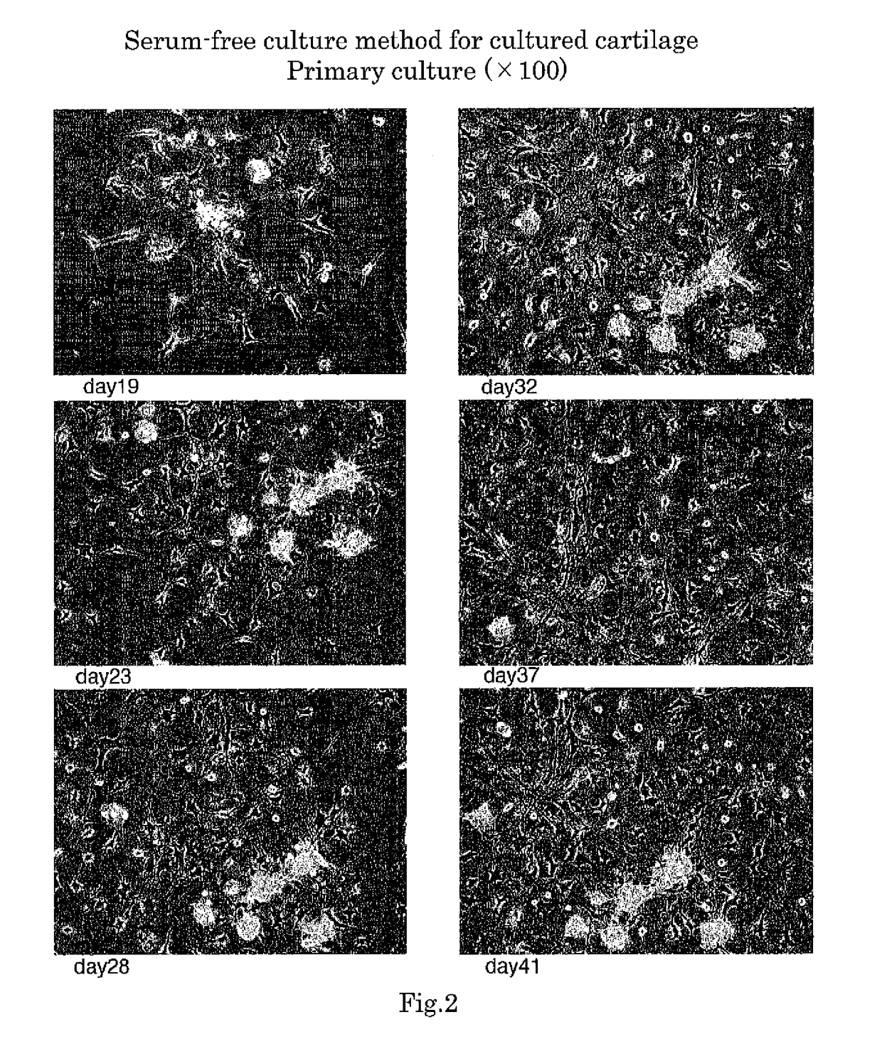
[0067]FIG. 2 is a photograph substituted for drawings showing the cultured chondrocytes from the 19t...
example 2
[0077]A mixture obtained by adding 100 ng / mL FGF2, 0.5 μM SAG, 400 ng / mL HC, 5 ng / mL IGF and 5 μg / mL insulin to DMEM medium was used as a culture medium for subculture. The cultured cells in Example 1 were seeded at a density of 10000 cells / cm2 and cultured in an environment of 37° C. and 5% CO2.
[0078]FIG. 4 is a photograph substituted for drawings showing the cultured chondrocytes from the first day to 13th day of culture in Example 2 (subculture). It was observed that after about 10 days from the onset of culture, viscosity was developed in the culture medium and, when removing the culture medium with an aspirator, slim threads were generated. The viscosity of the culture medium had increased from the 13th day.
[0079]FIG. 5 is a photograph substituted for drawings showing the cultured chondrocytes from the 18th day to 28th day of culture in Example 2. On the 18th day of culture, 50 μg / mL ascorbic acid was added to a culture medium. As a result of trial and error, it was found that ...
example 4
[0108]In order to examine the optimum concentration of SAG added to a culture medium, the concentration of SAG in a serum-free culture medium was changed to 0.05 uM, 0.01 uM or 0.5 uM, and an influence on proliferation of cultured chondrocytes was examined. Specifically, chondrocytes prepared in the same procedure as in Example 1 were seeded at a density of 10000 cells / well, and the number of cells on the 4th, 7th, 14th and 21st day after seeding was measured. The results were shown in FIG. 12.
[0109]In all the SAG concentrations, a difference in cell proliferation up to the 14th day of culture was not hardly observed, and an obvious difference which had been expected was not observed also on the 21st day of culture.
[0110]Because SAG is a very expensive reagent, it is preferred that SAG be able to be used at as low concentration as possible. This was verified because the inventors obtained knowledge that a low concentration of SAG can sufficiently promote the proliferation of chondro...
PUM
| Property | Measurement | Unit |
|---|---|---|
| immersion time | aaaaa | aaaaa |
| concentration | aaaaa | aaaaa |
| concentration | aaaaa | aaaaa |
Abstract
Description
Claims
Application Information
 - R&D
- Intellectual Property
- Life Sciences
- Materials
- Tech Scout
- Unparalleled Data Quality
- Higher Quality Content
- 60% Fewer Hallucinations
Browse by: Latest US Patents, China's latest patents, Technical Efficacy Thesaurus, Application Domain, Technology Topic, Popular Technical Reports.
© 2025 PatSnap. All rights reserved.Legal|Privacy policy|Modern Slavery Act Transparency Statement|Sitemap|About US| Contact US: help@patsnap.com


