
Method for producing therapeutic corneal endothelial substitute cell sphere

a technology of endothelial cells and stem cells, which is applied in the field of producing stem cells for producing corneal endothelial cells, can solve the problems of reducing the transparency of the cornea, edema of the corneal stroma, and serious influence on the visual function, so as to achieve efficient production, prevent the epithelial-mesenchymal transition, and improve the effect of corneal transparency

Active Publication Date: 2017-11-30
KEIO UNIV
View PDF5 Cites 3 Cited by
  • Summary
  • Abstract
  • Description
  • Claims
  • Application Information

AI Technical Summary

Benefits of technology

The present invention provides a more efficient way to produce therapeutic corneal endothelial cells from stem cells. The spherical cells have tight junctions, which prevent epithelial-mesenchymal transition and allow for rapid construction of a corneal endothelium tissue after transplantation. The cells can also be more effectively retained and adhered to the transplantation site using a viscoelastic substance. Overall, the invention offers a more effective and reliable approach to treat corneal endothelial disease.

Problems solved by technology

Therefore, the turbidity in the cornea caused by damage and the like has a serious influence on the visual function.
The above-mentioned function of corneal endothelial cell is impaired by a damage to the corneal endothelium such as a decrease in the corneal endothelial cell and the like, thus resulting in the edema of the corneal stroma.
This decreases transparency of the cornea, and reduces the visual acuity.
While the penetrating keratoplasty is an established technique, the supply of cornea is short in Japan as the situation stands, and the rejection reaction poses a problem.
Since the number of donor of cornea is limited, the problem of donor shortage cannot be overcome, like penetrating keratoplasty.
Furthermore, since corneal endothelial cell is difficult to culture, preparation of cultured cells in a number sufficient for transplantation places a large burden in terms of time and cost.

Method used


Image

  • Method for producing therapeutic corneal endothelial substitute cell sphere
  • Method for producing therapeutic corneal endothelial substitute cell sphere
  • Method for producing therapeutic corneal endothelial substitute cell sphere

Examples


example 1

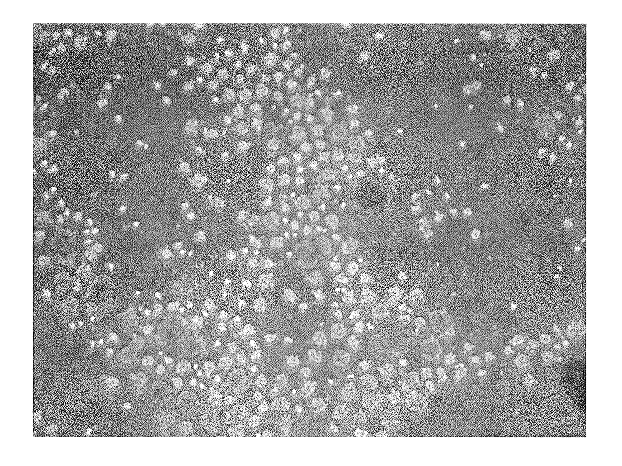
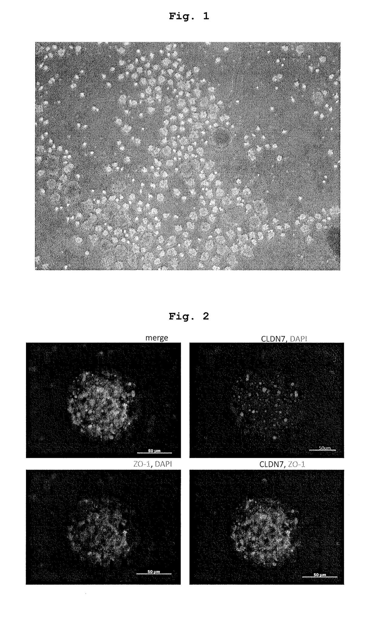
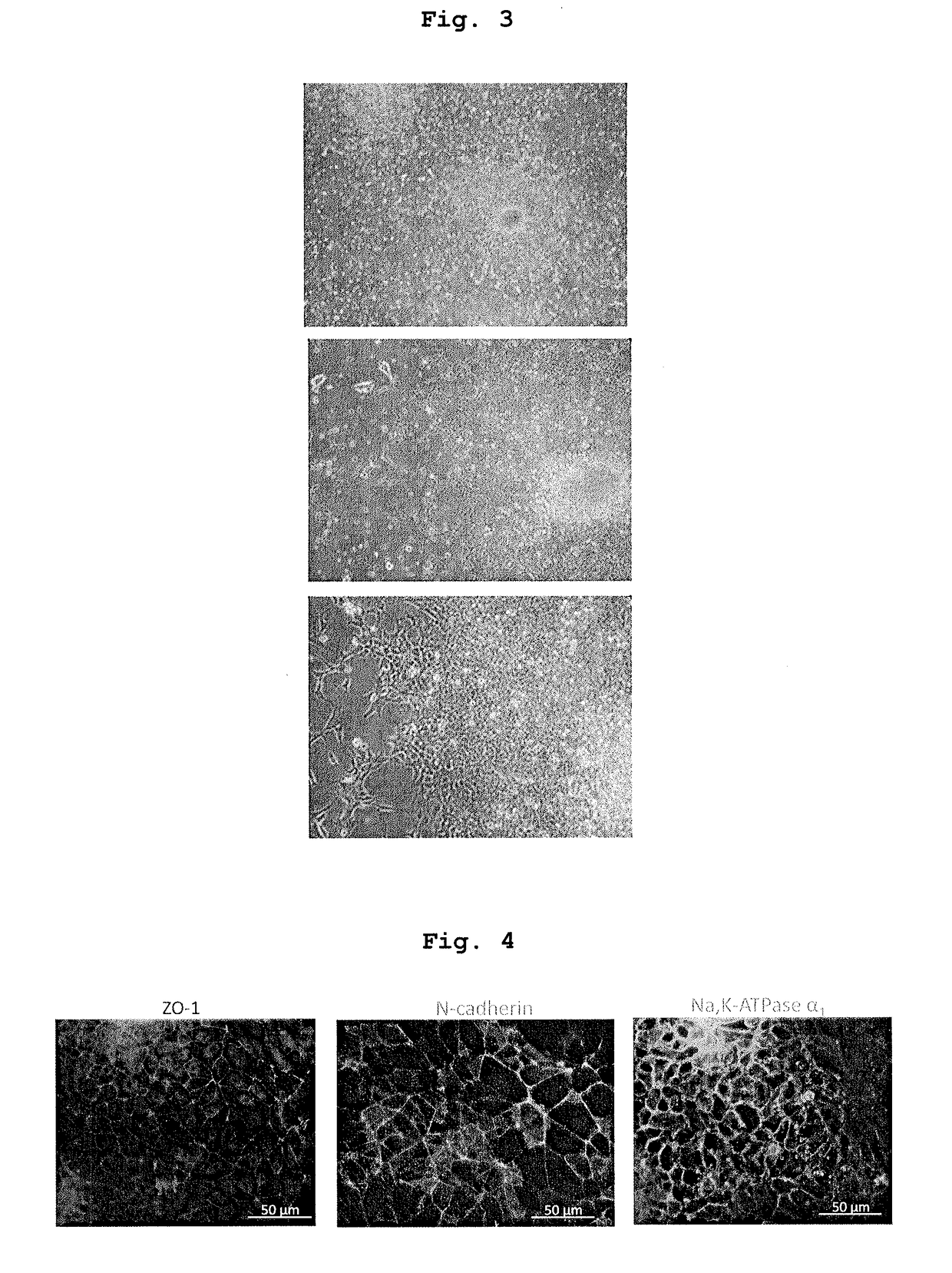
Production and Functional Evaluation of Therapeutic Alternative Corneal Endothelial Cell Sphere

[0102]1. Preparation of human iPS cell-derived neural crest stem cell Based on a publication (Nature, 2010 vol. 463, 958-964), neural crest stem cells were obtained from human iPS cell. This Example is different from the above-mentioned publication in that suspension culture was employed without using Matrigel for culturing iPS cells. Suspension culture enabled more efficient induction of differentiation into neural crest stem cells (iPS-NCC). The human iPS cells used were 201B7 (provided by Prof. Shinya Yamanaka (Kyoto University) and Prof. Hideyuki Okano (Keio University)).

2. Production of Therapeutic Alternative Corneal Endothelial to Cell Sphere

[0103]iPS-NCC obtained in the above-mentioned 1. was subjected to an enzyme treatment with Accutase to give single cells. The cells were cultured in a low adhesive plate (or dish) (Nunc, Corning etc.) at a cell density of 1×10⁴-105 cells / cm2.

[01...

example 2

Sphere Transplantation Experiment

1. Pharmaceutical Composition for Transplantation Containing Therapeutic Alternative Corneal Endothelial Cell Sphere

[0127]Using the sphere obtained in the above-mentioned 2. and by the following procedure, a pharmaceutical composition for transplantation of the composition described in Table 1 was prepared.[0128](1) DMEM / F12 added with ATRA, Y27632, insulin and bFGF is prepared as a medium.[0129](2) Sphere is mixed with the medium obtained in (1) to a cell density of 1×10⁷ cells / ml to give a cell suspension (×2).[0130](3) Viscoat (registered trade mark) 0.5 (200 μl) was added to and mixed with the cell suspension (200 μl) obtained in (2).

[0131]Viscoat (registered trade mark) 0.5 contains the Japanese Pharmacopoeia purified sodium hyaluronate (30 mg / ml), and chondroitin sulfate ester sodium (40 mg / ml).

TABLE 1(in DMEM / F12)sodium hyaluronate15 mg / mlchondroitin sulfate ester sodium20 mg / mlATRA500 nMY276325 μMinsulin5 μg / mlbFGF5 ng / mldensity of cell const...

example 3

Cultured Corneal Endothelial Cell Transplantation Experiment

[0145]1. Pharmaceutical Composition for Transplantation, which to Contains Cultured Corneal Endothelial Cell

[0146]Established cells of cultured corneal endothelial cell (B4G12 cells) were treated with an enzyme to give single cells.

[0147]A single cell suspension added with sodium hyaluronate (15 mg / ml) and chondroitin sulfate ester sodium (20 mg / ml) (suspension 1) and a single cell suspension not added with these but added with PBS instead (suspension 2) were prepared. As the additives other than sodium hyaluronate and chondroitin sulfate ester sodium, those similar to the additives described in Example 2, Table 1, were used at similar concentrations. The final cell density of suspensions 1 and 2 was adjusted to 1.0×10⁵ cells / 100 μl.

2. Transplantation of Cultured Corneal Endothelial Cells

(Procedure)

[0148]Using the pharmaceutical composition for transplantation prepared in the above-mentioned 1., cultured therapeutic alterna...


PUM

PropertyMeasurementUnit
concentrationaaaaaaaaaa
diameteraaaaaaaaaa

Abstract

The problem of the present invention is to provide a method of efficiently producing therapeutic alternative corneal endothelial cells, particularly, a method capable of stably producing them in a condition suitable for intraocular transplantation (in the anterior chamber) in a large amount. Furthermore, the present invention aims to provide a composition for transplantation, which is preferable for intraocular administration, particularly, into the anterior chamber. A therapeutic alternative corneal endothelial cell sphere can be produced by culturing stem cells in suspension in a differentiation induction medium containing a GSK3 inhibitor, retinoic acid and a ROCK inhibitor. Addition of a viscoelastic substance during intraocular (into the anterior chamber) transplantation of the sphere or cultured corneal endothelial cells dispersed into single cells can increase the number of adherent cells after transplantation.

Description

TECHNICAL FIELD[0001]The present invention relates to a method of producing a therapeutic alternative corneal endothelial cell from a stem cell, particularly, a method of inducing a stem cell into a therapeutic alternative corneal endothelial cell via a cell aggregate (sphere) by suspension culture. Furthermore, the present invention relates to a pharmaceutical composition for transplantation, which contains a sphere of therapeutic alternative corneal endothelial cells, and a pharmaceutical composition for transplantation, which contains cultured corneal endothelial cells.BACKGROUND ART[0002]The visual information is recognized when the light that entered from the cornea, which is a transparent tissue on the frontmost surface of the eyeball, reaches the retina to excite the retinal nerve cell, and the developed electric signals reach the visual field in the cerebrum via optic nerve. In other words, the cornea is positioned in front of a path where the light passes when a living orga...

Claims


Application Information

Patent Timeline
Patent Type & Authority Applications(United States)
IPC IPC(8): A61K35/30A61K48/00C12N5/079A61F9/00A61K31/203A61K35/44A61K38/22
CPCA61K35/30C12N5/0621A61K48/0091A61K38/22A61K31/203A61K35/44A61F9/0017A61K38/28A61K47/36C12N5/10A61P27/02
Inventor SHIMMURA, SHIGETOHATOU, SHINTSUBOTA, KAZUO
Owner KEIO UNIV
Learn More