
Device-based motion-compensated digital subtraction angiography

a digital subtraction and motion compensation technology, applied in the field of image processing method, can solve the problems of affecting image quality, affecting image quality, and affecting image quality,

Active Publication Date: 2017-11-30
KONINKLJIJKE PHILIPS NV
View PDF0 Cites 15 Cited by
  • Summary
  • Abstract
  • Description
  • Claims
  • Application Information

AI Technical Summary

Benefits of technology

The proposed method is a way to make pictures look smoother by only tracking the movement of a specific part of the picture. This helps to focus the operator on the most important information, even if there are other objects in the picture that are moving too. This is useful when trying to visualize complex anatomy and motion patterns in real-time.

Problems solved by technology

In the above two exemplary interventions and also in others, the image quality is often compromised by poor contrast in one or more motion layers viewed against a complex and cluttered background (spine, ribs, medical devices, etc.).
However due to complex motion patterns during the intervention, subtraction artifacts may be introduced by DSA which again undermines image quality.

Method used


Image

  • Device-based motion-compensated digital subtraction angiography
  • Device-based motion-compensated digital subtraction angiography
  • Device-based motion-compensated digital subtraction angiography

Examples


Embodiment Construction

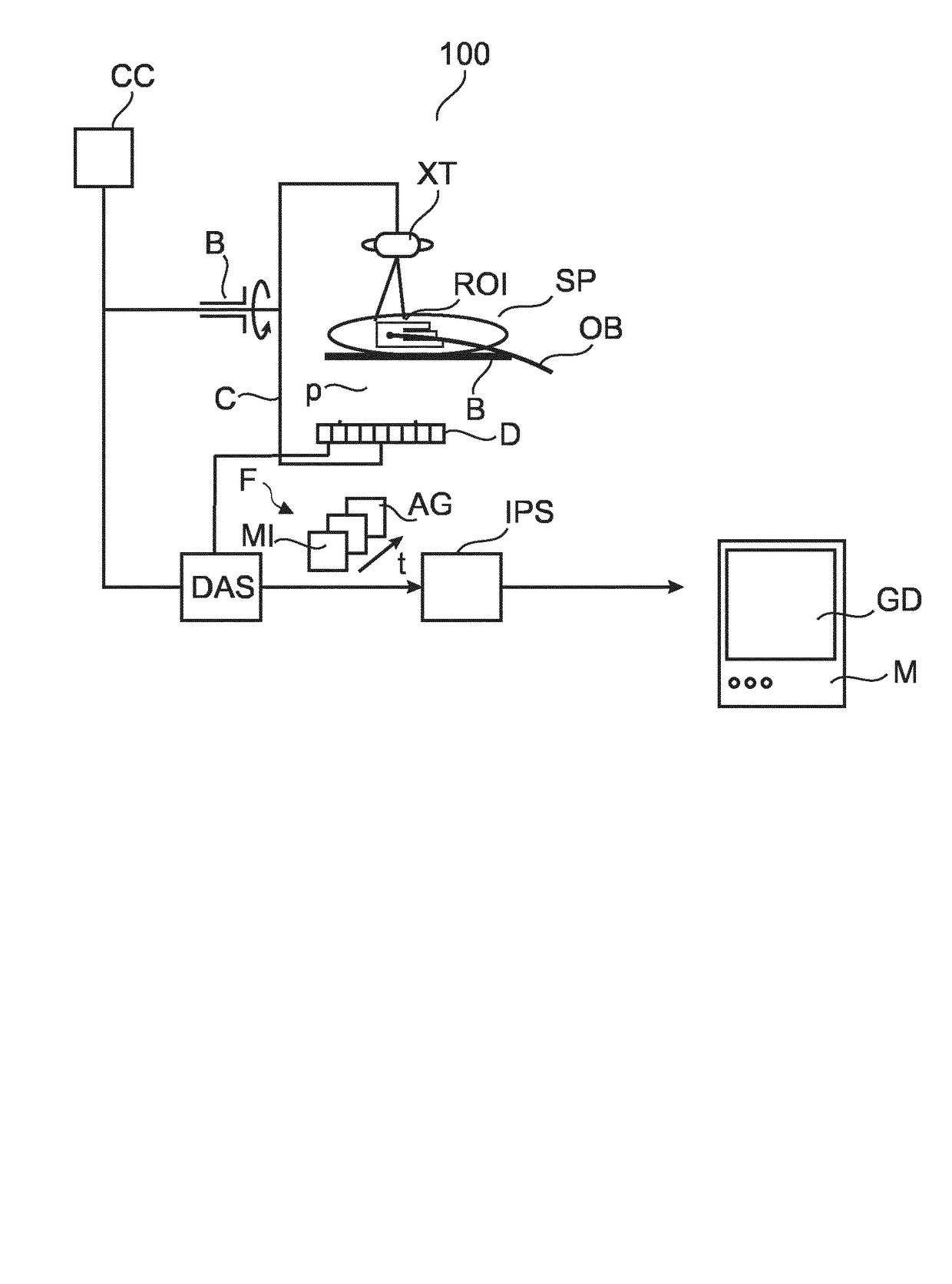
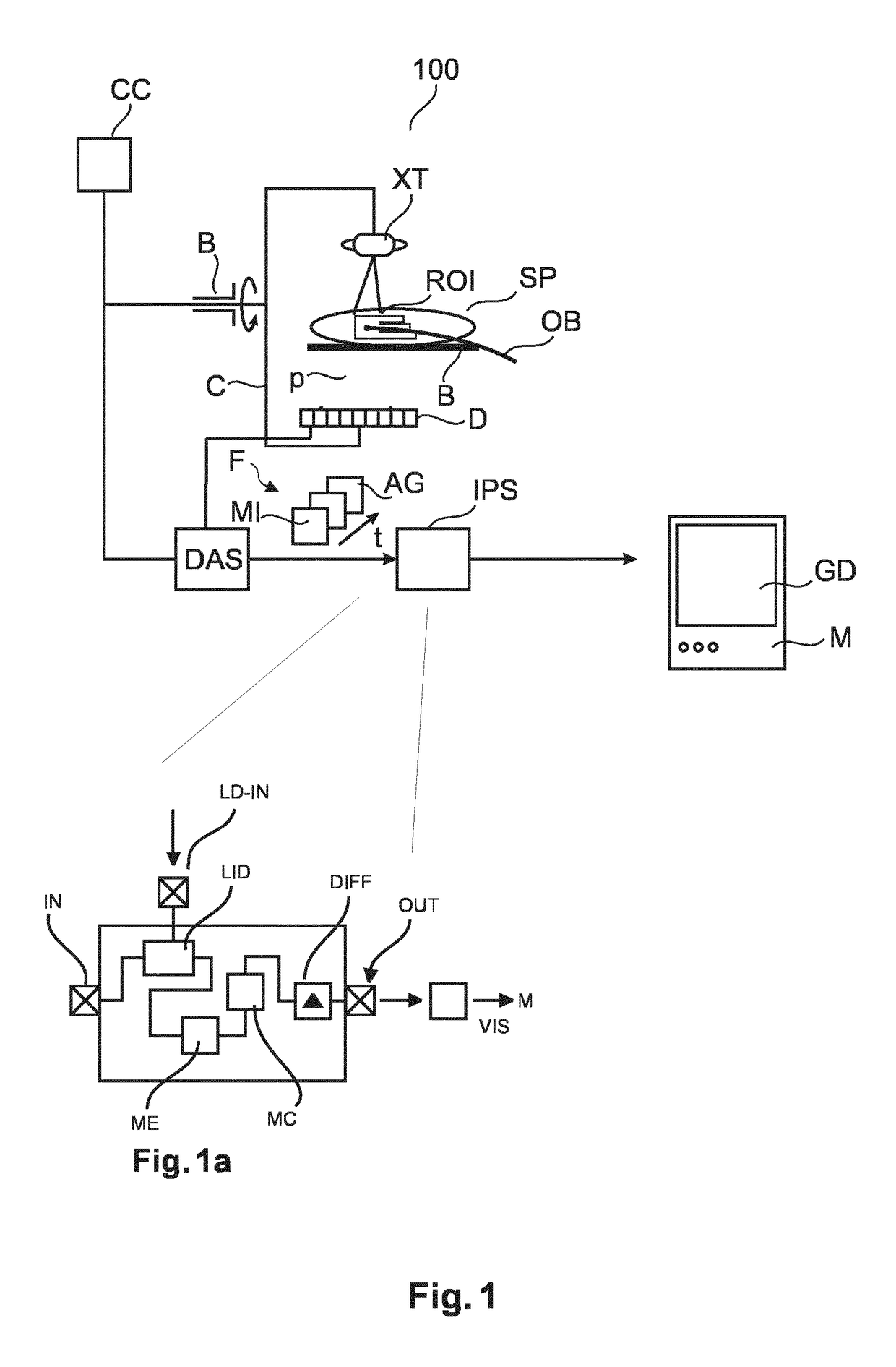
[0036]With reference to FIG. 1, the basic components of a fluoroscopic or angiographic imaging arrangement are shown that can be used to support interventional procedures.

[0037]A patient SP may suffer from a malfunctioning heart valve. During a TAVI interventional procedure, medical personnel introduces a guide wire into the femoral artery of patient SP and then guides a delivery catheter OB to the diseased aortic valve ROI to be repaired or replaced As guidewire progresses through patient's P cardiac vasculature, a series of sequential fluoroscopic images F are acquired by an x-ray imager 100. Another example is an embolization procedure where a catheter OB for embolization agent administration is guided to an AVM or cancer (as in TACE) site.

[0038]During the intervention, patient SP is deposed on a bed B between an x-ray imager 100's x-ray tube XT and detector D. X-ray tube XT and detector D are attached to rigid frame C rotatably mounted on a bearing B. The fluoroscopic image oper...


PUM


Abstract

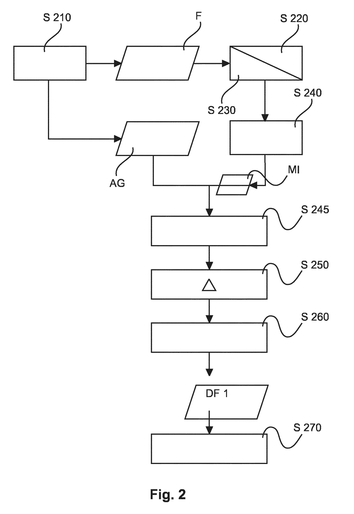
An image processing method and related system to register projection images (AG, MI) only with respect to a motion of a landmark across said images. The motion of the landmark relates to a motion of a region of interest, ROI. The so registered images (AG, MI) are then subtracted from each other to arrive at a difference image that is locally motion compensated and that represents the ROI at good contrast whilst subtraction artifacts can be avoided.

Description

FIELD OF THE INVENTION[0001]The invention relates to an image processing method, to an image processing system, to a computer program element and to a computer readable medium.BACKGROUND OF THE INVENTION[0002]There are a number of medical interventions that are carried under fluoroscopy or angiography. In other words, real-time projection X-ray images are acquired to image the internals of the patient along with medical tools or devices introduced into the patient to achieve the intervention.[0003]For example, in order to stem growth of cancerous tissue or of an AVM (arteriovenous malformation) its arterial feeders may be intentionally embolized to shut down the blood supply (AVM) and / or stop nutrient supply to the cancer or growth (such as in transcatheter arterial chemoembolization (TACE)). This type of intervention, called medical embolization, may be brought about by administering an embolization agent at a desired location (region of interest (ROI)) in the human body by way of ...

Claims


Application Information

Patent Timeline
IPC IPC(8): G06T7/00G06T7/254G06T11/00G06T7/11G06T7/246
CPCG06T7/0012G06T7/254G06T7/246G06T7/11G06T11/006G06T2211/428G06T2207/20224G06T2207/10121G06T2207/30021G06T2207/30048G06T2211/404G06T5/50G06T2207/10072G06T2207/20201G06T2207/20104G06T2207/30101
Inventor NEMPONT, OLIVIER PIERREDEN HARTOG, MARKUS JOHANNES HARMENNACHABE, RAMIPIZAINE, GUILAUME JULIEN JOSEPHRADAELLI, ALESSANDRO GUIDOFLORENT, RAOUL
Owner KONINKLJIJKE PHILIPS NV
Learn More