High-throughput drug and genetic assays for cellular transformation

a high-throughput, cellular transformation technology, applied in the direction of genetically modified cells, instruments, skeletal/connective tissue cells, etc., can solve the problems of difficult to disaggregate individual cells, difficult to achieve rapid and quantitative, and difficult to detect cells grown on low attachment surfaces. to individual cells, the effect of increasing cellular transformation or tumorigenicity

Pending Publication Date: 2018-02-22
THE BROAD INST INC +3
View PDF0 Cites 3 Cited by
  • Summary
  • Abstract
  • Description
  • Claims
  • Application Information

AI Technical Summary

Benefits of technology

The patent describes a method for detecting and measuring the growth of cancer cells by culturing them in a low attachment condition, permeabilizing them, and measuring markers of cell viability. This assay can be used for high-throughput drug screening and genetic screening for oncogenic growth and tumorigenicity. The method can also be used to detect transformed cells in a sample, and to identify drugs that selectively inhibit the growth of transformed cells. Overall, the patent provides a powerful tool for studying cancer cell growth and inhibition, which can be useful in personalized medicine.

Problems solved by technology

While pharmacological perturbation is imminently applicable, success of genome editing approaches in primary patient derived ex vivo cultures remain challenging.
Yet, unlike traditional attachment conditions for adherent cells (on surfaces that allow cell attachment) cells that are grown on low attachment surfaces cannot be detected properly solely by visualization.
Also, once the cells grow as an aggregate it is very hard to disaggregate them to individual cells.
A clear plate, for example, is not appropriate for this technique.
Also, many informative reagents were overlooked due to minimal effect shown on high-attachment surfaces, yet these reagents were largely effective in low-attachment conditions (Rotem et al., 2015).
Yet, some experiments involve over-expression of oncogenic genes (Rotem et al., 2015) or inhibition of tumor suppressors, which might result in higher viability than the control group.
Yet, some of the plates that we have examined had a compromised ability to support low-attachment growth and cells managed to adhere to the surfaces.

Method used


Image

  • High-throughput drug and genetic assays for cellular transformation
  • High-throughput drug and genetic assays for cellular transformation
  • High-throughput drug and genetic assays for cellular transformation

Examples


example 1

[0085]A Replacement for the Soft-Agar Assay that Permits High-Throughput Drug and Genetic Screens for Cellular Transformation

[0086]This example describes the development of the assays for measuring oncogenic growth and viability, and / or degree of cellular transformation of a sample, including a patient sample or a cell line, without the need for soft agar plates.

Results

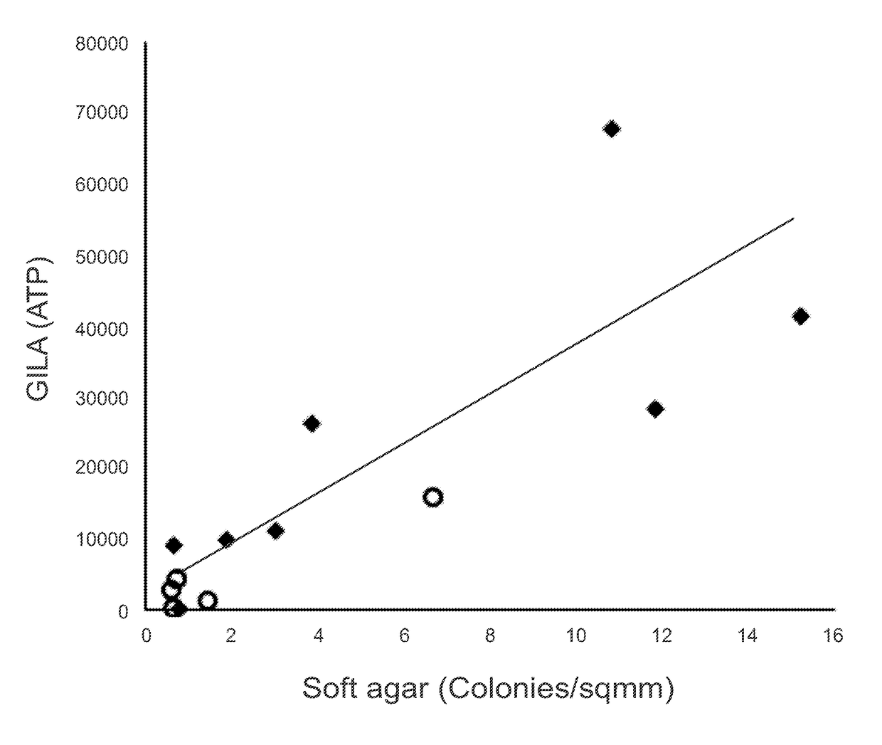
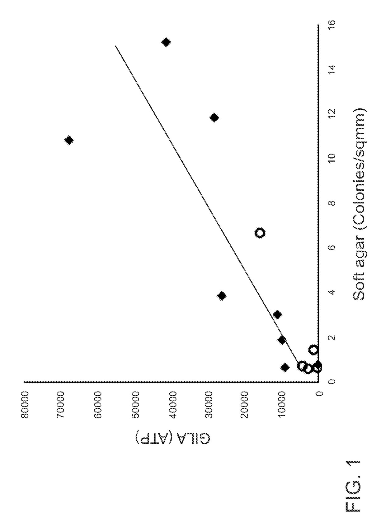
[0087]The GILA Assay is Comparable to the Soft-Agar Assay.

[0088]The principle of GILA is that transformed cells can grow on low-attachment plates, whereas nontransformed cells cannot. A critical parameter for the GILA assay is cell density (number of cells / well), and for these experiments, this was optimized at 1000 cells / well in 100 μl medium for 96-well plates or 50 cells / well in 30 μl growth medium for 384-well plates. 13 cell lines originating from 4 different tissues-fibroblast, breast, ovary and prostate were examined (FIG. 6). An equal number of cells from each line were seeded into wells of an ultra-low attach...

example 2

[0118]This example describes a method for the assessment of tumorigenicity of cell, in vitro and ex vivo, and the steps towards optimizing the method for individual applications. This protocol presents one main method with three optional applications. The main method is GILA assay using cell lines, and an optional method is GILA assay using primary patient-derived cells. The two additional applications described in this protocol are pharmacological and genetic perturbations. While the unit describes the pharmacological approach for both cultured and patient derived-cells cells, the genetic screen has been used only for cultured cells. This protocol uses appropriate cell number and reagent volume for 96-well plates; some adjustment will be necessary if using another format.

Materials List

[0119]Solutions and reagents: Cell line, growth medium (DMEM, FCS, antibiotics), trypsin, PBS, cell counter device / hemocytometer, ATP quantification reagent (CellTiter-Glo kit, CTC, Promega), plate re...

example 3

ample Assay

[0195]This embodiment demonstrates the application of the growth in low attachment technology to detect drug sensitivity of tumor cells in a clinical environment.

[0196]Fresh tumors were dissociated and treated to exclude red blood cells. Tumor cells were cultured on high- and low-attachment surfaces and drugs were added after 1 day. At day 4 the cells were lysed and cellular ATP was measured as a surrogate of cell viability. Drug sensitivity of cells to a specific drug was compared between cells that grew on high- and low-attachment surfaces.

[0197]Several metastatic melanoma patients were sampled and fresh cells were treated with 4 targeted drugs specific for the RAF / MEK pathway; Dabrafenib (BRAF inhibitor), Trametinib (MEK1 / 2 inhibitor), SCH772984 (ERK1 / 2 inhibitor) and VX-11e (ERK2 inhibitor).

[0198]Patient number 1—GILA assay revealed that this specific individual might benefit from ERK1 / 2 inhibitors. The GILA assay was 10 times more sensitive at predicting this drug se...


PUM

PropertyMeasurementUnit
Concentrationaaaaaaaaaa
Cell viabilityaaaaaaaaaa

Abstract

A method for detecting oncogenic growth and viability, and / or degree of cellular transformation and / or identifying an agent that inhibits cellular transformation is disclosed. The method including: providing a cellular sample, such as a sample of cells obtained from a subject or a cell line; culturing the cellular sample in low attachment conditions; and detecting growth and7or cell viability of the sample, wherein increased growth relative and / or viability relative to a control or control level indicative of basal growth and / or viability indicates cellular transformation. In some embodiments, the method includes introducing a n expression vector into cells of the cellular sample, wherein the expression vector comprises a gene product expression sequence being tested for transformation ability. In some embodiments the cellular sample is contacted with a test agent and growth and / or cell viability of the sample is determined to determine if the agent inhibits transformation.

Description

FIELD OF THE DISCLOSURE[0001]The present disclosure relates to methods of and assays for high-throughput drug and cellular transformation screening and specifically for assays based on conditions of cellular non-attachment as a replacement for soft-agar. The subject matter disclosed herein is directed to methods and materials for genome-wide screening of genetic perturbations combined with imaging assays for complex phenotypes to identify relationships between genotypes and phenotypes.BACKGROUND[0002]For nearly 50 years, the gold-standard assay for cellular transformation / tumorigenicity has been the soft-agar assay (see Macpherson & Montagnier (1964) Agar suspension culture for the selective assay of cells transformed by polyoma virus. Virology 23:291-294; Shin S I, Freedman V H, Risser R, & Pollack R (1975); Tumorigenicity of virus transformed cells in nude mice is correlated specifically with anchorage independent growth in vitro. Proc. Natl. Acad. Sci. U.S.A. 72(11):4435-4439). T...

Claims


Application Information

Patent Timeline
IPC IPC(8): C12Q1/68G01N33/574G01N33/50
CPCC12Q1/68G01N33/574G01N33/5011C12M33/00G01N33/5008C12N5/0631C12N5/0656C12N2510/04C12N2539/00
Inventor ROTEM, ASAFSTRUHL, KEVINBLAINEY, PAULXU, LIYI
Owner THE BROAD INST INC
Why Eureka
Try Eureka