Devices, systems, and methods to optimize annular orientation of transcatheter valves

a technology of annular orientation and transcatheter valve, which is applied in the field of devices, systems and methods to optimize the annular orientation of the transcatheter valve, can solve the problems of loss of elasticity and efficiency, increased risk of stroke or pulmonary embolism, and other complications

Active Publication Date: 2018-03-22
CEDARS SINAI MEDICAL CENT
View PDF5 Cites 4 Cited by
  • Summary
  • Abstract
  • Description
  • Claims
  • Application Information

AI Technical Summary

Benefits of technology

[0007]To solve these problems, described herein is a Device to Optimize aNnUlar orientation of Transcathether valves (DONUT). DONUT can correct large differences between a native valve's anatomical dimensions and the external dimensions of a transcatheter valve stent frame. DONUT may act as a mould for the anatomy of a native valve externally, and its central hole may match a transcatheter replacement valve, for example, a standardized valve or those commercially available ones including but not limited to the MEDTRONIC COREVALVE device and the EDWARDS SAPIEN device. The outside perimeter of the DONUT conforms to the anatomy of a native valve, cardiovascular structure or chamber, and its center hole fits a transcatheter valve. Also described herein is a method of using DONUT for transcatheter heart valve implantation. DONUT is first deployed to a diseased heart valve (e.g., the aortic valve and mitral valve) and serves as a consistent landing zone and stable anchor point. Next, a transcatheter valve is implanted into the center hole of DONUT, even when the native valve's anatomy and stenosis are not favorable, suitable or safe for the direct deployment of a transcatheter valve.

Problems solved by technology

To compensate for poor pumping action, the heart muscle enlarges and thickens, thereby losing elasticity and efficiency.
In addition, in some cases, blood pooling in the chambers of the heart has a greater tendency to clot, increasing the risk of stroke or pulmonary embolism.
In mild cases there may be no symptoms, while in advanced cases, valvular heart disease may lead to congestive heart failure and other complications.
However, the native valve's shape, size and stenosis vary considerably, and hence are not always favorable, suitable or safe for the deployment of transcatheter valves.
If an implanted transcatheter valve does not conform to the native valve's anatomy or is not stably anchored, paravalvular leakage as well as device embolization or malposition may occur.
To fill the gaps between the transcatheter valve and the native valve's anatomical structures, an external cuff (either fabric or hydrogel) may be added, but this cannot correct large differences in dimension.

Method used


Image

  • Devices, systems, and methods to optimize annular orientation of transcatheter valves
  • Devices, systems, and methods to optimize annular orientation of transcatheter valves
  • Devices, systems, and methods to optimize annular orientation of transcatheter valves

Examples


example 1

Self-Expandable DONUT

For Aortic Valve

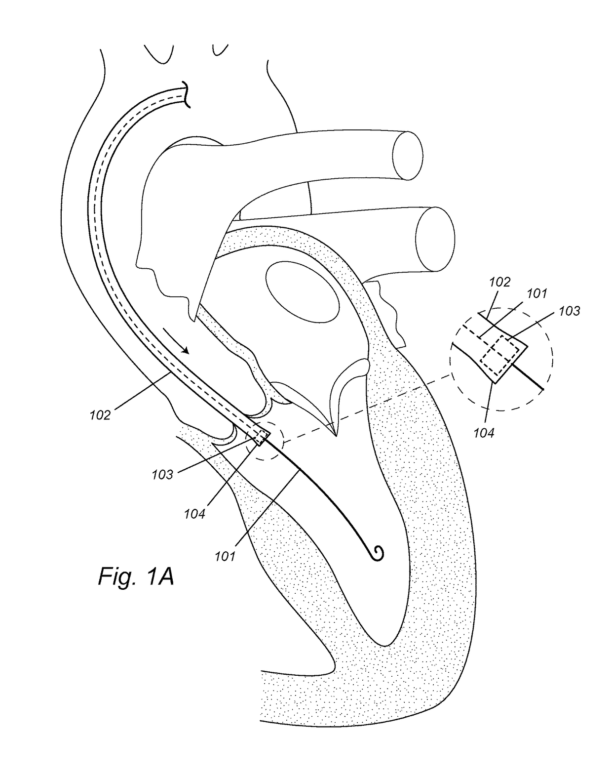
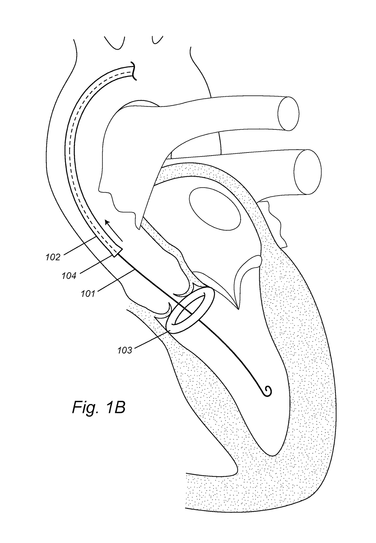
[0059]FIGS. 1A-1B illustrate an example of an implantation procedure for a DONUT device as disclosed herein. After induction of anesthesia, and sterile preparation, an incision is performed at the femoral artery. Alternatively, percutaneous femoral access may be performed with or without pre-closure. A guide wire 101 is inserted via the femoral artery and the ascending aorta through the aortic valve into the left ventricle (FIG. 1A). A self-expandable DONUT device 103 is compressed and enclosed in the enclosing sheath 104 at the distal end of a first delivery catheter 102. The loaded first delivery catheter 102 is advanced over the guide wire 101 and inserted through the aortic valve into the left ventricle, and places the compressed DONUT device 103 on the ventricular side of the aortic valve and near the valve annulus (FIG. 1A). The first delivery catheter 102 is retracted and the compressed DONUT device 103 is hence released out of the enclosi...

example 2

Balloon-Expandable DONUT

For Aortic Valve

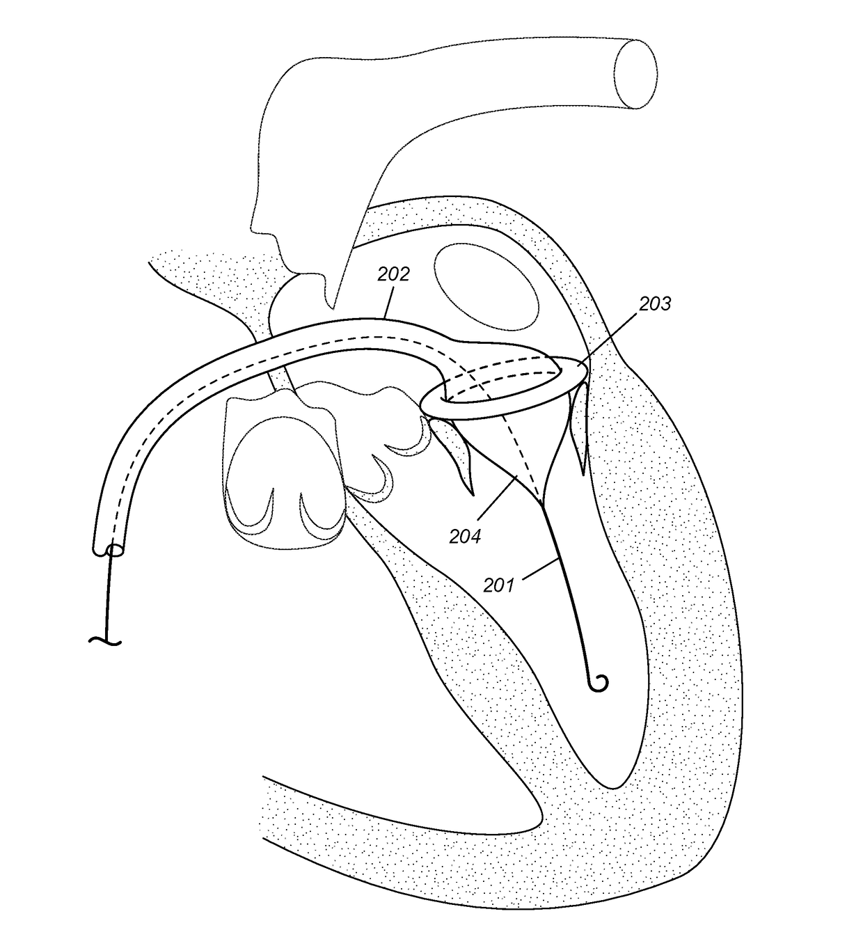
[0061]FIGS. 2A-2C illustrate an example of an implantation procedure for a DONUT device as disclosed herein. After induction of anesthesia, and sterile preparation, an incision is performed at the femoral artery. Alternatively, percutaneous femoral access may be performed with or without pre-closure. A guide wire 201 is inserted via the femoral artery and the ascending aorta through the aortic valve into the left ventricle (FIG. 2A). A balloon-expandable DONUT device 203 is compressed and mounted over the inflatable balloon 204 at the distal end of a first delivery catheter 202. The loaded first delivery catheter 202 is advanced over the guide wire 201 and inserted through the aortic valve into the left ventricle, and places the compressed DONUT device 203 on the ventricular side of the aortic valve and near the valve annulus (FIG. 2A). The inflatable balloon 204 is inflated to expand the compressed DONUT device 203 (FIG. 2B). The expanded DON...

example 3

Delivery of Self-Expandable Replacement Valve

[0063]This is performed after the DONUT device is deployed to the aortic valve peri-annular region. A self-expandable replacement valve 105 is compressed and enclosed in the enclosing sheath 107 at the distal end of a second delivery catheter 106. The loaded second delivery catheter 106 is advanced over the guide wire 101 and inserted through the aortic valve into the left ventricle, and places the compressed replacement valve 105 across the aortic valve and the center hole of the DONUT device (FIG. 3A). The second delivery catheter 106 is retracted and the compressed replacement valve 105 is hence released out of the enclosing sheath 107 to expand (FIG. 3B). The expanded replacement valve anchors to the center hole of the DONUT device, thereby replacing the native aortic valve (FIG. 3B).

[0064]In various embodiments, the same or similar steps may be used for other heart valves, such as the mitral valve, pulmonary valve and tricuspid valve...


PUM


Abstract

In various embodiments, provided herein are methods, devices and systems to optimize annular orientation of transcatheter valves and thereby facilitate transcatheter aortic valve replacement in the setting of challenging cardiovascular anatomy. These methods, devices and systems are used to treat patients with valvular diseases. To solve these problems, described herein is a Device to Optimize aNnUlar orientation of Transcathether valves (DONUT). DONUT can correct large differences between a native valve's anatomical dimensions and the external dimensions of a transcatheter valve stent frame.

Description

FIELD OF INVENTION[0001]This invention relates to devices, systems, and methods to optimize annular orientation of transcatheter valves, which may be used to treat valvular heart diseases.BACKGROUND OF THE INVENTION[0002]All publications cited herein are incorporated by reference in their entirety to the same extent as if each individual publication or patent application was specifically and individually indicated to be incorporated by reference. The following description includes information that may be useful in understanding the present invention. It is not an admission that any of the information provided herein is prior art or relevant to the presently claimed invention, or that any publication specifically or implicitly referenced is prior art.[0003]Valvular heart disease is characterized by damage to or a defect in one of the four heart valves: the mitral, aortic, tricuspid or pulmonary. The mitral and tricuspid valves control the flow of blood between the atria and the ventr...

Claims


Application Information

Patent Timeline
Patent Type & Authority Applications(United States)
IPC IPC(8): A61F2/24A61M25/09
CPCA61F2/2409A61F2/2433A61M25/09A61F2/2436A61F2/2418A61F2250/006A61F2250/007A61F2210/0014A61F2230/0065A61F2250/0003
Inventor AL-JILAIHAWI, HASANIAN
Owner CEDARS SINAI MEDICAL CENT
Why Eureka
Try Eureka