
Method and apparatus to measure tissue displacement and strain

a tissue displacement and strain technology, applied in the field of displacement and strain reconstruction, can solve the problems of inability to access the raw radiofrequency (rf) ultrasound signals of low-to-mid-end and old machines, inconvenient use, and inability to provide tissue mechanical characteristics or stiffness information in imaging mode, etc., to achieve low computation complexity, high quality of estimated elastogram, and cost-effective

Active Publication Date: 2018-05-17
MAHMOUD AHMED M EHAB +1
View PDF3 Cites 5 Cited by
  • Summary
  • Abstract
  • Description
  • Claims
  • Application Information

AI Technical Summary

Benefits of technology

This patent describes an innovative method and system for reconstructing displacement and strain images of tissues or objects in response to mechanical or thermal stimulation using ultrasound imaging. The method is flexible, cost-effective, and accurate, and can be used with different types of imaging machines and data transfer methods. It involves a computationally efficient method that uses block matching and machine learning techniques to measure displacements and creates high quality strain images with high signal-to-noise ratios. The method can be used to evaluate the effectiveness of therapies or treatments that aim to modify the tissue's motion or response to stimulation. Overall, this invention offers a more accurate and reliable method for analyzing tissue motion and deformation.

Problems solved by technology

However, conventional ultrasound B-mode does not provide information about the mechanical characteristics, or stiffness, of tissue that is essential for diagnosing several diseases including liver disease, atherosclerosis, and tumors.
Although some ultrasound companies have developed this new mode (elastography) on few of their premium and high-end ultrasound machines, this imaging mode is still not available on most ultrasound machines and ultrasound machines developed before year 2003.
This elastography mode is only compatible with these specific machines and cannot be used on other machines from the same manufacturer, i.e. until now it is a machine dependent mode and there is not machine independent elastography processing unit.
Both low- to medium-end and old machines do not provide access to raw radiofrequency (RF) ultrasound signals that is commonly used to reconstruct elastography in all machines.

Method used


Image

  • Method and apparatus to measure tissue displacement and strain
  • Method and apparatus to measure tissue displacement and strain
  • Method and apparatus to measure tissue displacement and strain

Examples


Embodiment Construction

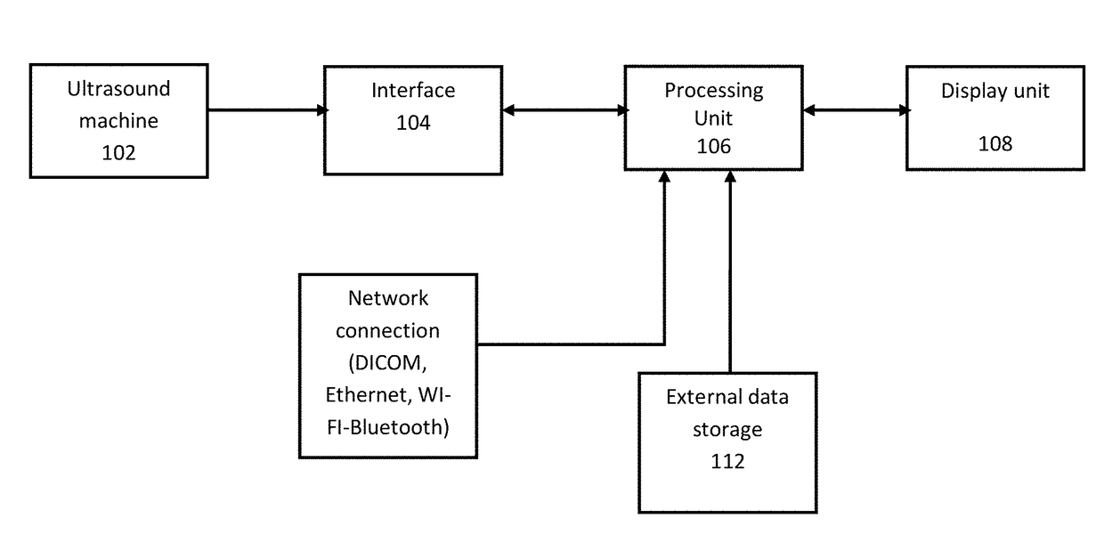
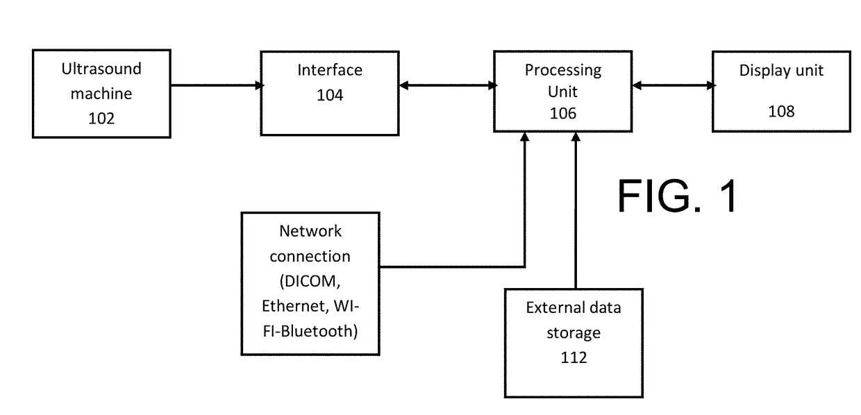
[0019]Referring to FIG. 1 images are captured from the ultrasound machine (102) using a hardware interface (104) such as video capture device then they are processed by our method in the processing unit (106) where displacements and strains between different frames are measured and displayed on the display unit (108). Images can be transferred to the processing unit by means of network connections (110) through Ethernet or WI-FI and / or external data storage (112).

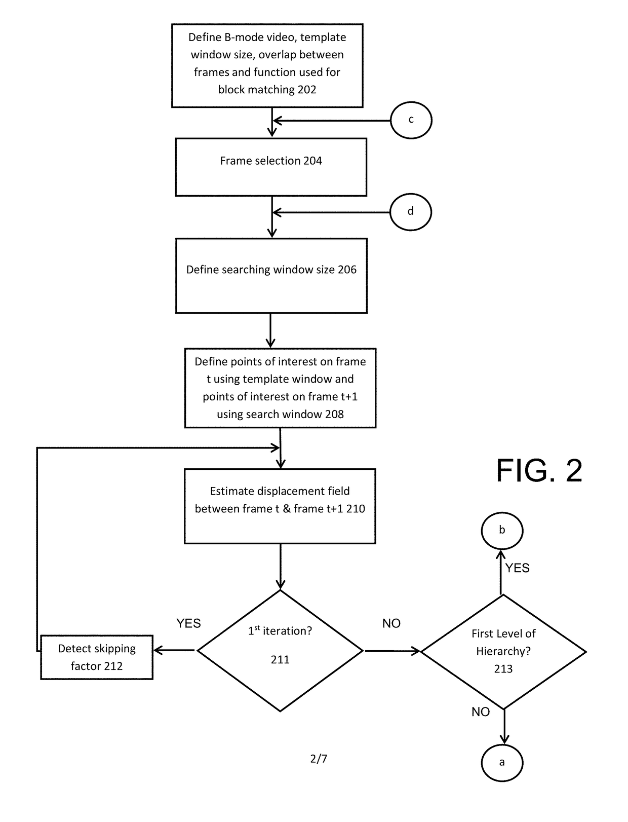
[0020]FIG. 2 refers to the first part of the displacement estimation algorithm where ultrasound images, cine-loop, or videos the region of interest (ROI) is automatically segmented at which there are imaging data (scan lines) acquired by the transducer. In the first block (202) the imaging depth is entered or determined automatically for processing successive or selected frames (204) acquired after time t. Template and search window size (206) are determined automatically by taking a ratio from the imaging frame size (such ...


PUM


Abstract

A method for reconstructing displacement and strain maps of human tissue of a subject in vivo or other objects includes applying a mechanical deformation for a target area, imaging tissues while deformation is applied on the target area, measuring the axial and lateral displacements and axial, lateral, and shear strains of tissue in the target area, differentiating between tissues of different stiffness and strain responses, and labeling tissues of different stiffness and strain responses. In some embodiments, displacement and strain imaging are performed using a high-resolution, high-speed, highly-accuracy hierarchy recursive displacement tracking from conventional ultrasound brightness mode images. Particularly, this invention relates to the reconstruction of displacement and strain images from conventional ultrasound B-mode images acquired using any ultrasound machine of different manufacturers without the need to use carrier signals.

Description

CROSS-REFERENCE TO RELATED APPLICATIONS[0001]This application claims the benefit of U.S. Provisional Application No. 62 / 274, 265, which was filed on Jan. 2, 2016.FIELD OF THE INVENTION[0002]This invention relates to a method and system for the reconstruction of displacement and strain images from conventional ultrasound brightness mode (B-mode) images. Particularly, this invention relates to the reconstruction of displacement and strain images from conventional ultrasound B-mode images acquired using any ultrasound machine of different manufacturers without the need to use carrier signals. More particularly, the invention relates to a manufacturer-independent method and system for the acquisition of conventional ultrasound B-mode images from ultrasound machines and the generation of displacement and strain images from those images. Specifically, the invention relates to a constrained recursive motion tracking technique with a feature extraction algorithm that ensures a real-time hig...

Claims


Application Information

Patent Timeline
Patent Type & Authority Applications(United States)
IPC IPC(8): A61B8/08A61B8/00A61B5/00A61B6/00G01R33/563
CPCA61B8/485A61B8/5223A61B8/565A61B5/0059A61B6/4258A61B5/0048G01R33/56358A61B5/0077A61B5/055A61B6/00A61B6/5217G16H50/30
Inventor MAHMOUD, AHMED M EHABALI, MOHAMED TAREK MOHAMED
Owner MAHMOUD AHMED M EHAB
Learn More