Fixation devices, systems and methods for heart valve leaf repair

a technology of fixation device and heart valve leaf, which is applied in the field of repair of valves, can solve the problems of reducing the pumping efficiency of the heart, affecting the repair effect of the valve leaf, and putting the patient at risk of severe heart failure, and achieves the effects of minimizing tissue injury, enhancing retention, and high retention for

Active Publication Date: 2018-05-17
ABBOTT CARDIOVASCULAR
View PDF3 Cites 18 Cited by
  • Summary
  • Abstract
  • Description
  • Claims
  • Application Information

AI Technical Summary

Benefits of technology

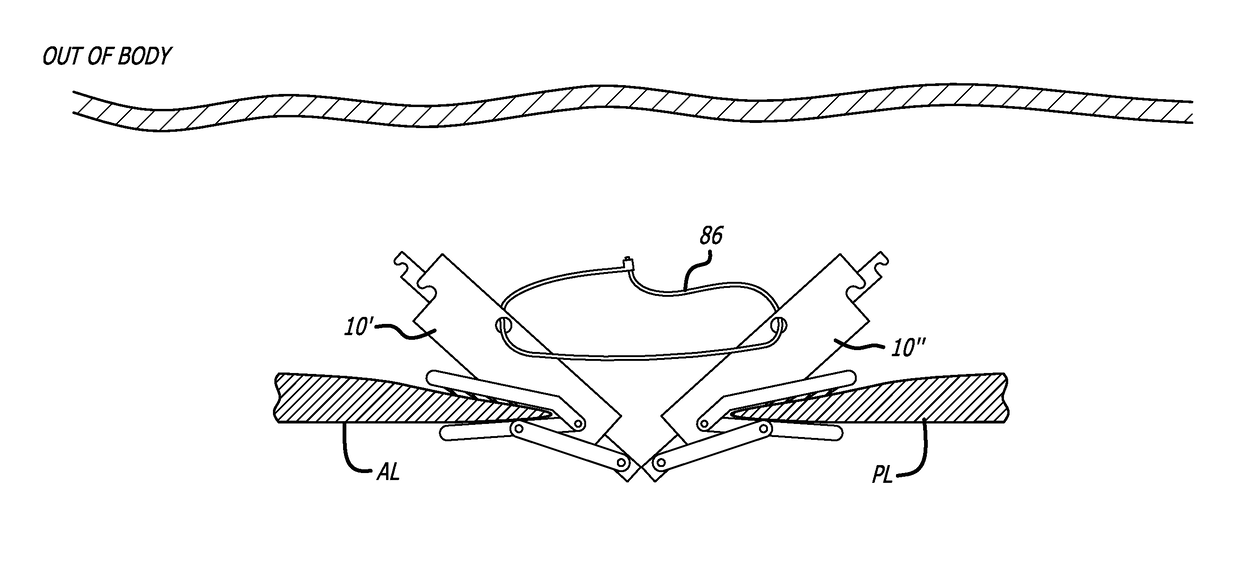
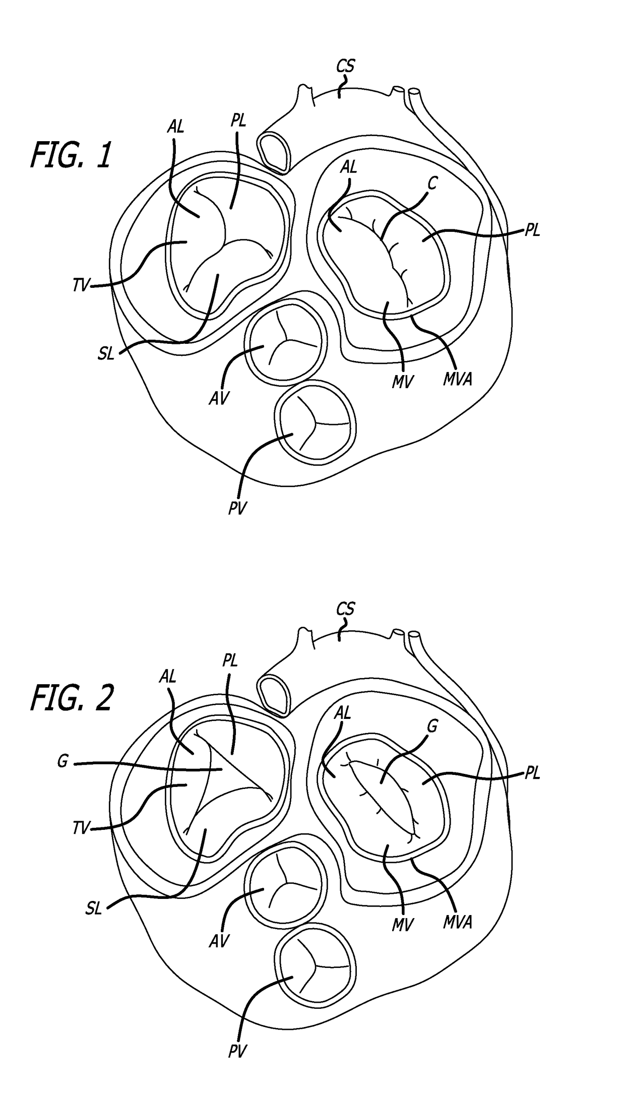
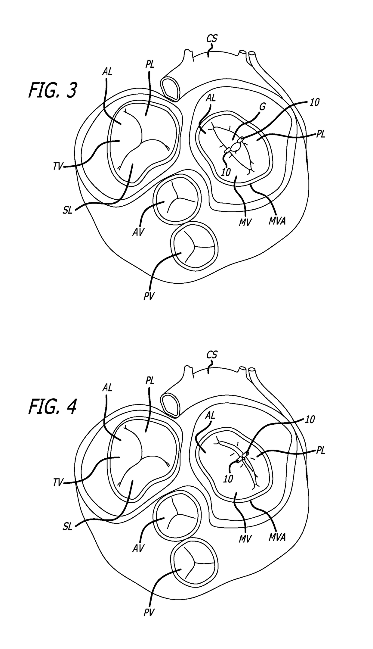
[0014]The devices, systems and methods of the present invention are centered on the use of at least two separate fixation devices which grasp and position different portions of target tissue in order to alter the physiological positioning of the target tissue. In such a manner, gaps formed between normally adjacent tissue structure can be reduced or eliminated by initially attaching a first fixation device to one of the targeted tissue and then attaching a second fixation to a second target tissue. In another aspect, additional fixation devices could be further utilized if additional target tissue is needed to be grasped. These two or more fixations devices are coupled to each other which allows the interventionalist to re-position the fixation devices relative to each other which, in turn, manipulates or re-positions the target tissue which are gripped or grasped by each fixation device. For example, the present invention can be used to reduce the size of a gap formed between the free ends of the posterior leaflet and anterior leaflet of the mitral valve to minimize regurgitation. Initially, a first fixation device is inserted into the left atrium and is actuated to physically grasp the free end of either the posterior or anterior leaflet of the mitral valve. Another fixation device is then advanced into the left atrium and is attached to the other leaflet. The two or more fixation devices have an associated coupling device / system which couples the fixation devices together and which can be used to move each fixation in closer proximity to one another in vivo. As a result, the target tissue can be drawn closer to each other allowing the valve leaflets to better coapted using a modified “edge-to-edge” or “bow-tie” technique. Accordingly, any space or gap formed between the damaged leaflets of the heart valve can be reduced causing the damaged valve to function more like a normal valve which reduces unwanted regurgitation.
[0015]In these preferred embodiments, the present invention allows each leaflet to be grasped individually, eliminating the need to capture both leaflets simultaneously using a single fixation device, as is presently used in systems employing “edge-to-edge” or “bow-tie” techniques. Additionally, the use of individual fixation devices to grasp the target tissue allows the interventionist to grasp the target tissue at a location which might not directly across from the other target tissue. In other words, the target tissue to be grasped by each fixation device need not be located directly across from the other target tissue, which is usually required when utilizing a single fixation clip system. For example, one fixation device could be used to grasp target tissue located some distance away from the other target tissue. Since the fixation devices remain coupled to each other, the associated coupling device / system of the present invention allows the fixation devices to be drawn closer together after being initially attached to the respective target tissue irrespective of the fixation devices' initial location. Accordingly, the devices can be moved or drawn towards each other allowing the target tissue to be simultaneously drawn closer together thus reducing the size of any gap formed between the leaflets of the valve. These features are particularly useful when repairing a tricuspid valve since it is often difficult to grasp all three leaflets simultaneously using a single fixation device, as is currently being implemented in conventional repair systems.
[0017]In an exemplary embodiment, the invention provides a fixation device including a stud and a pair of gripping elements (first and second gripping elements) for engaging tissue which are operatively coupled to the stud, each gripping element having a first end and a free end opposite the first end. The first and second gripping elements are moveable between a first open position wherein the free ends of the gripping elements are spaced apart and a second closed position wherein the free ends of the gripping elements are spaced close together to engage or “grip” the tissue. Preferably, the gripping elements are sufficiently spaced apart in the first open position to allow the target tissue to be positioned between the first and second gripping elements and are then brought much closer together to engage or grip the tissue. The gripping elements are movable to positions between the first open position and the second closed position to allow the gripping elements to grip tissue having different thicknesses. In this regard, the ability of the gripping elements to move to various positions allows the space between the gripping elements to be varied which allows the gripping elements to grasp tissue having different thicknesses.
[0020]The gripping elements may be configured to provide high retention force so that the fixation device remains securely fastened to the target tissue throughout the cardiac cycle. At the same time, these gripping elements should be configured to minimize trauma to the tissue engaged by them. This allows the fixation device to be removed from the tissue after initial application without creating clinically significant injury to the tissue. In order to enhance retention without creating significant trauma, the gripping elements may have friction-enhancing features on their surfaces that engage the target tissue. Such friction-enhancing features may include barbs, bumps, grooves, openings, channels, surface roughening, coverings, and coatings, among others. Preferably the friction-enhancing features and the magnets will be configured to increase the retention force of the gripping elements on the target tissue, while not leaving significant injury or scarring if the fixation device is removed. The gripping elements may further have a shape and flexibility to maximize retention force and minimize trauma to the target tissue. The gripping elements may also be somewhat flexible so that they deflect to some degree in response to forces against the tissue engaged thereby, reducing the chances that the tissue will tear or bruise in response to such forces.
[0021]The fixation devices of the invention preferably are associated with a coupling device / system which allows any number of fixation devices to be coupled to each other at the site of the target tissue. In this regard, the coupling device / system of the present invention not only couples the various fixation devices together, but also provides a device / system for drawing the fixation devices closer to each other once the device is attached to the target tissue. Again, such a coupling device / system allows multiple fixation devices to be utilized, eliminating the need for the interventionalist to grasp all of the target tissue simultaneously with a single fixation device. The present invention further allows fixation devices to be attached to non-adjacent target tissue since the coupling device / system can be used to draw each fixation device in close proximity to each other.

Problems solved by technology

Regurgitation can significantly decrease the pumping efficiency of the heart, placing the patient at risk of severe, progressive heart failure.
Mitral valve regurgitation can result from a number of different mechanical defects in the mitral valve or the left ventricular wall.
The valve leaflets, the valve chordae which connect the leaflets to the papillary muscles, the papillary muscles or the left ventricular wall may be damaged or otherwise dysfunctional.
Commonly, the valve annulus may be damaged, dilated, or weakened, limiting the ability of free ends of the leaflets of the mitral valve to close adequately against each other due to the high fluid pressures that can develop in the left ventricle during systole.
The need to both open the chest and place the patient on bypass is traumatic and has associated high mortality and morbidity.
However, valves that are heavily calcified or significantly compromised by disease may need to be replaced.
Tricuspid regurgitation, like mitral regurgitation, is typically caused by defective coaptation of the leaflets of the valve against each other and results in reduced pumping efficiency.
Methods for repairing the mitral valve normally do not apply conveniently to a method for repairing the tricuspid valve.
The mechanical problems involved in grasping all three leaflets simultaneously at a single point are far more complex than with the mitral valve because the operator is not presented with two elongated edges to grasp, but rather, with three triangulated points that must be grasped simultaneously.

Method used


Image

  • Fixation devices, systems and methods for heart valve leaf repair
  • Fixation devices, systems and methods for heart valve leaf repair
  • Fixation devices, systems and methods for heart valve leaf repair

Examples


Embodiment Construction

I. Cardiac Physiology

[0054]The human heart has four chambers, namely, the left atrium, the left ventricle, the right atrium and the right ventricle. The right atrium and the right ventricle together are sometimes referred to as the right heart. Similarly, the left atrium and the left ventricle together are sometimes referred to as the left heart. The left side of the heart provides the pumping action which supplies oxygenated blood to the arteries. The right side of the heart receives spent blood from the veins and pumps such blood to the lungs for oxygenation. The left atrium receives the oxygenated blood from the lungs via one of the four pulmonary veins. Blood from the left atrium is initially pumped into the chamber of the left ventricle which then pumps blood to the arteries. The left atrium is connected to the left ventricle by the mitral valve which is configured as a “check valve” which prevents back flow when pressure in the left ventricle is higher than that in the left at...


PUM


Abstract

Devices, systems and methods for repairing a valve in a patient's heart includes a two or more fixations devices, each fixation device being attachable to the free end of a target tissue, such as a valve leaflet, the fixation devices being coupled together by a coupling device capable of repositioning and coupling the fixation devices in order to re-position the free ends of the valve leaflets.

Description

BACKGROUND[0001]The present invention relates generally to medical methods, devices, and systems for the endovascular, percutaneous or minimally invasive surgical treatment of bodily tissues, such as tissue approximation or valve repair. More particularly, the present invention relates to repair of valves of the heart and venous valves.[0002]Surgical repair of bodily tissues often involves tissue approximation and fastening of such tissues in an approximated arrangement. Tissue approximation includes coapting the leaflets of the valves in a therapeutic arrangement which may then be maintained by fastening or fixing the leaflets via the use of an implanted fixation device. Such coaptation can be used to treat regurgitation which most commonly occurs in the mitral valve and tricuspid valve of the human heart.[0003]During normal operation of the heart, the valve leaflets of the mitral valve open during diastole when the left atrium of the heart is filled with blood allowing the blood t...

Claims


Application Information

Patent Timeline
Patent Type & Authority Applications(United States)
IPC IPC(8): A61F2/24
CPCA61F2/2442A61B17/00234A61F2/2463A61B17/122A61B17/1285A61B17/29A61B2017/00243A61B2017/00292A61B2017/2946A61B17/0401A61B2017/0464A61B2017/0417A61B2017/0409
Inventor KIZUKA, KOJI J.CHONG, MICHAEL Z.
Owner ABBOTT CARDIOVASCULAR
Why Eureka
Try Eureka