
Innervation Of Engineered Structures

Inactive Publication Date: 2018-09-13
WAKE FOREST UNIV HEALTH SCI INC
View PDF1 Cites 3 Cited by
  • Summary
  • Abstract
  • Description
  • Claims
  • Application Information

AI Technical Summary

Benefits of technology

The patent text discusses the discovery of neural progenitor cells in both the central nervous system and enteric nervous system of rodents and humans. These cells can differentiate into various mature enteric neuronal subtypes and may provide a potential source of autologous neuronal cells for re-engineering the gut's nerve cells. The text also describes recent advances in cell culture techniques for isolating these progenitor cells and the successful differentiation of them into enteric phenotype. Additionally, the text mentions the reliable and reproducible isolation of enteric neuronal progenitor cells from adult human gut up to 84 years of age.

Problems solved by technology

The regeneration of stratified smooth muscle layers with the proper orientation, however, remains a challenge.
Moreover, restoration of functional motility was not demonstrated in prior studies, highlighting the biggest challenge yet in functional tissue engineering of the GI neuromusculature.
Reconstruction of the stomach by tissue engineering is also a challenge.
The major problem encountered with these grafts, however, was shrinkage.
Unfortunately, these techniques did not regenerate the enteric neuronal layers, and the smooth muscle cells demonstrated a phenotypic switch to their non-contractile forms.
Tissue engineered small intestinal constructs, likewise, have not achieved the alignment of the smooth muscle cells or their innervation that appears to be crucial to generating appropriate force and motility to facilitate nutrient absorption.
Disruption of colonic motility alters transit time, resulting in constipation or diarrhea.
They demonstrated that the tissue engineered conduits have significant absorptive capacity when implanted into animals, but there was no direct measurement of peristalsis or motility.
Although significant advances have been made in tissue engineering of phasic neuromuscular structures, many gaps exist in proposed techniques for regeneration of functional smooth muscle and enteric neuronal plexuses.

Method used


Image

  • Innervation Of Engineered Structures
  • Innervation Of Engineered Structures
  • Innervation Of Engineered Structures

Examples


example 1

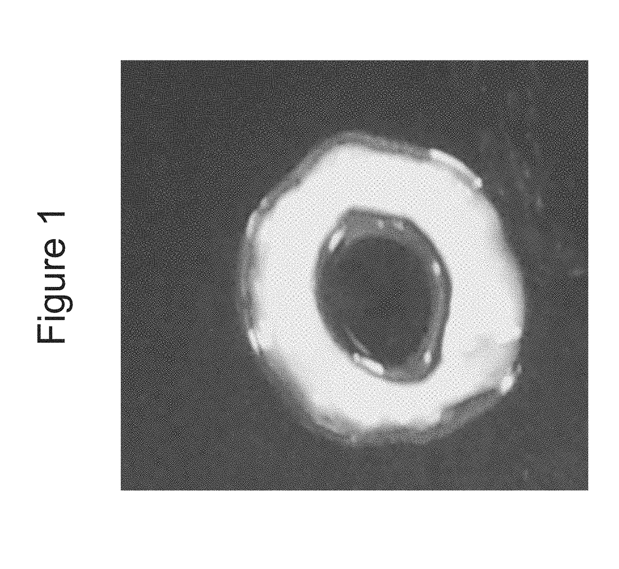
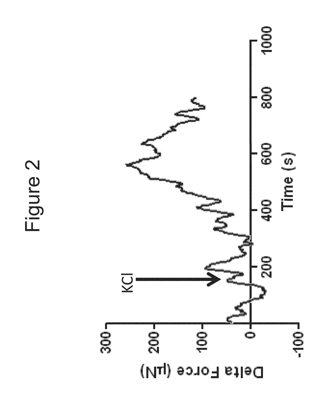
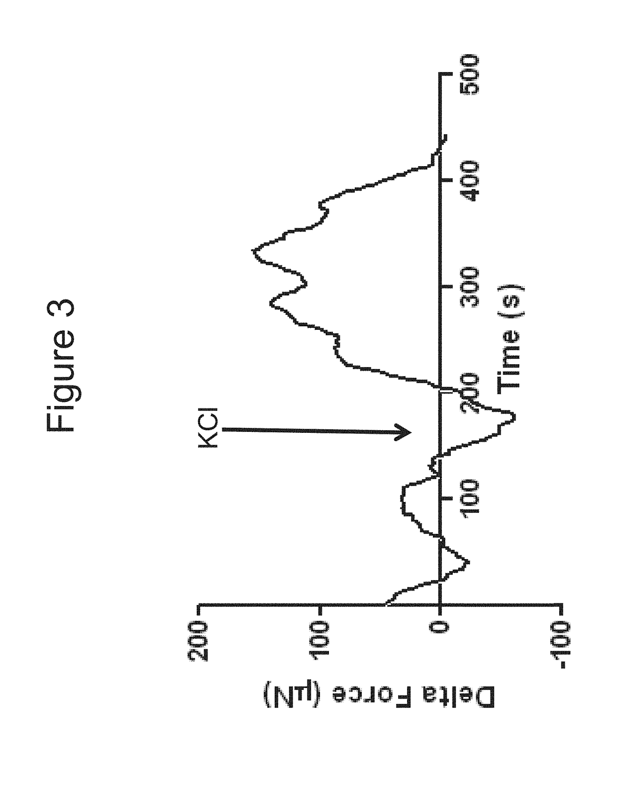
[0104]Bioengineered innervated and non-innervated internal anal sphincter (IAS) constructs were made using autologous rabbit IAS smooth muscle and enteric neuronal progenitor cells. After 4 days in culture, the constructs were placed around a biodegradable composite chitosan tubular scaffold. A non-innervated muscle construct (lacking neuronal cells) was placed abutting an innervated construct (smooth muscle cells with neuronal cells) on one side. Another non-innervated muscle construct was placed 1 mm away from the innervated construct on the other side. Physiological functionality of the constructs was assessed in vitro.

[0105]Positive NADPH Diaphorase staining of the bioengineered innervated construct demonstrated the presence of nitrergic neurons.

[0106]Microscopic images showed cellular processes bridging the gap between the innervated and the non-innervated construct as early as day 4 after placing the constructs on the scaffold. At day 10, a network of differentiated neurons wa...

example 2

[0109]In this example, intrinsically innervated three-dimensional rabbit colon constructs were bioengineered and characterized.

[0110]Smooth muscle cells were trypsinized and neurospheres were accutased. 500 k smooth muscle cells and 200 k enteric neuronal progenitor cells were centrifuged to form a cell pellet.

[0111]The enteric neuronal progenitor cells were resuspended in a 0.4 mg / ml collagen (type I rat tail) and laminin (5 ug / ml) solution. The solution was pipetted onto Sylgard coated 35 mm dishes around a central post. When placed in the 37 C incubator, solution gelled in 15-30 minutes.

[0112]Smooth muscle cell pellet was resuspended in a 0.4 mg / ml collagen solution and pipetted over the first gel layer and returned to the incubator.

[0113]After 2-4 hours, gels were released from the edge of the plate using a sterile 22 g needle.

[0114]1 ml of Neuronal Differentiation Medium (Neurobasal A+B27) was added and the plates were returned to the 37 C / 7% CO2 incubator for differentiation.

[...


PUM

PropertyMeasurementUnit
Structureaaaaaaaaaa

Abstract

Methods of generating an innervated muscle structures are disclosed as well as bioengineered structures for tissue repair or regeneration. The methods can include the steps of obtaining populations of smooth muscle cells and neuronal progenitor cells and then seeding the cells together onto a matrix material, followed by culturing the seeded cells to form an innervated smooth muscle cell construct of directionally oriented smooth muscle cells. In one embodiment, the neuronal progenitor cells can be seeded first as neurospheres in a biocompatiable solution, e.g., a collagen / laminin solution, and allowed to gel. Next, a second suspension of smooth muscle cells can be deposited as separate layer. Multiple layer structures of alternating muscle or neuron composition can also be formed in this manner. Differentiation of the neuronal progenitor cells can be induced by exposure to a differentiation medium, such as Neurobasal A medium and / or exposure to a differentiating agent, such as B-27 supplement. The innervated muscle structures can be disposed around a tubular scaffold, e.g., a chitosan-containing tube and further cultured to form tubular, bioengineered structures and two or more innervated muscle structures can be joined together to form an elongate composite structure.

Description

REFERENCE TO RELATED APPLICATIONS[0001]This application is based upon and claims the priority of U.S. Provisional Patent Application No. 61 / 592,890, entitled “Innervation of Engineered Structures, and U.S. Provisional Patent Application No. 61 / 592,871 filed Jan. 31, 2012, entitled “Tubular Bioengineered Smooth Muscle Structures,” which are both hereby incorporated in its entirety by reference.GOVERNMENT SUPPORT[0002]This invention was made with government support under grants NIH RO1DK071614 and NIH RO1DK042876 awarded by The National Institute of Health. The United States Government has certain rights in the invention.FIELD OF THE INVENTION[0003]The present invention concerns tissue engineering and in particular, innervation of tissue engineered structures.BACKGROUND OF THE INVENTION[0004]Innervation is extremely important for maintaining the functionality of almost every part of the human body. Innervation of the gastrointestinal (GI) tract is extremely important for smooth muscle...

Claims


Application Information

Patent Timeline
IPC IPC(8): A61K35/34A61F2/08C12N5/077A61L27/20A61L27/38C12N5/00A61F2/04A61L27/50C12N5/0797
CPCA61F2002/045A61F2/04C12N5/0623A61K35/34A61F2/08C12N5/0068A61L2430/30A61L27/50A61L27/3873A61L27/3826C12N2533/72C12N2533/54C12N5/0661A61L27/20C12N2501/999A61K35/30A61L27/225A61L27/227A61L27/24A61L27/383A61L27/3886C12N5/0697C12N2502/081C12N2502/1347C08L5/08
Inventor BITAR, KHALIL
Owner WAKE FOREST UNIV HEALTH SCI INC
Learn More