Blood Preparation and Profiling

a blood preparation and profiling technology, applied in the field of haematology, can solve the problems of adversely affecting the accuracy of known protein profiling and inadequate current analyses, and achieve the effects of increasing the ability to detect proteins, increasing the detection level, and being easy to d

Active Publication Date: 2018-10-25
SANGUI BIO PTY LTD
View PDF0 Cites 6 Cited by
  • Summary
  • Abstract
  • Description
  • Claims
  • Application Information

AI Technical Summary

Benefits of technology

[0008]The present inventors have surprisingly identified that RBCs, a major component of the blood that is routinely excluded from blood protein profile analyses seeking to determine blood protein profiles, are nonetheless a source of a number of different proteins (e.g., cytokines, chemokines, growth factors) at substantial levels. Moreover, the present inventors have found that the levels of various proteins detected within individual blood compartments (e.g., red blood cells, plasma, leukocytes) may differ significantly depending on the manner in which the blood and / or the components are collected and processed. Thus, the present inventors have created a new and useful laboratory technique for producing a protein profile of an enriched red blood cell sample by evaluating the presence or level of proteins newly-identified in RBCs. The new and useful laboratory technique is a significant improvement over current techniques for producing a protein profile from blood, increasing the ability to detect proteins (e.g., increasing the level of detection). Proteins are more easily detected in an enriched RBC sample due to their higher concentration in RBCs, a unique process to further increase their detectable levels in RBCs, and RBC isolation methods requiring less cell processing. Moreover, the high concentration of proteins in RBCs allows them to be detected in a small volume of blood. Without being bound to theory, it is postulated that RBCs may act as a reservoir for various proteins (e.g., cytokines, chemokines, and growth factors), and the manner in which blood is processed may influence the release and / or sequestration of these proteins by RBCs. This in turn may alter and / or enhance the protein profile of blood and / or other blood compartment(s).

Problems solved by technology

RBCs are routinely removed prior to conducting protein analyses on plasma / serum and / or white blood cells (leukocytes) because they may complicate processing / assaying and / or are not considered to provide a significant contribution to the overall protein profile of the blood.
Accordingly, current analyses are inadequate in the case where RBCs make a substantial contribution and / or otherwise influence the protein profile of blood.
It has also become evident that inconsistencies in blood collection techniques and / or the way in which blood is processed may adversely affect the accuracy of known protein profiling.

Method used


Image

  • Blood Preparation and Profiling
  • Blood Preparation and Profiling
  • Blood Preparation and Profiling

Examples


example 1

ting the Presence of the MIF Receptor on the Surface of RBCs

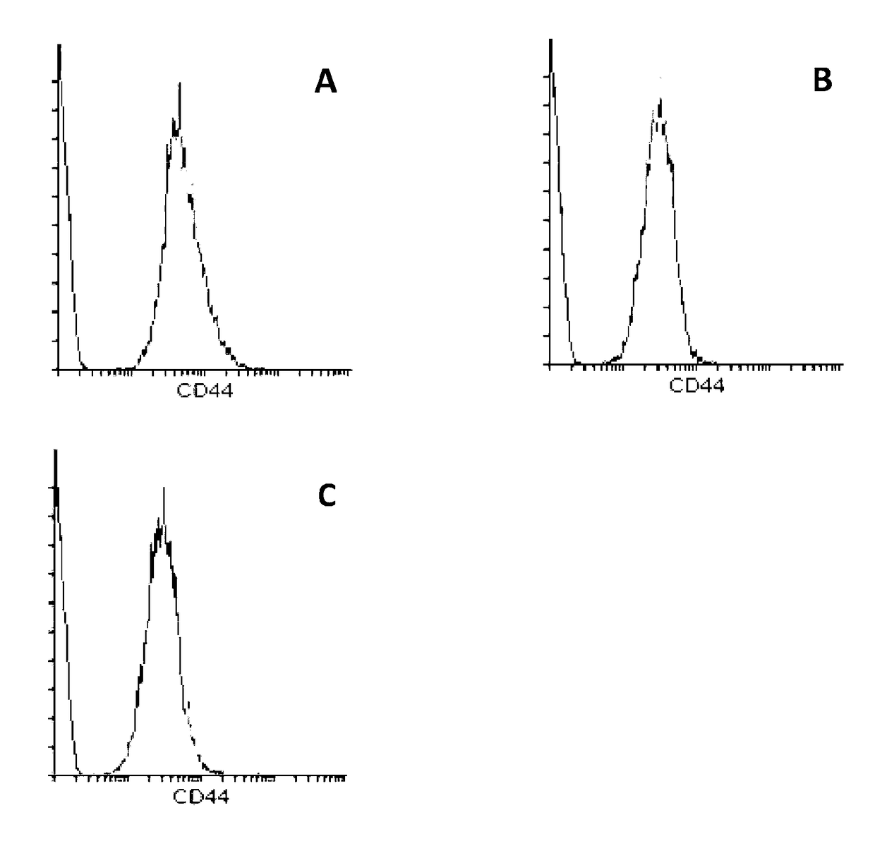
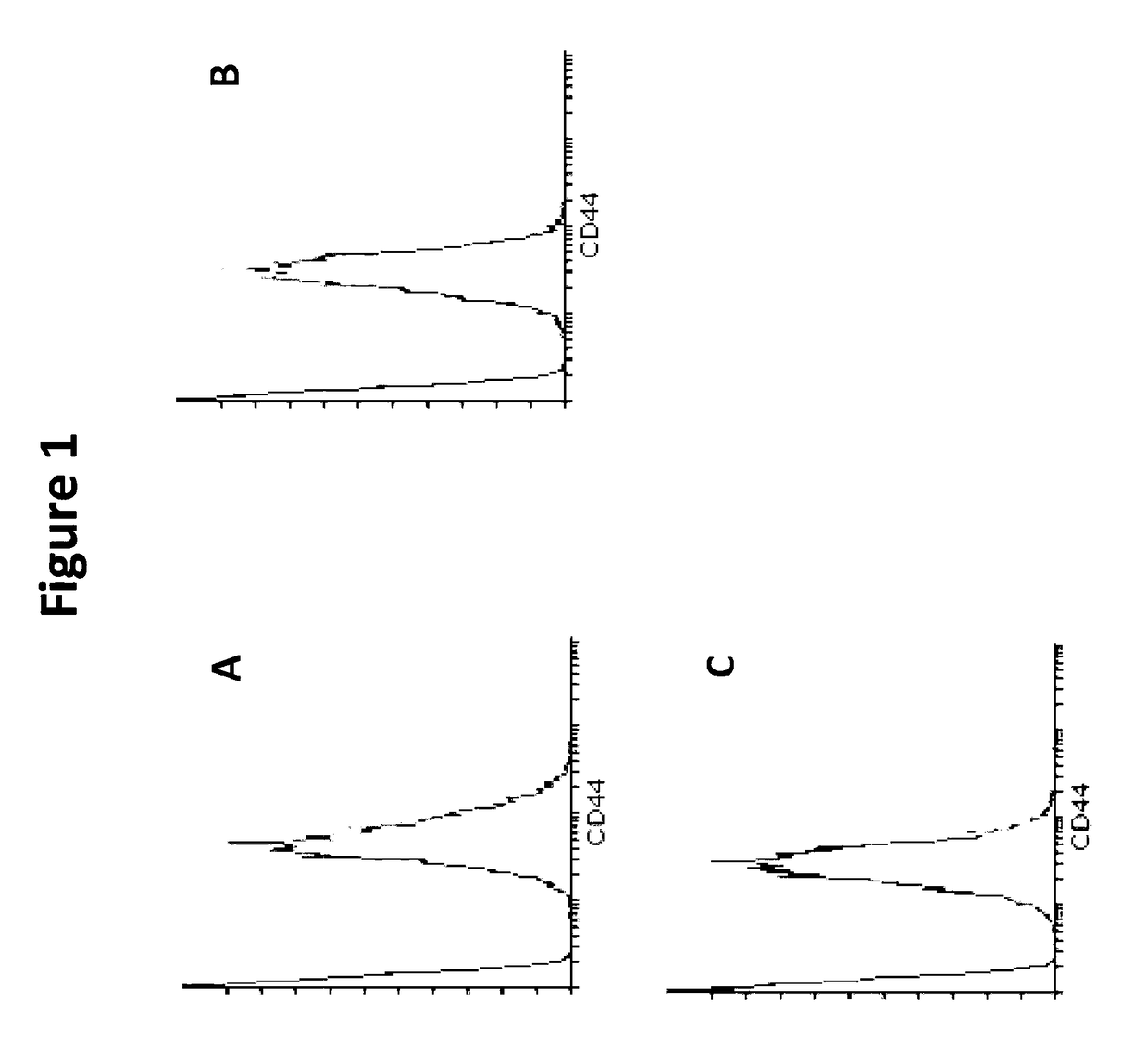
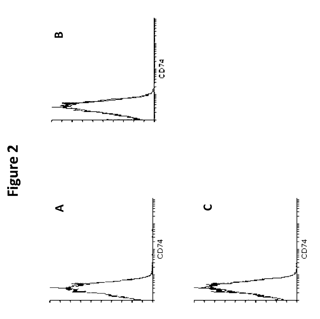
[0209]RBCs contain MIF. To determine whether RBCs have a receptor to bind MIF they were analyzed for the most well-known receptor for MIF, a complex formed between CD44 and CD74 on the cell surface. Immunophenotyping was used to identify the absence or presence of CD44 and CD74 on RBCs. Whole blood (WB) was collected into an EDTA vacutainer, washed twice in PBS+FBS (2%) (centrifuged at 500 g, for 5 minutes) and finally resuspended to 1 mL of solution. 5 uL of solution was added to each respective tube. Cells were then stained with 5 μL or 20 μL, of each antibody (anti-CD44, anti-CD74, anti-CD45, and an IgG control) and 50 μL of PBS+FBS (2%). Cell solutions were incubated in the dark at room temperature for 15 minutes and after incubation, cells were washed three times in 1×PBS (500 g, 5 minutes). Cells were resuspended in 200 uL of PBS and were analysed by flow cytometry using quadrant statistics and overlay histograms

[0210...

example 2

ization in RBCs

[0211]RBCs were analyzed for the localization of MIF using immunocytochemistry. Whole blood was collected into a syringe and washed twice in 1×PBS (spun in a centrifuge at 500 g, for 5 minutes). The cell pellet was the used to prepare blood smears (10 μL per slide) and the blood slides left to dry at room temperature for at least 2 hours. Once the slides were completely dried, the smears were fixed in 100% methanol for 5 minutes at room temperature. After the slides were fixed, they were removed from the methanol and were left to air dry for ˜10 minutes. Slides were washed thoroughly with 1×PBS and blood smears were then blot dried. The smears were blocked with PBS+5% BSA for 1 hour at room temperature. After blocking, the samples were incubated with or without primary MIF antibody (anti-human, rabbit MIF antibody, 1 μg / mL) overnight at 4° C. After incubation, slides were washed well with 1×PBS and then incubated with secondary antibody (anti-rabbit, AP conjugated ant...

example 3

ting the Presence of MIF in RBCs

[0216]Because MIF levels were ˜1000 times higher in RBCs than in plasma, RBCs were analyzed for their intracellular levels of MIF. Whole blood was collected from the capillary bed (fingertip collection). Blood was anti-coagulated (1:1) with EDTA (30 mg / mL) solution and washed twice with PBS+FBS (2%). Cells were stained with CD45-FITC (5 μL) for 15 min at room temperature in the dark. After incubation, cells were washed twice with 1×PBS and RBCs were then isolated using negative sorting by flow cytometry (FACS). RBCs and WB were pelleted by centrifugation (2000 g, 10 minutes) and were resuspended to 250,000 cells / 50 μL. Cells were frozen at −80° C., and samples were subjected to 3 freeze / thaw cycles to lyse all the cells. Samples were then analysed on a Hu MIF ELISA. For fingertip and venous blood, whole blood was collected from either the capillary bed (fingertip collection) or from a vein. Blood was anti-coagulated (1:1) with EDTA (30 mg / mL) solution...


PUM

PropertyMeasurementUnit
volumeaaaaaaaaaa
volumeaaaaaaaaaa
volumeaaaaaaaaaa

Abstract

The present disclosure relates to methods for generating blood protein profiles in red blood cell-enriched blood samples. The disclosed methods represent a new and improved laboratory technique for producing a protein profile from blood, increasing protein detection.

Description

TECHNICAL FIELD[0001]The present disclosure relates generally to the field of haematology. The present disclosure also relates to protein profiling in the blood and, in particular, methods for producing or generating blood protein profiles, including cytokine and / or chemokine profiles, in red blood cell-enriched blood samples.CROSS REFERENCE[0002]This applications claims priority to and is related to Australian Application No. 2015904075 entitled “Blood Preparation and Profiling” filed on 7 Oct. 2015. This application is incorporated herein by reference in its entirety. In addition, the other references or publications referred to in the present disclosure are also hereby incorporated by reference in their entirety.BACKGROUND[0003]Protein profiling of blood is used for a variety of purposes. For example, the profiling of indicative proteins in peripheral blood mononuclear cells (PBMC) and serum / plasma is commonly used in disease diagnosis. Additionally, monitoring protein profiles w...

Claims


Application Information

Patent Timeline
Patent Type & Authority Applications(United States)
IPC IPC(8): G01N33/80G01N33/574
CPCG01N33/80G01N33/57484G01N2800/368G01N33/56972G01N33/6863G01N33/6869G01N33/6866G01N33/6893G01N33/581G01N33/57488G01N2333/525G01N2333/495G01N2333/57G01N2333/5421G01N2333/5412G01N2333/5406G01N2333/5409G01N2333/5428G01N2333/545G01N2333/5434G01N33/49G01N33/5002G01N2570/00
Inventor KARSTEN, ELISABETHHERBERT, BENLIDDLE, ALANHILL, CAMERON
Owner SANGUI BIO PTY LTD
Why Eureka
Try Eureka