

Magnetic resonance apparatus and method for vascular imaging
a magnetic resonance and vascular imaging technology, applied in the field of vascular imaging, can solve the problems of t2*-based signal loss, prolonging the acquisition time, double the scanning time and the need for an approach, etc., and achieves good detection ability, good contrast of mr images, and high accuracy.
- Summary
- Abstract
- Description
- Claims
- Application Information
AI Technical Summary
Benefits of technology
Problems solved by technology
Method used
Image

Examples
Embodiment Construction
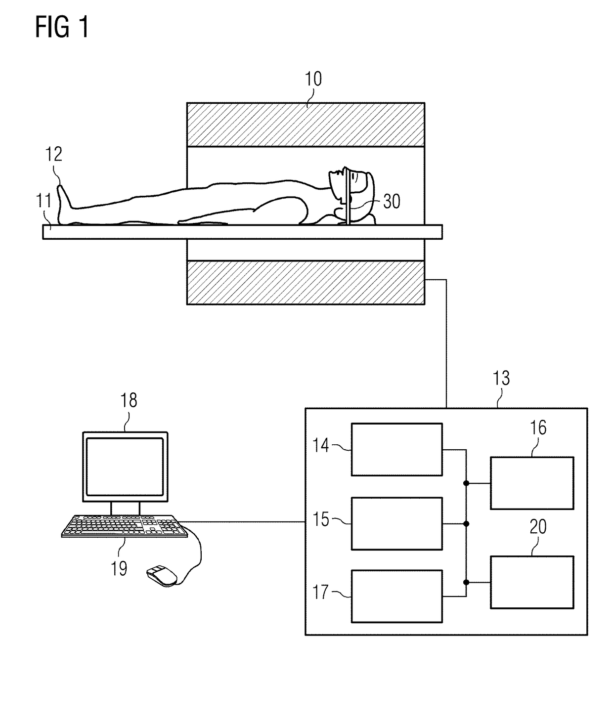
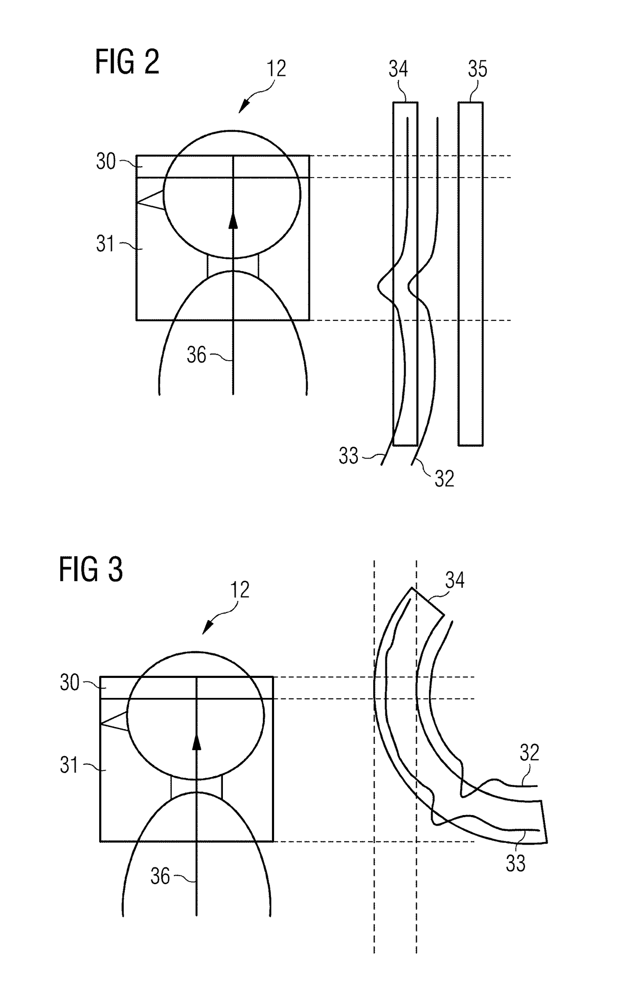
[0042]The present invention relates to a method of vascular imaging with the use of an MR system. With reference to FIG. 2, in a vascular imaging based on the TOF technique, a fixed magnetization, in particular of brain tissue, spinal fluid and fat, which generate an undesirable background signal in an imaging volume 30, is saturated by an RF pulse 40 with simultaneously applied inventive magnetic field. The RF pulse 40 and the applied magnetic field are designed so as to suppress the background signals without exciting the spins of the vessel fluid flowing into the imaging volume.
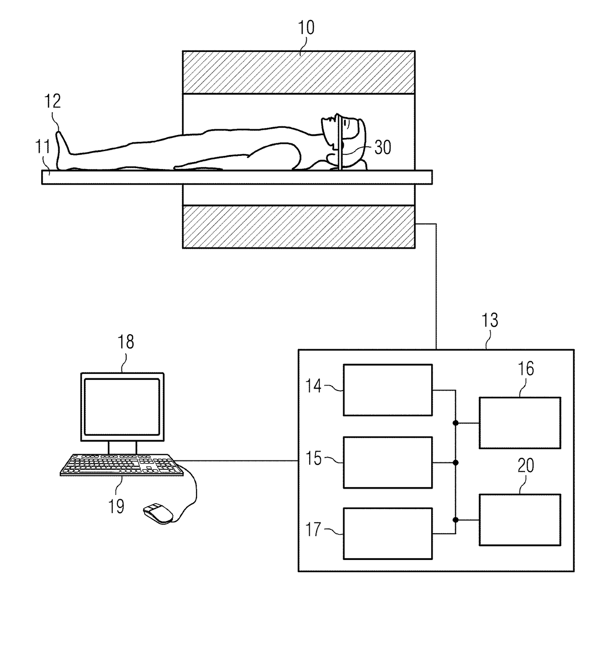
[0043]FIG. 1 schematically shows an MR apparatus with which such a method of vascular imaging can be inventively carried out.
[0044]An examination person 12, or, more generally, an examination object, is moved into the tunnel of the an MR data acquisition scanner 10 that has a magnet that generates a basic field B0, with the examination person 12 on a bed 11 being moved into the center of the scanner 10 in ...
PUM
 Abstract
Description
Claims
Application Information